

Enhancing surgical planning for abdominal tumors in children through advanced 3D visualization techniques: a systematic review of future prospects
- PMID: 38863523
- PMCID: PMC11166126
- DOI: 10.3389/fped.2024.1386280
Enhancing surgical planning for abdominal tumors in children through advanced 3D visualization techniques: a systematic review of future prospects
Abstract
Introduction: Preoperative three-dimensional (3D) reconstruction using sectional imaging is increasingly used in challenging pediatric cases to aid in surgical planning. Many case series have described various teams' experiences, discussing feasibility and realism, while emphasizing the technological potential for children. Nonetheless, general knowledge on this topic remains limited compared to the broader research landscape. The aim of this review was to explore the current devices and new opportunities provided by preoperative Computed Tomography (CT) scans or Magnetic Resonance Imaging (MRI).
Methods: A systematic review was conducted to screen pediatric cases of abdominal and pelvic tumors with preoperative 3D reconstruction published between 2000 and 2023.
Discussion: Surgical planning was facilitated through virtual reconstruction or 3D printing. Virtual reconstruction of complex tumors enables precise delineation of solid masses, formulation of dissection plans, and suggests dedicated vessel ligation, optimizing tissue preservation. Vascular mapping is particularly relevant for liver surgery, large neuroblastoma with imaging-defined risk factors (IDRFs), and tumors encasing major vessels, such as complex median retroperitoneal malignant masses. 3D printing can facilitate specific tissue preservation, now accessible with minimally invasive procedures like partial nephrectomy. The latest advancements enable neural plexus reconstruction to guide surgical nerve sparing, for example, hypogastric nerve modelling, typically adjacent to large pelvic tumors. New insights will soon incorporate nerve plexus images into anatomical segmentation reconstructions, facilitated by non-irradiating imaging modalities like MRI.
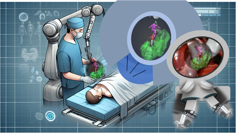
Conclusion: Although not yet published in pediatric surgical procedures, the next anticipated advancement is augmented reality, enhancing real-time intraoperative guidance: the surgeon will use a robotic console overlaying functional and anatomical data onto a magnified surgical field, enhancing robotic precision in confined spaces.
Keywords: augmented reality; minimally invasive surgery; pediatric oncology; three-dimensional printing; virtual reality.
© 2024 Lopez, Belgacem, Sarnacki, Arnaud, Houari, Piguet, Baudouin, Fourcade, Lauvray and Ballouhey.
Conflict of interest statement
The authors declare that the research was conducted in the absence of any commercial or financial relationships that could be construed as a potential conflict of interest.
Figures


Similar articles
-
Virtual Reality for Preoperative Planning and Education in Pediatric Surgery: Preliminary Results for the Treatment of Congenital Malformations and Tumors.World J Surg. 2025 Jun;49(6):1497-1507. doi: 10.1002/wjs.12594. Epub 2025 Apr 17. World J Surg. 2025. PMID: 40246557 Free PMC article.
-
Exploring the Potential of Three-Dimensional Imaging, Printing, and Modeling in Pediatric Surgical Oncology: A New Era of Precision Surgery.Children (Basel). 2023 May 3;10(5):832. doi: 10.3390/children10050832. Children (Basel). 2023. PMID: 37238380 Free PMC article.
-
Minimally invasive and invasive liver surgery based on augmented reality training: a review of the literature.J Robot Surg. 2023 Jun;17(3):753-763. doi: 10.1007/s11701-022-01499-2. Epub 2022 Nov 28. J Robot Surg. 2023. PMID: 36441418 Review.
-
Three-dimensional Printing and Augmented Reality: Enhanced Precision for Robotic Assisted Partial Nephrectomy.Urology. 2018 Jun;116:227-228. doi: 10.1016/j.urology.2017.12.038. Epub 2018 May 22. Urology. 2018. PMID: 29801927
-
Virtual and augmented reality systems and three-dimensional printing of the renal model-novel trends to guide preoperative planning for renal cancer.Asian J Urol. 2024 Oct;11(4):521-529. doi: 10.1016/j.ajur.2023.10.004. Epub 2024 Mar 11. Asian J Urol. 2024. PMID: 39534007 Free PMC article. Review.
Cited by
-
Primary Adenocarcinoma of the Lesser Omentum: A Case Report.Clin Case Rep. 2025 Apr 8;13(4):e70408. doi: 10.1002/ccr3.70408. eCollection 2025 Apr. Clin Case Rep. 2025. PMID: 40206575 Free PMC article.
-
The Role of 3D Printing and Augmented Reality in the Management of Hepatic Malignancies.Technol Cancer Res Treat. 2025 Jan-Dec;24:15330338251323138. doi: 10.1177/15330338251323138. Technol Cancer Res Treat. 2025. PMID: 39980434 Free PMC article. Review.
References
Publication types
LinkOut - more resources
Full Text Sources