. 2024 Jun 11;6(2):lqae065.
doi: 10.1093/nargab/lqae065. eCollection 2024 Jun.

Critical cis-parameters influence STructure assisted RNA translation (START) initiation on non-AUG codons in eukaryotes

Affiliations

Critical cis-parameters influence STructure assisted RNA translation (START) initiation on non-AUG codons in eukaryotes

Antonin Tidu et al. NAR Genom Bioinform. .

Abstract

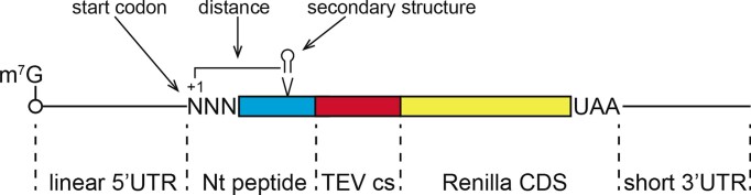
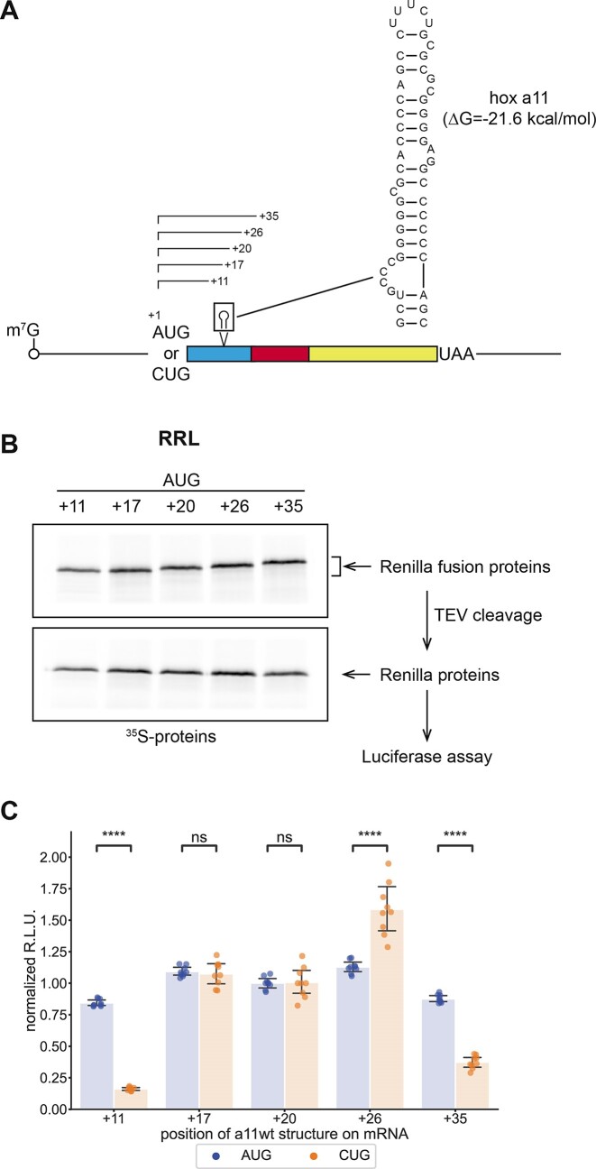
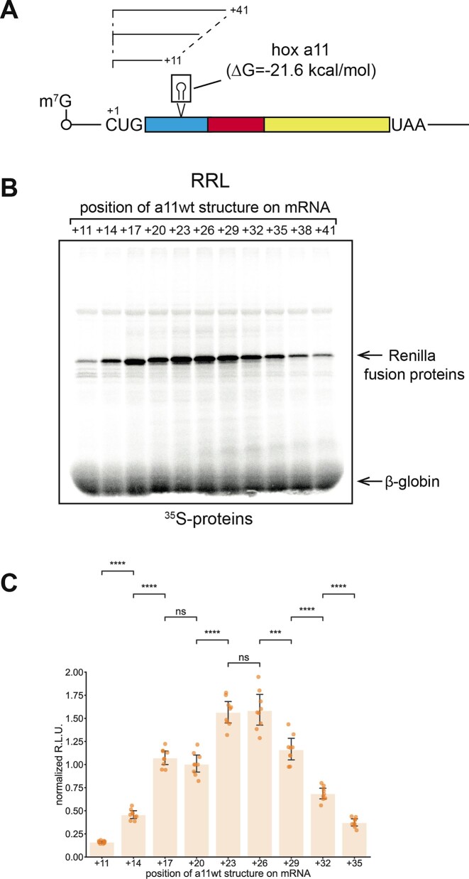
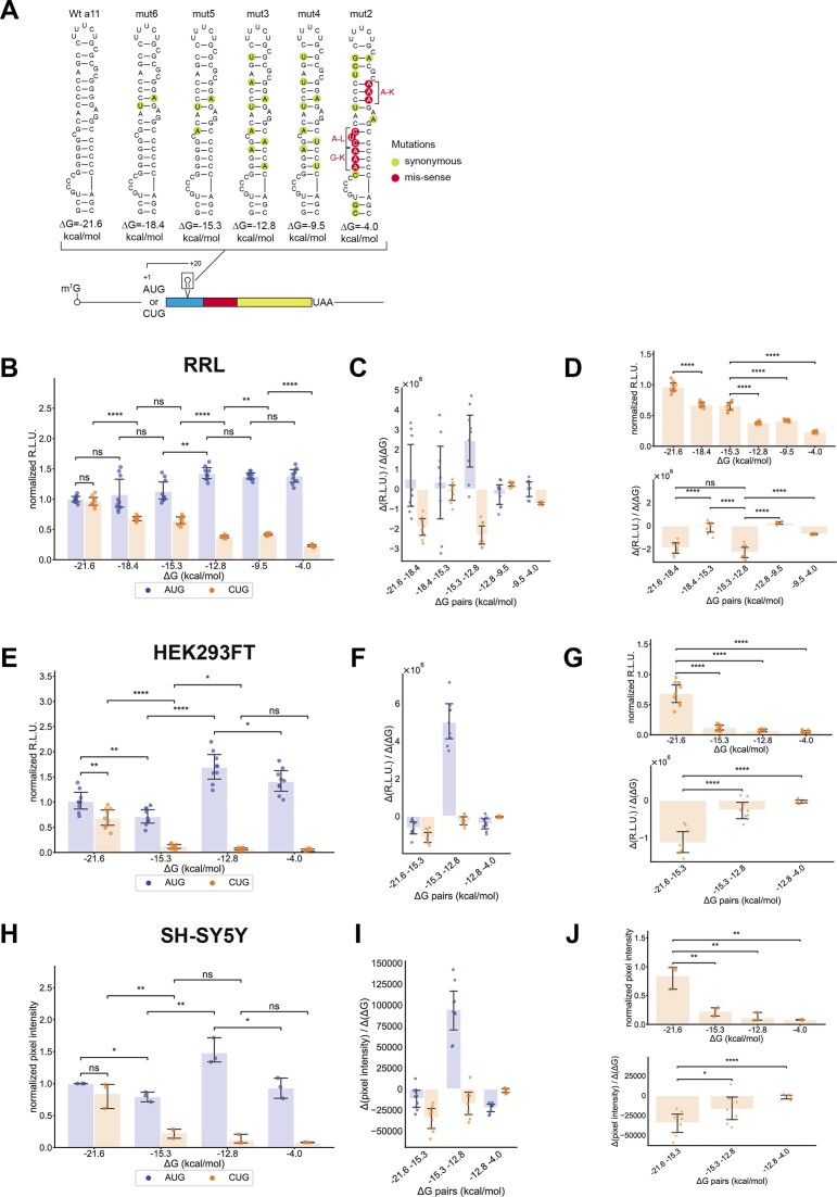
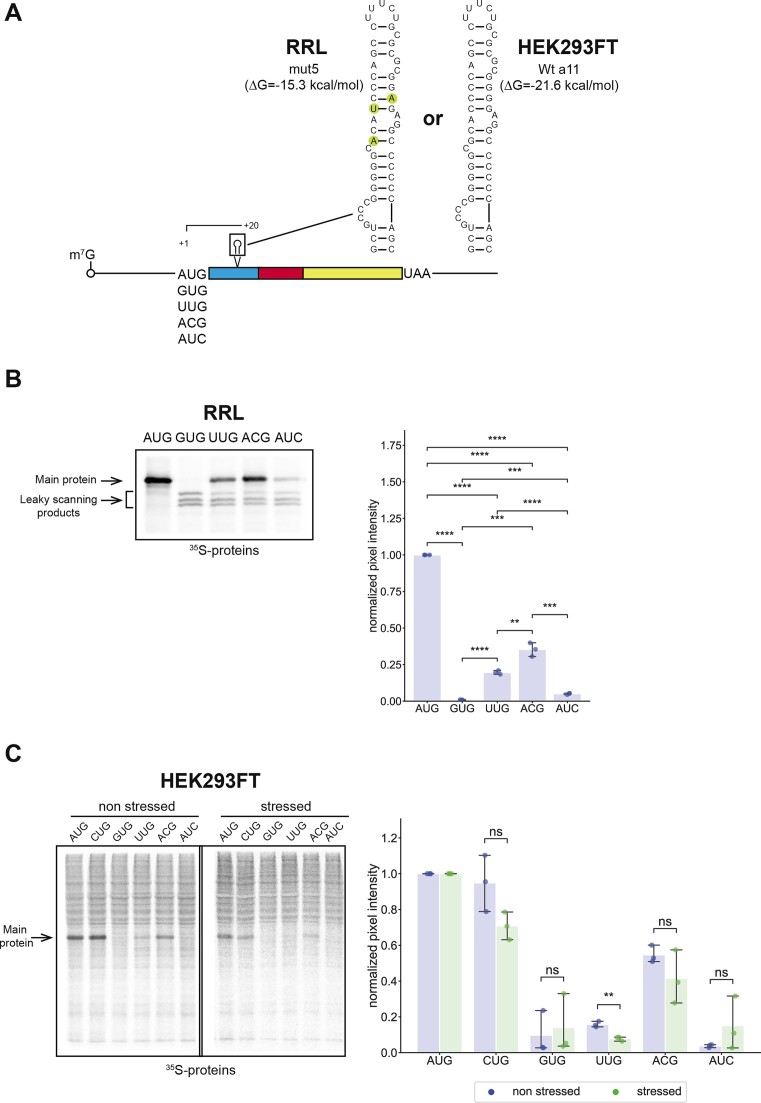
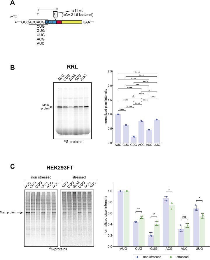
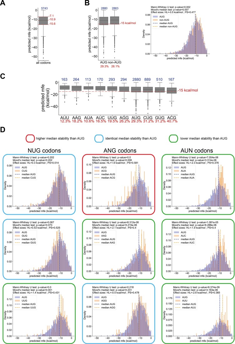
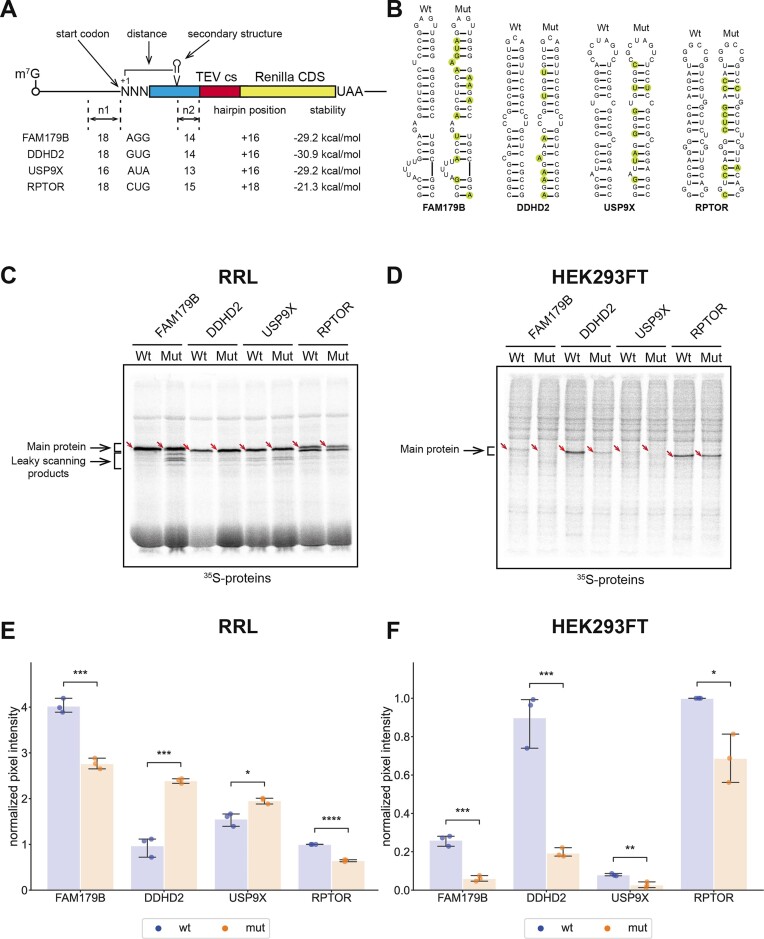
In eukaryotes, translation initiation is a highly regulated process, which combines cis-regulatory sequences located on the messenger RNA along with trans-acting factors like eukaryotic initiation factors (eIF). One critical step of translation initiation is the start codon recognition by the scanning 43S particle, which leads to ribosome assembly and protein synthesis. In this study, we investigated the involvement of secondary structures downstream the initiation codon in the so-called START (STructure-Assisted RNA translation) mechanism on AUG and non-AUG translation initiation. The results demonstrate that downstream secondary structures can efficiently promote non-AUG translation initiation if they are sufficiently stable to stall a scanning 43S particle and if they are located at an optimal distance from non-AUG codons to stabilize the codon-anticodon base pairing in the P site. The required stability of the downstream structure for efficient translation initiation varies in distinct cell types. We extended this study to genome-wide analysis of functionally characterized alternative translation initiation sites in Homo sapiens. This analysis revealed that about 25% of these sites have an optimally located downstream secondary structure of adequate stability which could elicit START, regardless of the start codon. We validated the impact of these structures on translation initiation for several selected uORFs.


Figures

Figure 1.
Figure 1.
Schematic representation of the RNA reporters used in the study. The three cis-acting parameters studied are (i) the distance between a downstream structure and the start codon, (ii) the stability of the downstream structure, (iii) the nature of the start codon. TEV cs: Tobacco Etch Virus protease cleavage site, Nt peptide: N-terminal fusion peptide, Renilla CDS: Renilla luciferase coding sequence.
Figure 2.
Figure 2.
Translation initiation on CUG is sensitive to the position of the downstream secondary structure. (A) Schematic representation of the RNA reporters highlighting the two AUG or CUG initiation codons used and the wild-type a11 hairpin, which is inserted at +11, +17, +20, +26 or +35 positions. (B) Representative SDS-PAGE of in vitro translation products obtained with AUG reporters in RRL. The upper panel shows the 35S-labelled products before TEV cleavage, the lower panel shows the 35S-labelled products after TEV cleavage. Proteins produced by TEV cleavage were used for Luciferase assays. (C) Quantification of Renilla luciferase luminescence produced in RRL from a11-RNA reporters initiating with an AUG codon (blue) or with a CUG codon (orange). Relative luminescence units (R.L.U.) were normalized to those obtained with the +20 AUG reporter for all AUG reporters or with the +20 CUG reporter for all CUG reporters. Bar heights show mean normalized ratios and error bars represent the corresponding 99% confidence interval using the t-distribution. P-values were calculated using a Student t-test for independent samples. *: 0.01 < P < 0.05, **: 0.001 < P < 0.01, ***: 0.0001 < P < 0.001, ****: P < 0.0001
Figure 3.
Figure 3.
The optimal position of the secondary structure for initiation on a CUG codon is between +23 to +26. (A) Schematic representation of the RNA reporters initiating on a CUG codon upstream of the wild-type a11 hairpin inserted at positions +11, +17, +20, +23, +26, +29, +32, +35, +38 or +41. (B) Representative SDS-PAGE of in vitro35S radiolabelled translation products obtained with AUG reporters in RRL. (C) Quantification of Renilla luciferase luminescence produced in RRL from a11 RNA reporters initiating with a CUG codon. Relative luminescence units (R.L.U.) were normalized to those obtained with the +20 CUG reporter. Bar heights show the mean normalized ratios and error bars show the 99% confidence intervals calculated with the t-distribution. P-values were calculated using a Student t-test for independent samples. *: 0.01 < P < 0.05, **: 0.001 < P < 0.01, ***: 0.0001 < P < 0.001, ****: P < 0.0001.
Figure 4.
Figure 4.
Secondary structure stability determines translation initiation efficiency in different cell extracts. (A) Schematic representation of the RNA reporter and sequences of the stem–loops used during the study. The initiation codons were AUG or CUG. Wild-type and mutant a11 hairpins were inserted at +20 position. Silent mutations introduced to lower the a11 structure stability are shown in green circles whereas missense mutations are shown in red circles. (B) Quantification of Renilla luciferase luminescence produced in RRL from variable a11 RNA reporters initiating with AUG or CUG codon. Relative luminescence units (R.L.U.) were normalized to those obtained with the reporter containing the wild-type a11 structure and initiating from an AUG codon. Bar heights show the mean normalized ratios and error bars show the 99% confidence intervals calculated with the t-distribution. P-values were calculated using a student t-test for independent samples. *: 0.01 < P < 0.05, **: 0.001 < P < 0.01, ***: 0.0001 < P < 0.001, ****: P < 0.0001. (C) The ratio Δ(R.L.U.)/Δ(ΔG) quantifies the effect of a ΔG variation (Δ(ΔG)) on translation efficiency for AUG and CUG codons in RRL. (D) Stability threshold determination for initiation on a CUG codon in RRL. The top graph corresponds to the luminescence data for the CUG codon from (B), and the lower graph corresponds to the data for the CUG codon from (C). The set of pairwise comparisons presented enable to define two stability thresholds for CUG initiation at –21.6 kcal/mol and –15.3 kcal/mol. P-values were calculated using a Student t-test for independent samples. *: 0.01 < P < 0.05, **: 0.001 < P < 0.01, ***: 0.0001 < P < 0.001, ****: P < 0.0001. (E) Quantification of Renilla luciferase luminescence produced in HEK293FT cell extracts from variable a11 RNA reporters initiating with AUG or CUG codon. Results are presented as in (B). (F) Effect calculation of a ΔG variation on translation efficiency for the AUG and CUG reporters as in (C) in HEK293FT cell extracts. (G) Stability threshold determination for initiation on a CUG codon in HEK293FT cell extracts as in (D). The set of pairwise comparisons presented enable to define the stability threshold for CUG initiation at –21.6 kcal/mol. (H) Quantification of 35S incorporation in Renilla luciferase proteins produced in SH-SY5Y cell extracts. Results are presented as in (B) and (E). (I) Effect calculation of a ΔG variation on translation efficiency for the AUG and CUG reporters as in (C) in SH-SY5Y cell extracts. (J) Stability threshold determination for initiation on a CUG codon in SH-SY5Y cell as in (D). The set of pairwise comparisons presented enable to define the stability threshold for CUG initiation at –21.6 kcal/mol.
Figure 5.
Figure 5.
Stable secondary structures do not allow the initiation of translation for all AUG-like codons. (A) Schematic representation of the RNA reporters and sequences of the stem-loops used during the study. The initiation codons are AUG, GUG, UUG, ACG or AUC codons upstream of the mut5 a11 hairpin for RRL and wt a11 hairpin for HEK293FT which are inserted at +20 position. The nucleotide context of the initiation codons is CGUAAUNNNGAC. (B) Representative SDS-PAGE of in vitro35S radiolabeled translation products obtained with RRL and quantification of pixel intensities of the corresponding gels. Pixel intensities were normalized to those obtained with the AUG reporter. Bar heights show the mean normalized ratios and error bars show the 99% confidence intervals calculated with the t-distribution. P-values were calculated using a Student t-test for independent samples. *: 0.01 < P < 0.05, **: 0.001 < P < 0.01, ***: 0.0001 < P < 0.001, ****: P < 0.0001. (C) Representative SDS-PAGE of in vitro35S radiolabeled translation products obtained with unstressed and stressed HEK293FT extracts and quantification of pixel intensities of the corresponding gels. Pixel intensities were normalized to those obtained with the AUG reporter for each extract separately and results are presented as in (B).
Figure 6.
Figure 6.
Changing the start-codon nucleotide context influences the efficiency of initiation rescue by START. (A) Schematic representation of the RNA reporters used in the study. The initiation codons are AUG, CUG, GUG, UUG, ACG or AUC codons upstream of wt a11 hairpin for RRL and HEK293FT which is inserted at +20. The nucleotide context of the tested initiation codons is GCCACCNNNGCG (boxed). (B) Representative SDS-PAGE of in vitro35S radiolabeled translation products obtained with RRL and quantification of pixel intensities of the corresponding gels. Pixel intensities were normalized to those obtained with the AUG reporter. Bar heights show the mean normalized ratios and error bars show the 99% confidence intervals calculated with the t-distribution. P-values were calculated using a Student t-test for independent samples. *: 0.01 < P < 0.05, **: 0.001 < P < 0.01, ***: 0.0001 < P < 0.001, ****: P < 0.0001. (C) Representative SDS-PAGE of in vitro35S radiolabeled translation products obtained with unstressed and stressed HEK293FT extracts and quantification of pixel intensities of the corresponding gels. Pixel intensities were normalized to those obtained with the AUG reporter for each extract separately and are presented as in (B).
Figure 7.
Figure 7.
Analysis of △G distributions of downstream secondary structures within the G (or mfe = minimum free energy) distributions of downstream secondary structures within +16/+65 for all codons (A), for AUG and non-AUG codons (B) and for each codon individually (C). Top blue numbers show the population size of each boxplot. Numbers in red show the percentage of smORFs starting with the corresponding start codon that contain a structure more stable than –15 kcal/mol (i.e. mfe inferior to –15 kcal/mol). Histograms in (B) and (D) show the density distributions corresponding to the boxplots. Histograms have a bin width of 0.5 kcal/mol, and the total area of each histogram is normalized to 1. The Mann–Whitney U test was used to determine if the AUG and the non-AUG distributions are equal. The Mood's median test was used to determine if the AUG and the non-AUG distributions have the same median. The probability of superiority (PS) and the Hodges–Lehmann estimator (HL) were used for effect size assessment (see Methods section).
Figure 8.
Figure 8.
In vitro translation of RNA reporters carrying secondary structures from four identified human smORFs using a non-AUG codon. (A) Schematic representation of the four RNA reporters FAM179B, DDHD2, USP9X and RPTOR. The stability of the respective structures is indicated as well as the number of nucleotides upstream of the start codon that originates from the human transcript (n1), the start codon, the position of the secondary structure, the predicted stability of the secondary structure and the number of nucleotides downstream the structure that originates from the human transcript (n2). (B) Representations of the secondary structures of each reporter studied. The silent mutations that were introduced to lower the structure stability are shown in green circles in the Mut stem–loops. (C) Representative SDS-PAGE of in vitro35S radiolabeled translation products obtained with FAM179B, DDHD2, USP9X, RPTOR wild-type and Mut reporters in RRL. The positions of the full-length fusion proteins are shown by red arrows. Lower bands correspond to alternative translation initiation sites resulting from a leaky scanning of the main start codon. In the case of the RPTOR reporter, there is an in-frame AUG at +34. (D) Same as (C) but in HEK293FT extracts. (E, F) Quantification of 35S incorporation in Renilla luciferase proteins produced in RRL and HEK293FT extracts. Bar heights show the mean values and error bars show the 99% confidence intervals calculated with the t-distribution. P-values were calculated using a Student t-test for independent samples. *: 0.01 < P < 0.05, **: 0.001 < P < 0.01, ***: 0.0001 < P < 0.001, ****: P < 0.0001.

Similar articles

Cited by

References

    1. Tidu A., Martin F. The interplay between cis- and trans-acting factors drives selective mRNA translation initiation in eukaryotes. Biochimie. 2023; 217:20–30. - PubMed
    1. Rogers G.W., Richter N.J., Lima W.F., Merrick W.C. Modulation of the helicase activity of eIF4A by eIF4B, eIF4H, and eIF4F. J. Biol. Chem. 2001; 276:30914–30922. - PubMed
    1. Schütz P., Bumann M., Oberholzer A.E., Bieniossek C., Trachsel H., Altmann M., Baumann U. Crystal structure of the yeast eIF4A-eIF4G complex: an RNA-helicase controlled by protein–protein interactions. Proc. Natl. Acad. Sci. U.S.A. 2008; 105:9564–9569. - PMC - PubMed
    1. Passmore L.A., Schmeing T.M., Maag D., Applefield D.J., Acker M.G., Algire M.A., Lorsch J.R., Ramakrishnan V. The eukaryotic translation initiation factors eIF1 and eIF1A induce an open conformation of the 40S ribosome. Mol. Cell. 2007; 26:41–50. - PubMed
    1. Valášek L., Nielsen K.H., Zhang F., Fekete C.A., Hinnebusch A.G. Interactions of eukaryotic translation initiation factor 3 (eIF3) subunit NIP1/c with eIF1 and eIF5 promote preinitiation complex assembly and regulate start codon selection. Mol. Cell. Biol. 2004; 24:9437–9455. - PMC - PubMed