

Primary cervicothoracic melanoma of spinal cord: a case report and literature review
- PMID: 38863638
- PMCID: PMC11165122
- DOI: 10.3389/fonc.2024.1417268
Primary cervicothoracic melanoma of spinal cord: a case report and literature review
Abstract
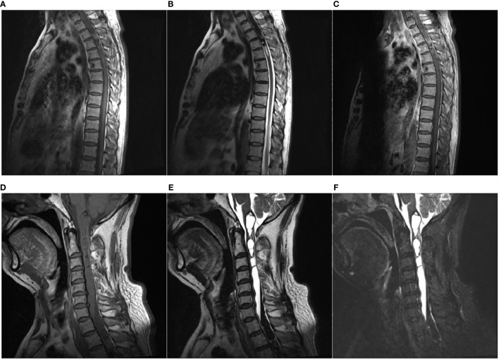
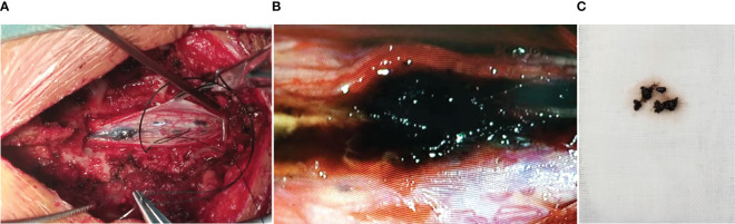
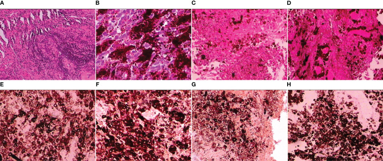
A 53-year-old male patient presented progressive numbness and weakness in the right limbs for a 2-year duration. Magnetic resonance imaging scans revealed an intramedullary lesion crossed over cervical and thoracic levels accompanied by syringomyelia at the proximal end of the lesion. The patient underwent subtotal resection of the neoplasm. The histological findings of the tumor were consistent with primary intramedullary malignant melanoma and not initial ependymoma after careful dermatologic and ophthalmologic re-examination. Primary melanoma of the spinal cord, particularly cervicothoracic localization with syringomyelia, is seldom reported in the literature. We report a case of this uncommon tumor and also discuss the clinical course, diagnosis, and treatment.
Keywords: CSEP; DSEP; cervicothoracic; electromyogram; primary; spinal cord.
Copyright © 2024 Dang, Du, Wei and Xue.
Conflict of interest statement
The authors declare that the research was conducted in the absence of any commercial or financial relationships that could be construed as a potential conflict of interest.
Figures
Similar articles
-
Primary intramedullary melanoma of lumbar spinal cord: A case report.World J Clin Cases. 2021 Apr 6;9(10):2352-2356. doi: 10.12998/wjcc.v9.i10.2352. World J Clin Cases. 2021. PMID: 33869613 Free PMC article.
-
Presurgical role of MRI tractography in a case of extensive cervicothoracic spinal ependymoma.Surg Neurol Int. 2017 Apr 26;8:56. doi: 10.4103/sni.sni_33_17. eCollection 2017. Surg Neurol Int. 2017. PMID: 28540122 Free PMC article.
-
Intramedullary clear cell ependymoma in the cervical spinal cord: case report.Neurosurgery. 2000 Dec;47(6):1434-7; discussion 1437-8. Neurosurgery. 2000. PMID: 11126915 Review.
-
Ten-Segment Intramedullary Ependymoma and Whole Spinal Syringomyelia.World Neurosurg. 2020 Jul;139:20-22. doi: 10.1016/j.wneu.2020.03.149. Epub 2020 Apr 3. World Neurosurg. 2020. PMID: 32251824
-
Pediatric intramedullary schwannoma with syringomyelia: a case report and literature review.BMC Pediatr. 2018 Nov 28;18(1):374. doi: 10.1186/s12887-018-1341-2. BMC Pediatr. 2018. PMID: 30486806 Free PMC article. Review.
Cited by
-
Primary spinal meningeal melanoma with intramedullary and intradural extramedullary components-a case report.BJR Case Rep. 2025 Mar 25;11(2):uaaf020. doi: 10.1093/bjrcr/uaaf020. eCollection 2025 Mar. BJR Case Rep. 2025. PMID: 40161969 Free PMC article.
-
Beyond surgical radicality in intramedullary spinal cord metastases: neurological function and systemic disease burden drive patient outcomes.J Neurooncol. 2025 Jun 30. doi: 10.1007/s11060-025-05119-5. Online ahead of print. J Neurooncol. 2025. PMID: 40587076
-
Intramedullary primary spinal cord melanoma: illustrative case.J Neurosurg Case Lessons. 2025 Mar 10;9(10):CASE24732. doi: 10.3171/CASE24732. Print 2025 Mar 10. J Neurosurg Case Lessons. 2025. PMID: 40063999 Free PMC article.
References
Publication types
LinkOut - more resources
Full Text Sources