. 2024 May 28:15:1260439.
doi: 10.3389/fimmu.2024.1260439. eCollection 2024.

Dengue virus exploits autophagy vesicles and secretory pathways to promote transmission by human dendritic cells

Affiliations

Dengue virus exploits autophagy vesicles and secretory pathways to promote transmission by human dendritic cells

Alexandra P M Cloherty et al. Front Immunol. .

Abstract

Dengue virus (DENV), transmitted by infected mosquitoes, is a major public health concern, with approximately half the world's population at risk for infection. Recent decades have increasing incidence of dengue-associated disease alongside growing frequency of outbreaks. Although promising progress has been made in anti-DENV immunizations, post-infection treatment remains limited to non-specific supportive treatments. Development of antiviral therapeutics is thus required to limit DENV dissemination in humans and to help control the severity of outbreaks. Dendritic cells (DCs) are amongst the first cells to encounter DENV upon injection into the human skin mucosa, and thereafter promote systemic viral dissemination to additional human target cells. Autophagy is a vesicle trafficking pathway involving the formation of cytosolic autophagosomes, and recent reports have highlighted the extensive manipulation of autophagy by flaviviruses, including DENV, for viral replication. However, the temporal profiling and function of autophagy activity in DENV infection and transmission by human primary DCs remains poorly understood. Herein, we demonstrate that mechanisms of autophagosome formation and extracellular vesicle (EV) release have a pro-viral role in DC-mediated DENV transmission. We show that DENV exploits early-stage canonical autophagy to establish infection in primary human DCs. DENV replication enhanced autophagosome formation in primary human DCs, and intrinsically-heightened autophagosome biogenesis correlated with relatively higher rates of DC susceptibility to DENV. Furthermore, our data suggest that viral replication intermediates co-localize with autophagosomes, while productive DENV infection introduces a block at the late degradative stages of autophagy in infected DCs but not in uninfected bystander cells. Notably, we identify for the first time that approximately one-fourth of DC-derived CD9/CD81/CD63+ EVs co-express canonical autophagy marker LC3, and demonstrate that DC-derived EV populations are an alternative, cell-free mechanism by which DCs promote DENV transmission to additional target sites. Taken together, our study highlights intersections between autophagy and secretory pathways during viral infection, and puts forward autophagosome accumulation and viral RNA-laden EVs as host determinants of DC-mediated DENV infection in humans. Host-directed therapeutics targeting autophagy and exocytosis pathways thus have potential to enhance DC-driven resistance to DENV acquisition and thereby limit viral dissemination by initial human target cells following mosquito-to-human transmission of DENV.

Keywords: autophagy; dendritic cells; dengue virus; extracellular vesicles; host-directed antivirals; secretory autophagy; viral evasion; viral transmission.


Conflict of interest statement

The authors declare that the research was conducted in the absence of any commercial or financial relationships that could be construed as a potential conflict of interest.

Figures
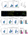
Figure 1
Figure 1
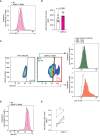
Dengue virus misuses host autophagy machinery to establish infection in human DCs. (A-C) Primary human DCs were infected with DENV-2/16681, with or without treatment of SDM25N replication inhibitor (10 µM) for 48 h. Representative flow cytometry plots (A) and quantification (B) of viral infection, determined by intracellular NS3 staining. Closed circles represent the mean of n=6 donors measured in duplicate; *P < 0.05, student’s t-test. (C) Confocal microscopy analyses of primary human DCs infected with DENV-2/16681 determined by staining for viral dsRNA (green), F-actin (yellow) and Nuclei (blue). Scale bar = 20 micron, Representative of n = 3. (D, E, G, H) Viral infection of DCs upon transfection with siATG5 (D, E) or siATG16L1 (G, H), or non-targeting control siRNA as control (D, E, G, H), followed by exposure to DENV-2/16681 for 48 h. Representative flow cytometry plots (D, G) and quantification of DENV-2 infection (E, H), determined by intracellular NS3 staining. (F, I) ATG5 (F) or ATG16L1 (I) silencing efficiency was determined by real-time PCR. mRNA expression was normalised to GAPDH and set at 1 in cells transfected with control siRNA. (E, F, H, I) Closed circles represent the mean of n=4-5 donors measured in duplicate; *P < 0.05, **P < 0.01, one-sample t-test.
Figure 2
Figure 2
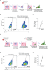
Productive infection of DCs results in increased autophagosome formation and colocalization of DENV replication intermediates with LC3+ autophagy vesicles. (A-D) Confocal microscopy analyses of primary human DCs infected with DENV-2/16681, or mock infected, for 48h, determined by staining for nuclei (blue), F-actin (yellow) LC3 (red), and viral dsRNA (green). Scale bar = 20 micron. (A) Representative confocal images. (B) Analysis of LC3 fluorescence; closed circles represent mean grey value across 10 fields of view for n = 3 donors. (C) Histograms of LC3 and viral dsRNA fluorescence intensities across three regions of interest (ROI) indicated in (A). (D) Pearson’s correlation coefficient for colocalization analyses on 10 ROIs per donor between viral dsRNA and LC3, n = 3 donors. (A-C) Data are representative of n = 3 donors.
Figure 3
Figure 3
Autophagy flux impairment in DENV-infected DCs. (A-C) Autophagosome levels in primary human DCs infected with DENV-2/16681 for 48 h. Representative flow cytometry plots (A) and quantification (B), determined by intracellular LC3 staining. Closed circles represent the mean of n = 6 independent donors measured in duplicate. (C) Autophagosomes levels in dengue-infected (NS3+) versus bystander (NS3-) DCs, using a concurrent intracellular NS3 and LC3 staining. Representative flow cytometry plots for n=2 donors measured in duplicate. (D, E) Intracellular p62 levels in primary human DCs infected with DENV-2/16681 for 48h. Representative flow cytometry plots (D) and quantification (E), determined by intracellular p62 staining. Closed circles represent the mean of n=5 donors measured in duplicate; *P < 0.05, student’s t-test.
Figure 4
Figure 4
Intrinsically higher number of autophagosomes correlates with increased dengue virus infection of primary human DCs. (A) ATG16L1 rs6861 (CC, blue, versus TT, red) genotyped blood-derived CD11c+ human DCs (52) were treated with bafilomycin A1 (50 nM) for 4 h prior to quantification of intracellular autophagosomes by multiparameter flow cytometry with intracellular LC3 staining combined with immune cell surface markers. DCs were defined as CD3-CD19-CD14-CD16-CD56-CD11c+ cells. Closed circles indicate quantification of intracellular LC3-II levels in n=4 donors for each ATG16L1 genotype, shown as LC3-II accumulation in bafilomycin-treated cells relative to untreated cells of the same genotype. (B, C) Primary human DCs were infected with DENV-2/16681 for 48 h, and viral infection was quantified by flow cytometric analysis of intracellular NS3 staining. (B) Data represent mean % NS3+ cells across n=11 donors measured in duplicate. (C) Differentiation of viral infection levels in genotyped DCs: ATG16L1 rs6861(CC) donors (blue-coloured circles) versus ATG16L1 rs6861(TT) donors (red coloured circles); closed circles represent the mean of n = 11 donors measured in duplicate. *P < 0.05, **P < 0.01, student’s t-test.
Figure 5
Figure 5
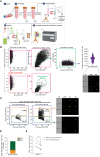
Cell-depleted supernatant derived from infected DCs mediate DENV transmission to target cells. (A) Graphical representation of the experimental strategy utilized to determine dengue virus transmission via cell-cell close contact in (B, C). (B, C) Primary human DCs were infected with DENV-2/16681, or mock infected, for 36 h. DCs were then extensively washed to remove input virus, replated for 30 h, then rigorously washed and co-cultured with the susceptible Vero cell line for 48 h. DENV-2/16681 transmission by DCs to Vero was assessed in DC-Vero co-culture, determined by intracellular NS3 staining by flow cytometer. DC marker CD11c was used to exclude DCs from analysis. Data are representative flow cytometry plots (B) and quantification (C) of n=3 donors measured in duplicate, represented by open squares. (D) Graphical representation of the experimental strategy utilized to determine dengue virus transmission via DC-derived cell-free supernatant in (E, F). (E, F) DCs were infected with DENV-2/16681, or mock infected, for 36 h. DCs were then extensively washed to remove input virus, and replated for 30 h. CM was then harvested, centrifuged to discard remaining DCs and cellular debris, and DC-free CM was transferred to Vero cell line culture. DENV-2/16681 transmission by DC-derived CM was assessed in DC-free Vero co-culture for 48 h, determined by intracellular NS3 staining by flow cytometer. Data are representative flow cytometry plots (E) and quantification (F) of n=3 individual donors, represented by open squares.
Figure 6
Figure 6
EVs derived from infected DCs facilitate dengue virus transmission. (A) Graphical representation of the experimental strategy utilized to determine dengue virus transmission via DC-derived EVs in (B, C). DCs were infected with DENV-2/16681, or mock infected, for 36 h. DCs were then extensively washed to remove input virus, and replated for 30 h. CM was then harvested, centrifuged to remove remaining DCs and cellular debris, and thereafter either left intact, or subjected to positive immunomagnetic depletion of CD9/CD81/CD63+ EVs, or incubated with magnetic beads only. Subsequently, DC-derived complete CM, or EV-depleted CM, or beads only incubated-CM was co-cultured for 48h with the Vero cell line. DENV-2/16681 transmission to Vero cells was determined by intracellular NS3 staining and flow cytometry analysis. (B, C) Data are representative flow cytometry plots (B) and quantification (C) of n=4 donors, represented by closed circles. ns, non-significant; *P < 0.05, ANOVA.
Figure 7
Figure 7
Extracellular autophagy vesicles released by human DCs support dengue virus transmission. (A) Graphical representation of the experimental protocol and rationale utilized for phenotypic characterization and quantification of DC-derived CD9/CD81/CD63+ singlet EVs using imaging flow cytometry, as presented in (B-D). Following overnight culture of DCs in FCS-depleted culture medium supplemented with bafilomycin A1 (100 nM), conditioned medium (CM) was collected and pre-cleared by serial centrifugation. EVs were immunomagnetically isolated using a pan-extracellular vesicle positive selection kit, and thereafter stained and analysed by imaging flow cytometry (62). General membrane labelling was performed using carboxyfluorescein succinimidyl ester (CFSE; 300 µM). Samples were additionally stained with anti-LC3 antibody, or left only CFSE-stained as a control, and thereafter EVs were washed twice more to remove residual dye and antibody prior to imaging flow cytometry analysis. (B) Gating strategy for analyses of DC-derived EVs, following immunomagnetic isolation and membrane labelling as outlined in (A). To reduce swarming or coincident event detection (“large events”) during individual EV analysis, EV samples were serially diluted in PBS to determine an operational range for analysis by imaging flow cytometry, at which event rate increase was proportional to sample dilution [89].To confirm selection for membrane-bound particles, sub-samples were lysed with 1% Triton X-100 for 60 minutes (63, 64). Closed circles represent the total number of CD9/CD81/CD63+ CFSE+ singlet EVs measured from n=6 donors. (C) Representative imaging flow cytometry plots and (D) quantification of a subset of DC-derived CD9/CD81/CD63+ CFSE+ singlet EVs that co-express LC3, determined by immunostaining followed by imaging flow cytometry analysis. (D) Data represent the percentage of LC3+ (orange) versus LC3- (green) CD9/CD81/CD63+ CFSE+ singlet EVs measured in samples derived from n=6 DC donors. (E) DCs were infected with DENV-2/16681 for 36 h. DCs were then extensively washed to remove input virus, and replated for 30 h. CM was then harvested, centrifuged to remove remaining DCs and cellular debris, and thereafter either subjected to positive immunomagnetic depletion of LC3+ EVs (LC3+ EVs-depleted CM), or incubated with magnetic beads only (beads only incubated-CM). Subsequently, each CM sample was co-cultured for 48h with the Vero cell line and DENV-2/16681 transmission to Vero cells was determined by intracellular NS3 staining and flow cytometry analysis. n = 4 donors, *P < 0.05, Paired t-test.

References

    1. World Health Organization . Dengue and severe dengue factsheet(2022). Available at: https://www.who.int/news-room/fact-sheets/detail/dengue-and-severe-dengue.
    1. Dengue vaccine. WHO position paper, September 2018 - recommendations(2018). Available at: https://www.who.int/publications/i/item/who-wer9335-457-476. - PubMed
    1. Ivanescu LM, Bodale I, Grigore-Hristodorescu S, Martinescu G, Andronic B, Matiut S, et al. . The risk of emerging of dengue fever in Romania, in the context of global warming. Trop Med Infect Dis. (2023) 8:65. doi: 10.3390/tropicalmed8010065 - DOI - PMC - PubMed
    1. Cochet A, Calba C, Jourdain F, Grard G, Durand GA, Guinard A, et al. . Autochthonous dengue in mainland France, 2022: geographical extension and incidence increase. Euro Surveill.. (2022) 27(44):pii=2200818. doi: 10.2807/1560-7917.ES.2022.27.44.2200818 - DOI - PMC - PubMed
    1. Bhatt P, Sabeena SP, Varma M, Arunkumar G. Current understanding of the pathogenesis of dengue virus infection. Curr Microbiol. (2021) 78:17–32. doi: 10.1007/s00284-020-02284-w - DOI - PMC - PubMed