Review
. 2024 May 28:15:1380063.
doi: 10.3389/fimmu.2024.1380063. eCollection 2024.

Emergence of the brain-border immune niches and their contribution to the development of neurodegenerative diseases

Affiliations
Review

Emergence of the brain-border immune niches and their contribution to the development of neurodegenerative diseases

Li Yang Tan et al. Front Immunol. .

Abstract

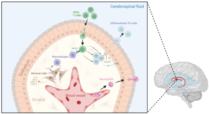
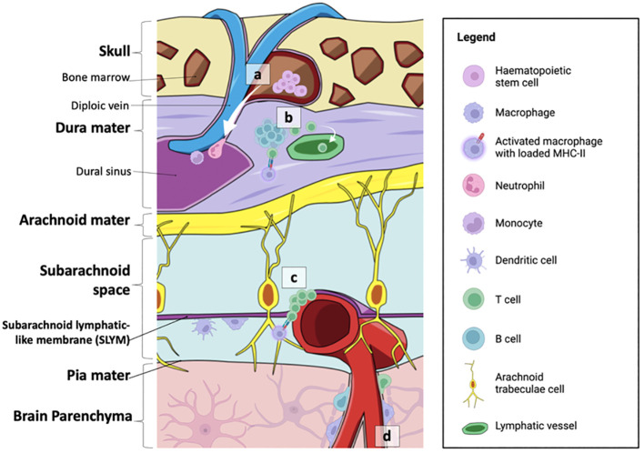
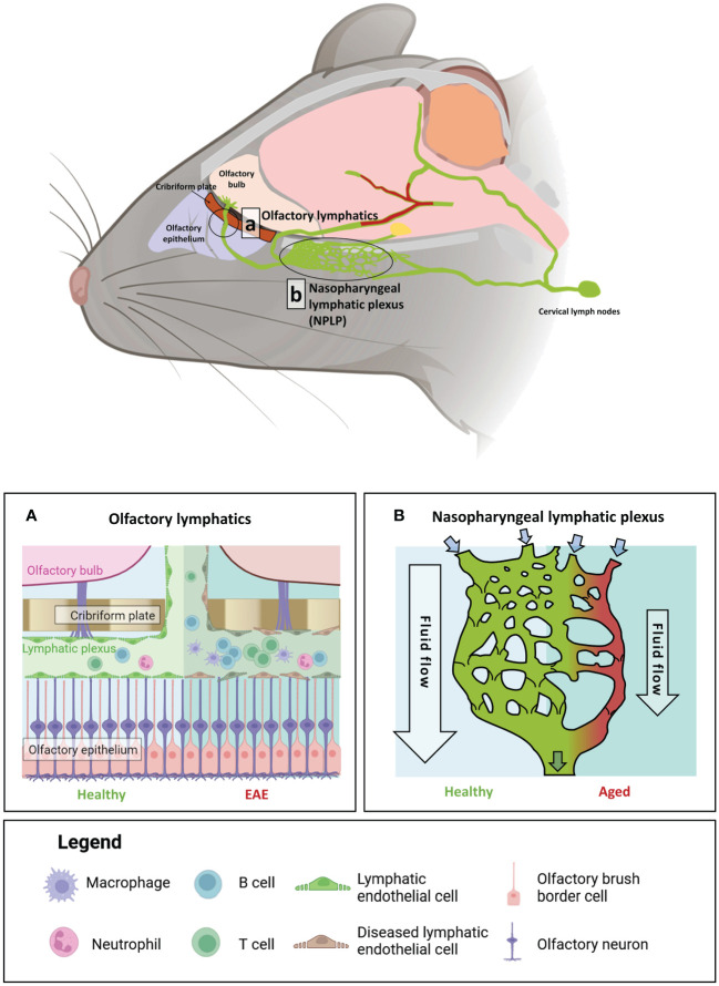
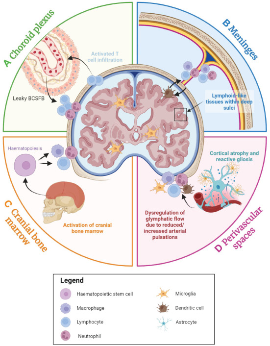
Historically, the central nervous system (CNS) was regarded as 'immune-privileged', possessing its own distinct immune cell population. This immune privilege was thought to be established by a tight blood-brain barrier (BBB) and blood-cerebrospinal-fluid barrier (BCSFB), which prevented the crossing of peripheral immune cells and their secreted factors into the CNS parenchyma. However, recent studies have revealed the presence of peripheral immune cells in proximity to various brain-border niches such as the choroid plexus, cranial bone marrow (CBM), meninges, and perivascular spaces. Furthermore, emerging evidence suggests that peripheral immune cells may be able to infiltrate the brain through these sites and play significant roles in driving neuronal cell death and pathology progression in neurodegenerative disease. Thus, in this review, we explore how the brain-border immune niches may contribute to the pathogenesis of neurodegenerative disorders such as Alzheimer's disease (AD), Parkinson's disease (PD), and multiple sclerosis (MS). We then discuss several emerging options for harnessing the neuroimmune potential of these niches to improve the prognosis and treatment of these debilitative disorders using novel insights from recent studies.

Keywords: Alzheimer’s disease; Parkinson’s disease; brain-border; multiple sclerosis; neurodegeneration; neuroimmune.


Conflict of interest statement

The authors declare that the research was conducted in the absence of any commercial or financial relationships that could be construed as a potential conflict of interest.

Figures

Figure 1
Figure 1
Dynamic neuroinflammatory responses at the choroid plexus. The choroid plexus is an active immune niche that monitors immune factors from the blood and the CSF. During the inflammatory response, surveilling CD4+ T cells infiltrate the choroid plexus and are presented with antigen-loaded MHC-II by resident macrophages. This triggers their differentiation into various T helper cell subtypes. Th17 cells secrete IL-17, which promotes choroid plexus epithelial CCL20 expression, therefore allowing for the adhesion of lymphocytes to the choroid plexus epithelium and aiding their transmigration into the CSF. Macrophages within the stroma secrete IL-1β due to interactions with activated CD4+ T cells. IL-1 β interacts with stromal cells, which in turn secrete pro-inflammatory cytokines and chemokines such as IL-6, CCL2, CXCL1, and CXCL2. Tight junctions between choroid plexus epithelial cells become compromised following inflammation, which may lead to increased movement of immune cells across the epithelium. Additionally, neutrophils from the blood stream accumulate in the choroid plexus stroma, allowing them to infiltrate the CSF. Figure created with BioRender.
Figure 2
Figure 2
Summary of the neuroimmune niches within the skull-meninges-brain axis. (A) The cranial bone marrow (CBM) provides a source of haematopoietic stem cells, which supply lymphoid and myeloid cells to the dural sinuses during inflammatory events. (B) Germinal-like centres in the dural-associated lymphatic tissues (DALTs) contain lymphocytes. T cell populations in the meninges are most likely derived from the blood or CSF. T helper cells can be activated by MHC-II-presenting macrophages within the dura mater, and subsequently activate B cells and cytotoxic T cells. Immune cell activity within lymphoid hubs may help prevent the entry of external infection into the brain. (C) T cell clusters and MHC-II-presenting macrophages are also found at the subarachnoid lymphatic-like membrane (SLYM), closely associated with pial blood vessels. The SLYM may therefore serve as an active immune niche that regulates immune exchanges between the blood stream and the CSF. (D) Perivascular spaces house resident immune cells, such as perivascular macrophages, as well as a smaller population of B cells and T cells. Figure created with BioRender.
Figure 3
Figure 3
The impact of age and auto-immune insult on brain-border lymphatics in Mus musculus. Brain-border lymphatics are integral for the removal of metabolic waste and immune cells from the CSF. However, their function can diminish with advancing age or inflammatory insult. Top panel: a schematic of the mouse brain displaying the locations of the olfactory lymphatics (A) and nasopharyngeal lymphatic plexus (NPLP) (B). (A) In healthy mice (left), lymphatics at the olfactory bulb drain CSF from the meninges and olfactory bulb. Mice with experimental autoimmune encephalomyelitis (EAE) display significant dilation of the lymphatic vessels with increased drainage of immune cells from the CSF (right). (B) The NPLP is a recently discovered anastomotic plexus found posterior to the olfactory lymphatics, present in both mice and humans. In healthy mice (left) the NPLP primarily serves to collect CSF that is drained from the ventral regions of the brain, before it is sent to the cervical lymphatics, which travel to deep cervical lymph nodes. During ageing (right), atrophy of the NPLP occurs, which can result in poorer drainage of lymphatic fluid that is likely to impair CSF flow. This age-related impaired lymphatic flow may contribute to the pathology of various neurodegenerative diseases. Figure created with BioRender.
Figure 4
Figure 4
An overview of the contributions of the brain-border niches to neurodegenerative diseases. The choroid plexus, cranial bone marrow (CBM), perivascular spaces and meninges all appear to play a role in neurodegenerative disease. (A) The choroid plexus serves as the primary brain-cerebrospinal fluid barrier (BCSFB) and actively responds to immune factors from both the CSF and the blood stream. With advancing age and during the inflammatory response, immune cell populations accumulate within the choroid plexus, and the BCSFB becomes leakier due to the loss of tight junctions, potentially facilitating greater peripheral immune cell activation and infiltration into the brain. (B) In many age-associated neurodegenerative diseases, high concentrations of inflammatory factors appear to result in the accumulation of myeloid and lymphoid cells in the dura, subarachnoid space, and even within the deep sulci of the cortex, which may perpetuate neurodegeneration. (C) The CBM is a brain-border niche containing haematopoietic stem cells, which can differentiate to myeloid and lymphoid cells in response to a range of immune factors. Evidence suggests that immune activation in the CBM is associated with various neurodegenerative diseases. (D) Cortical atrophy and gliosis associated with neurodegenerative disease may lead to a leaky blood-brain barrier (BBB), while inflammatory factors from the parenchyma may induce increased immune cell localisation in perivascular spaces. Furthermore, dysregulation of glymphatic flow due to factors such as diminishing arterial pulsations may affect clearance of metabolic waste and immune cells within the brain parenchyma, leading to the build-up of neurotoxic molecules within the brain. Figure created with Biorender.

Similar articles

Cited by

References

    1. Ehrlich P. Das Sauerstoff-Bedurfnisdes Organismus: eine farbenanalytischeStudie. Berlin: Hirschward. (1885).
    1. Bield A, Kraus R. U˙ber eine bisherunbekannte toxische Wirkung der Gal-lensauren auf das Zentralnervensystem. Zhl Inn Med. (1898) 19:1185–200.
    1. Lewandowsky M. Zur lehre der ze-rebrospinalflussigkeit. Z Klin Med. (1900) 40:480–4.
    1. Goldmann EE. Die aussere und innere Sekretion des gesunden und kranken Organismus im Liche der “vitalen Farbung”. Beitr Klin Chir. (1909) 64:192–265.
    1. Bentivoglio M, Kristensson K. Tryps and trips: cell trafficking across the 100-year-old blood-brain barrier. Trends Neurosci. (2014) 37:325–33. doi: 10.1016/j.tins.2014.03.007 - DOI - PMC - PubMed