. 2024 May 28:15:1388998.
doi: 10.3389/fimmu.2024.1388998. eCollection 2024.

Neuronal nicotinic acetylcholine receptor antibodies in autoimmune central nervous system disorders

Affiliations

Neuronal nicotinic acetylcholine receptor antibodies in autoimmune central nervous system disorders

Maria Pechlivanidou et al. Front Immunol. .

Abstract

Background: Neuronal nicotinic acetylcholine receptors (nAChRs) are abundant in the central nervous system (CNS), playing critical roles in brain function. Antigenicity of nAChRs has been well demonstrated with antibodies to ganglionic AChR subtypes (i.e., subunit α3 of α3β4-nAChR) and muscle AChR autoantibodies, thus making nAChRs candidate autoantigens in autoimmune CNS disorders. Antibodies to several membrane receptors, like NMDAR, have been identified in autoimmune encephalitis syndromes (AES), but many AES patients have yet to be unidentified for autoantibodies. This study aimed to develop of a cell-based assay (CBA) that selectively detects potentially pathogenic antibodies to subunits of the major nAChR subtypes (α4β2- and α7-nAChRs) and its use for the identification of such antibodies in "orphan" AES cases.

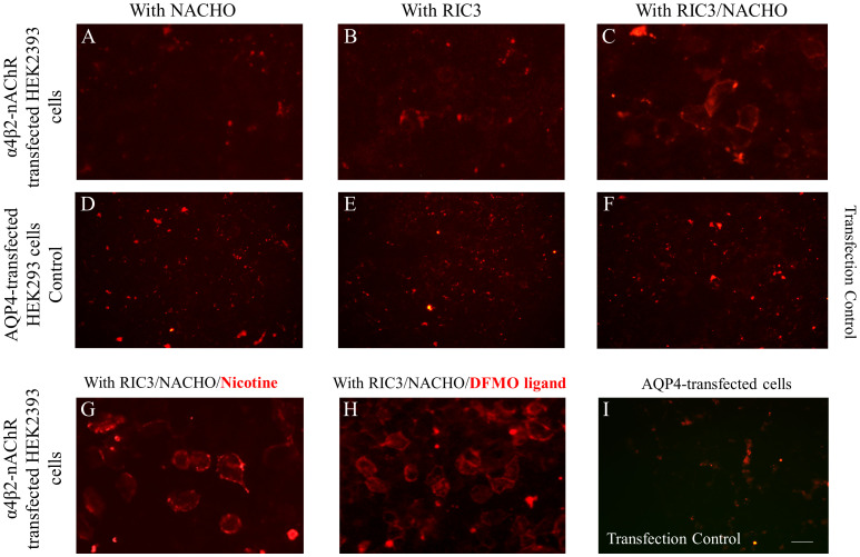
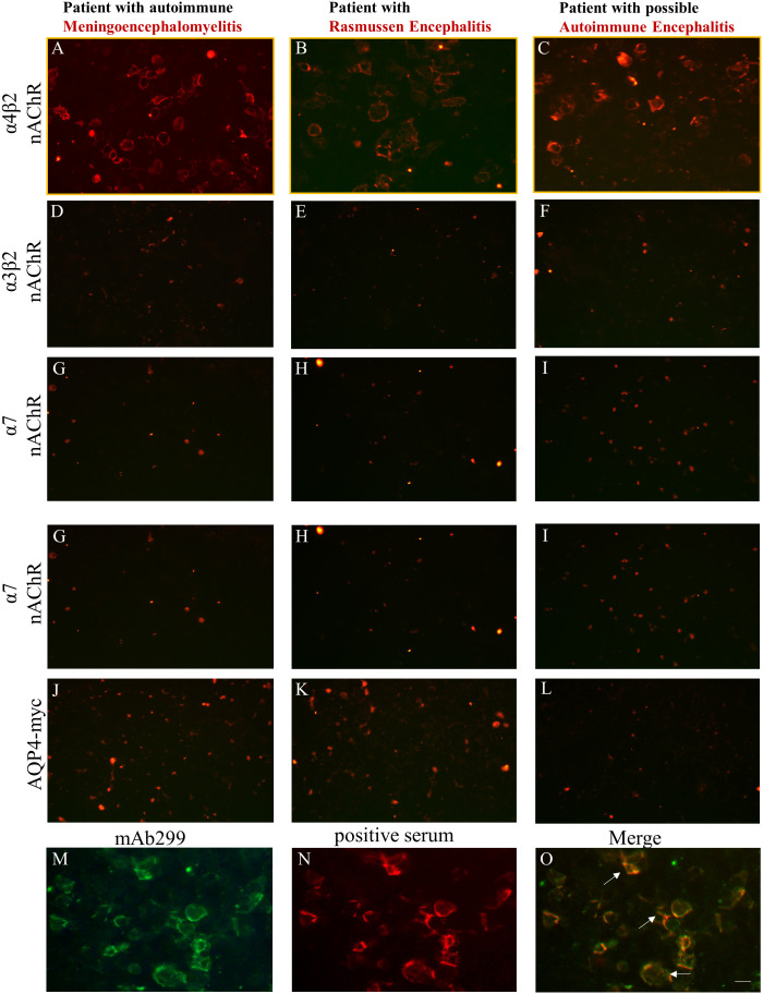
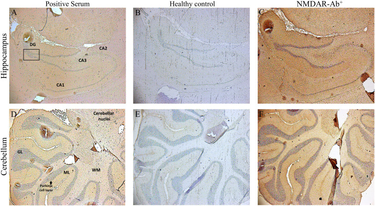
Methods: The study involved screening of sera derived from 1752 patients from Greece, Turkey and Italy, who requested testing for AES-associated antibodies, and from 1203 "control" patients with other neuropsychiatric diseases, from the same countries or from Germany. A sensitive live-CBA with α4β2-or α7-nAChR-transfected cells was developed to detect antibodies against extracellular domains of nAChR major subunits. Flow cytometry (FACS) was performed to confirm the CBA findings and indirect immunohistochemistry (IHC) to investigate serum autoantibodies' binding to rat brain tissue.

Results: Three patients were found to be positive for serum antibodies against nAChR α4 subunit by CBA and the presence of the specific antibodies was quantitatively confirmed by FACS. We detected specific binding of patient-derived serum anti-nAChR α4 subunit antibodies to rat cerebellum and hippocampus tissue. No serum antibodies bound to the α7-nAChR-transfected or control-transfected cells, and no control serum antibodies bound to the transfected cells. All patients positive for serum anti-nAChRs α4 subunit antibodies were negative for other AES-associated antibodies. All three of the anti-nAChR α4 subunit serum antibody-positive patients fall into the AES spectrum, with one having Rasmussen encephalitis, another autoimmune meningoencephalomyelitis and another being diagnosed with possible autoimmune encephalitis.

Conclusion: This study lends credence to the hypothesis that the major nAChR subunits are autoimmune targets in some cases of AES and establishes a sensitive live-CBA for the identification of such patients.

Keywords: autoimmune encephalitis syndromes; autoimmunity; autoimmunity-driven cognitive impairment; nAChR subunit α4; neuronal nicotinic acetylcholine receptor.


Conflict of interest statement

Authors MP, KK, EK, AT, MD, EN and ST were employed by company Tzartos NeuroDiagnostics. ST has shares in the research and diagnostic laboratory Tzartos NeuroDiagnostics. ST and JT have filed a patent on a cell-based method for detecting potentially pathogenic autoantibodies to neuronal nAChRs. RM received funding for travel, meeting attendance or Advisory Board participation from Alexion, Argenx, Biomarin, Catalyst, SANOFI, Regeneron and UCB. The remaining authors declare that the research was conducted in the absence of any commercial or financial relationships that could be construed as a potential conflict of interest. The author(s) declared that they were an editorial board member of Frontiers, at the time of submission. This had no impact on the peer review process and the final decision.

Figures

Figure 1
Figure 1
Optimization of the live α4β2-nAChR CBA. All panel images document immunostaining with anti‐nAChR α4 subunit monoclonal antibody (mAb299 at 1/20 dilution) followed by secondary anti-antibody Alexa Fluor 568. HEK293 cells were transfected with plasmids carrying genes encoding α4β2-nAChR subtype and chaperons NACHO (A) or RIC3 (B) individually, and in combination (C) The co-transfection of cells with α4β2-nAChR plasmids and both chaperons RIC and NACHO (C) significantly increased the expression of nAChRs which is demonstrated by the cell surface staining, indicative of anti‐α4 subunit antibody binding that correlates with receptor expression. HEK293 cells expressing the transfection control of the experimental conditions (A-C), which is the protein aquaporin-4 (AQP4), did not show any positive signal (D-F) as anti‐nAChR α4 subunit antibodies did not bind to the AQP4-expressing cells. Cells co-transfected with α4β2-nAChRs and chaperon plasmids and cultured in the presence of 1mM nicotine (G) or 5mM DFMO (H) for 24 hours, led to higher α4β2-nAChRs expression. The addition of nicotine or DFMO ligand similarly enhanced the signal compared to the plain medium. In the panel (I) with HEK293 cells expressing the transfection control AQP4 of the experimental conditions (G, H), the absence of a positive signal is displayed. Signal in panel (A, B, D-F, I) show non‐specific background staining. Scale bar: 20 μm.
Figure 2
Figure 2
Three patients in the AES spectrum were identified based on the live CBA as being positive for serum antibodies against nAChR α4 subunit epitopes. HEK293 cells expressing α4β2, α3β2, α7-nAChRs, or control AQP4 were incubated with sera derived from patients suspected for AES and found to be positive by the live α4β2-nAChR CBA, followed by secondary and third antibodies. Serum anti-nAChR α4 subunit antibodies were identified in a patient with autoimmune meningoencephalomyelitis (A), in a patient with Rasmussen (B), and in a patient with possible autoimmune encephalitis with movement disorder involving stiffness (C). No staining was observed for these patient serum antibodies when targets were α3β2‐ (D‐F) or α7‐nAChRs (G‐I) or AQP4 expressed in transfected cells (J‐L). Double immunostaining was performed in α4β2-transfected HEK293 cells using a monoclonal anti-α4 subunit (mAb299) (M) and positive sera antibodies to α4 subunit of the α4β2-nAChR derived from patients with AES (N). The overlapping immunoreactivity (white arrows) of the mΑb and the positive serum confirmed the specificity of our α4β2-nAChR CBA (O). Scale bar: 20 μm.
Figure 3
Figure 3
Binding of sera positive for autoantibodies to subunit α4 of the α4β2-nAChRs, to rat brain tissue by Indirect Immunostaining. Sagittal whole rat brain sections were incubated with sera antibodies from the three patients with AES (A, D), from NMDAR encephalitis patients (C, F) or from healthy controls (B, E). Representative images are shown the localization of rat α4β2‐nAChRs bound by specific serum antibodies from 1 out of the 3 patients with autoantibodies against subunit α4‐nAChRs in the hippocampus (A) and the cerebellum (D), a specific positive signal which was confirmed only by the patients’ sera bearing α4-nAChR-Abs, whereas no specific binding was detected in any region of the rat brain tissue by serum components originated from 1 out of the 10 healthy controls (B, E). Staining and images of rat brain sections with serum antibodies from one NMDAR encephalitis patient as positive control (C, F) are depicted. CA1–CA3, hippocampal area cornu ammonis 1–3; DG, dentate gyrus; cerebellar layers. GCl, granular cell layer; ML, molecular layer; WM, white matter.

References

    1. Hogg RC, Raggenbass M, Bertrand D. Nicotinic acetylcholine receptors: from structure to brain function. Rev Physiol Biochem Pharmacol. (2003) 147:1–46. doi: 10.1007/s10254-003-0005-1 - DOI - PubMed
    1. Gotti C, Clementi F. Neuronal nicotinic receptors: from structure to pathology. Prog Neurobiol. (2004) 74:363–96. doi: 10.1016/j.pneurobio.2004.09.006 - DOI - PubMed
    1. Changeux JP. The nicotinic acetylcholine receptor: the founding father of the pentameric ligand-gated ion channel superfamily. J Biol Chem. (2012) 287:40207–15. doi: 10.1074/jbc.R112.407668 - DOI - PMC - PubMed
    1. Gotti C, Zoli M, Clementi F. Brain nicotinic acetylcholine receptors: native subtypes and their relevance. Trends Pharmacol Sci. (2006) 27:482–91. doi: 10.1016/j.tips.2006.07.004 - DOI - PubMed
    1. Zoli M, Pucci S, Vilella A, Gotti C. Neuronal and extraneuronal nicotinic acetylcholine receptors. Curr Neuropharmacol. (2018) 16:338–49. doi: 10.2174/1570159X15666170912110450 - DOI - PMC - PubMed

LinkOut - more resources