. 2024 May 28:15:1391453.
doi: 10.3389/fmicb.2024.1391453. eCollection 2024.

Identification of Mycoplasma pneumoniae proteins interacting with NOD2 and their role in macrophage inflammatory response

Affiliations

Identification of Mycoplasma pneumoniae proteins interacting with NOD2 and their role in macrophage inflammatory response

Yongyu Wang et al. Front Microbiol. .

Abstract

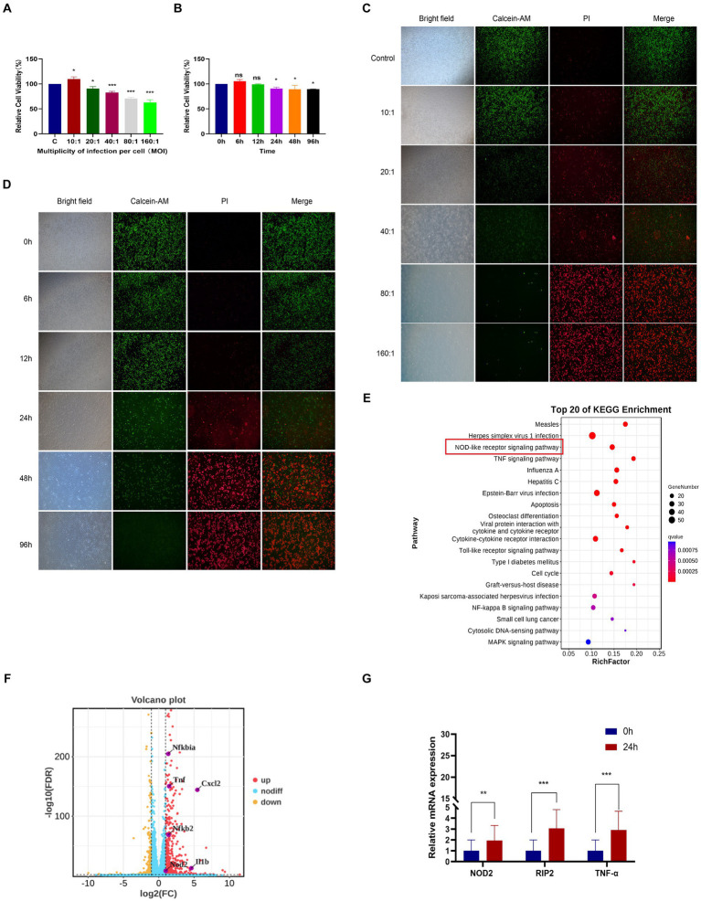
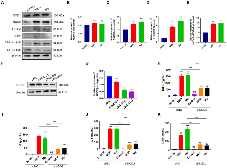
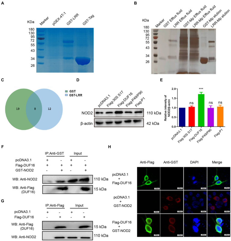
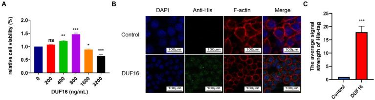
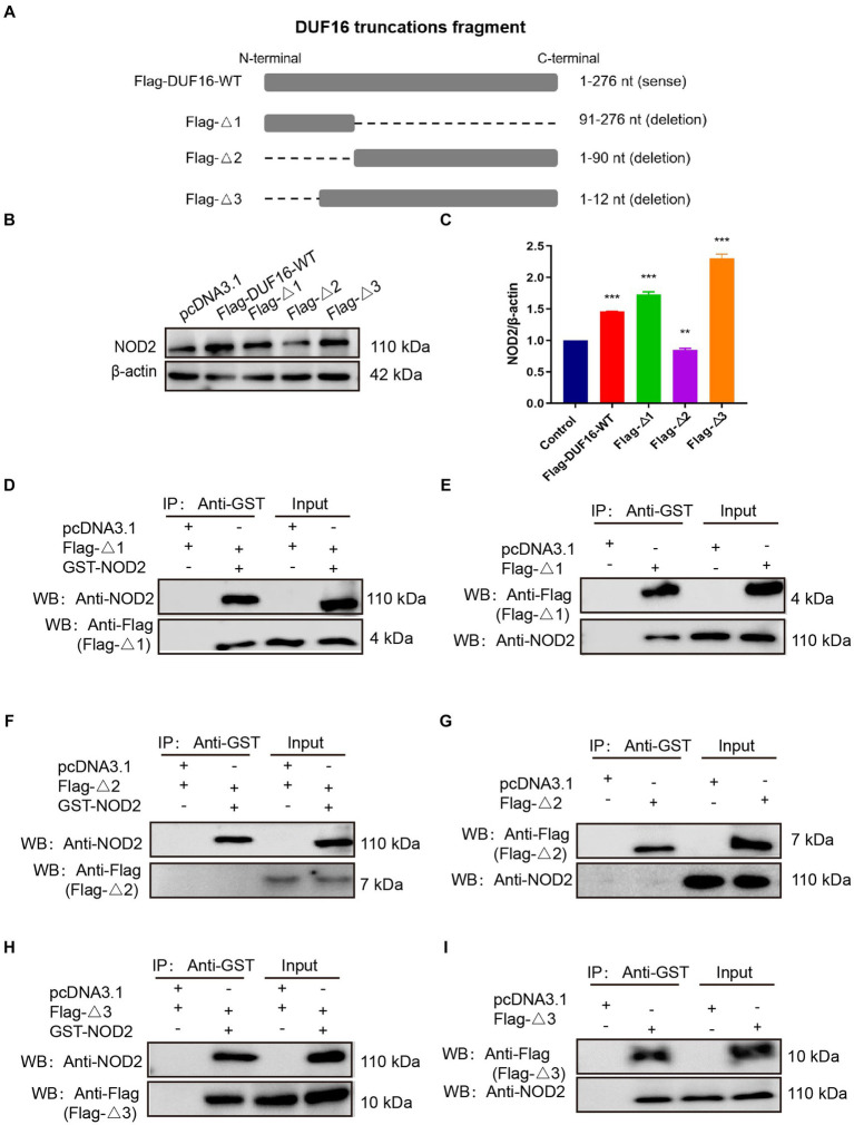
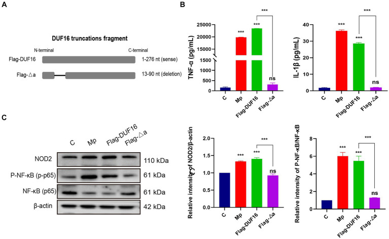
Mycoplasma pneumoniae (M. pneumoniae, Mp) is a cell wall-deficient microorganism known to cause chronic respiratory infections in both children and adults. Nucleotide-binding oligomerization domain-containing protein 2 (NOD2) is an intracellular pattern recognition receptor primarily responsible for identifying muramyl dipeptide (MDP) found in bacterial cell walls. Previous experiments have demonstrated that Mycoplasma ovipneumoniae induces macrophage autophagy through NOD2. In this study, we conducted RNA-seq analysis on macrophages infected with M. pneumoniae and observed an up-regulation in the expression of genes associated with the NOD2 signaling pathway. Mechanistic investigations further revealed the involvement of the NOD2 signaling pathway in the inflammatory response of macrophages activated by M. pneumoniae. We utilized GST pull-down technology in conjunction with liquid chromatography-tandem mass spectrometry (LC-MS/MS) to pinpoint the M. pneumoniae proteins that interact with NOD2. Additionally, co-immunoprecipitation (Co-IP) and immunofluorescence co-localization techniques were used to confirm the interaction between DUF16 protein and NOD2. We found that DUF16 protein can enter macrophages and induce macrophage inflammatory response through the NOD2/RIP2/NF-κB pathway. Notably, the region spanning amino acids 13-90 was identified as a critical region necessary for DUF16-induced inflammation. This research not only broadens our comprehension of the recognition process of the intracellular receptor NOD2, but also deepens our understanding of the development of M. pneumoniae infection.

Keywords: DUF16; Mycoplasma pneumoniae; NF-κB; NOD2; inflammation; macrophages.


Conflict of interest statement

The authors declare that the research was conducted in the absence of any commercial or financial relationships that could be construed as a potential conflict of interest.

Figures

Figure 1
Figure 1
RNA-seq analysis of macrophages from mice infected with M. pneumoniae. (A) CCK-8 detection results after Mp infected RAW264.7 cells at different MOIs for 24 h. (B) CCK-8 detection results of RAW264.7 cells infected with Mp (MOI = 10) for different times. (C) The results were observed by immunofluorescence after Mp infected RAW264.7 cells at different MOIs for 24 h. (D) Immunofluorescence observation results of RAW264.7 cells infected with Mp (MOI = 10) for different times. (E) KEGG enrichment analysis diagram of differentially expressed genes. (F) Volcano plot of differentially expressed genes between the infected group and the control group. (G) qPCR validation of NOD2 signaling pathway-related gene expression in RNA-Seq. The data represent three independent treatments, and p-values were calculated using the one-way ANOVA test. SD, error bars; ns, not significant; *p < 0.05, **p < 0.01, ***p < 0.001.
Figure 2
Figure 2
M. pneumoniae causes up-regulation of inflammatory factors in RAW264.7 cells through NOD2. (A) After treating RAW264.7 cells with 500 ng/mL MDP and Mp with MOI = 10 for 24 h, the expression of NOD1, NOD2, p-RIP2, RIP2, P-NF-κB (p65), NF-κB (p65) were detected in each treatment group using Western blot. Western blot was used to detect the expression of related proteins in the NOD2 signaling pathway in each treatment group: (B) NOD1, (C) NOD2, (D) p-RIP2/RIP2, (E) P-NF-κB (p65) / NF-κB (p65). (F,G) Western blot detects the knockdown efficiency of NOD2 small interfering RNA (siNOD2). ELISA analyzed the expression of inflammatory factors in each treatment group: (H) TNF-α, (I) IL-6, (J) IL-8, (K) IL-1β. The data represent three independent treatments, and p-values were calculated using the one-way ANOVA test. SD, error bars; ns, not significant; *p < 0.05, **p < 0.01, ***p < 0.001.
Figure 3
Figure 3
Screening and identification of M. pneumoniae proteins that interact with NOD2. (A) SDS-PAGE analysis of purified GST-tagged protein and GST-LRR protein. (B) Silver staining detection pictures of each group after GST pull-down. (C) Venn diagram of differential proteins in elution samples from the GST and GST-LRR groups. (D) After the eukaryotic plasmids of four Mp proteins (30S ribosomal protein S17, DUF16 family-like protein, P1 adhesin type 2 g2, and P40/P90 adhesin) were transfected into RAW264.7 cells for 48 h, the protein expression was identified by Western blot. (E) Histogram of NOD2 expression in each transfection group. (F) Co-precipitation of NOD2 protein with recombinant DUF16 in 293 T cell lysate. (G) Co-precipitation (Internal reverse IP) of NOD2 protein with DUF16 in RAW264.7 cell lysate. (H) Confocal microscopy analysis was carried out for demonstrating colocalization of NOD2 (red fluorescence) and DUF16 (green fluorescence). The data represent three independent treatments, and p-values were calculated using the one-way ANOVA test. SD, error bars; ns, not significant; ***p < 0.001.
Figure 4
Figure 4
DUF16 protein can be phagocytosed by RAW264.7 cells. (A) After RAW264.7 cells were treated with different concentrations of His-DUF16 recombinant protein for 24 h, Cell Counting Kit-8 was used to detect cell viability. (B) His-DUF16 protein is phagocytosed by RAW264.7 cells. (C) Histogram of His-DUF16 content in each treatment group. The data represent three independent treatments, and p-values were calculated using the one-way ANOVA test. SD, error bars; ns, not significant; *p < 0.05, **p < 0.01, ***p < 0.001.
Figure 5
Figure 5
DUF16 protein induces an inflammatory response in mouse macrophages through NOD2. ELISA analyzed the expression of inflammatory factors in each treatment group: (A) IL-1β, (B) IL-6, (C) IL-8, (D) TNF-α. (E) The expression changes of NOD2, p-RIP2, RIP2, P-NF-κB (p65), NF-κB (p65), p-IKB α, and IKB α. were detected using Western blot analysis in RAW264.7 cells induced by His-DUF16 (1,600 ng/mL) protein and MDP (500 ng/mL). Histogram of protein expression of NOD2 signaling pathway in cells of each treatment group detected by Western blot analysis: (F) NOD2, (G) p-RIP2/RIP2, (H) P-NF-κB (p65) / NF-κB (p65), (I) p-IKB α/IKB α. The data represent three independent treatments, and p-values were calculated using the one-way ANOVA test. SD, error bars; ns, not significant; *p < 0.05, **p < 0.01, ***p < 0.001.
Figure 6
Figure 6
DUF16 interacts with NOD2 through the 13–90 aa critical region. (A) Design expression vectors for different truncated bodies of DUF16. (B) Western blotting analysis of the expression levels of each protein in five transfection groups (pcDNA3.1, Flag-DUF16-WT, Flag-Δ1, Flag-Δ2, and Flag-Δ3). (C) Western blotting analysis of NOD2 protein levels in 5 transfection groups (pcDNA3.1, Flag-DUF16-WT, Flag-△1, and Flag-△3). pcDNA3.1-GST-NOD2 was co-transfected with pcDNA3.1-Flag-DUF16 different truncated expression vectors into 293 T (D,F,H) cells, or pcDNA3.1-Flag-DUF16 alone was transfected into RAW264.7 (E,G,I) After 48 h of transfection of cells, the cell lysates were verified by Co-IP using Flag tag and subjected to Western blot analysis. The data represent three independent treatments, and p-values were calculated using the one-way ANOVA test. SD, error bars; **p < 0.01, ***p < 0.001.
Figure 7
Figure 7
13-90 aa is a critical region for DUF 16 to trigger cellular NOD2-mediated inflammatory responses. (A) A truncated expression vector was designed and constructed based on the DUF16 interaction region. (B) After RAW264.7 cells were treated with Mp, DUF16 or Flag-△a, respectively, for 24 h, TNF-α and IL-1β in the cell supernatants of each group were detected by ELISA. (C) The expression changes of NOD2, RIP2, p-NF-κB p65, NF-κB p65 were detected using Western blot analysis in RAW264.7 cells induced by 4 treatment groups (Control, Mp, DUF16 and Flag-△a). The data represent three independent treatments, and p-values were calculated using the one-way ANOVA test. SD, error bars; ns, not significant; ***p < 0.001.

Similar articles

References

    1. Al Nabhani Z., Dietrich G., Hugot J. P., Barreau F. (2017). Nod2: the intestinal gate keeper. PLoS Pathog. 13:e1006177. doi: 10.1371/journal.ppat.1006177, PMID: - DOI - PMC - PubMed
    1. Alipoor S. D., Mirsaeidi M. (2021). Inborn errors in the Lrr domain of Nod2 and their potential consequences on the function of the receptor. Cells 10:2031. doi: 10.3390/cells10082031, PMID: - DOI - PMC - PubMed
    1. Caruso R., Warner N., Inohara N., Núñez G. (2014). Nod1 and Nod2: signaling, host defense, and inflammatory disease. Immunity 41, 898–908. doi: 10.1016/j.immuni.2014.12.010, PMID: - DOI - PMC - PubMed
    1. Chaudhry R., Ghosh A., Chandolia A. (2016). Pathogenesis of Mycoplasma Pneumoniae: an update. Indian J. Med. Microbiol. 34, 7–16. doi: 10.4103/0255-0857.174112, PMID: - DOI - PubMed
    1. Cloward J. M., Krause D. C. (2009). Mycoplasma Pneumoniae J-domain protein required for terminal organelle function. Mol. Microbiol. 71, 1296–1307. doi: 10.1111/j.1365-2958.2009.06602.x - DOI - PMC - PubMed