. 2024;5(1):138-164.
doi: 10.20517/evcna.2023.77. Epub 2024 Mar 28.

Non-invasive detection of orthotopic human lung tumors by microRNA expression profiling of mouse exhaled breath condensates and exhaled extracellular vesicles

Affiliations

Non-invasive detection of orthotopic human lung tumors by microRNA expression profiling of mouse exhaled breath condensates and exhaled extracellular vesicles

Megan I Mitchell et al. Extracell Vesicles Circ Nucl Acids. 2024.

Abstract

Aim: The lung is the second most frequent site of metastatic dissemination. Early detection is key to improving survival. Given that the lung interfaces with the external environment, the collection of exhaled breath condensate (EBC) provides the opportunity to obtain biological material including exhaled miRNAs that originate from the lung.

Methods: In this proof-of-principal study, we used the highly metastatic MDA-MB-231 subline 3475 breast cancer cell line (LM-3475) to establish an orthotopic lung tumor-bearing mouse model and investigate non-invasive detection of lung tumors by analysis of exhaled miRNAs. We initially conducted miRNA NGS and qPCR validation analyses on condensates collected from unrestrained animals and identified significant miRNA expression differences between the condensates of lung tumor-bearing and control mice. To focus our purification of EBC and evaluate the origin of these differentially expressed miRNAs, we developed a system to collect EBC directly from the nose and mouth of our mice.

Results: Using nanoparticle distribution analyses, TEM, and ONi super-resolution nanoimaging, we determined that human tumor EVs could be increasingly detected in mouse EBC during the progression of secondary lung tumors. Using our customizable EV-CATCHER assay, we purified human tumor EVs from mouse EBC and demonstrated that the bulk of differentially expressed exhaled miRNAs originate from lung tumors, which could be detected by qPCR within 1 to 2 weeks after tail vein injection of the metastatic cells.

Conclusion: This study is the first of its kind and demonstrates that lung tumor EVs are exhaled in mice and provide non-invasive biomarkers for detection of lung tumors.

Keywords: Extracellular vesicles; exhaled breath condensate; miRNAs; orthotopic lung tumor-bearing animal model.


Conflict of interest statement

Conflicts of Interest Olivier Loudig is a Junior Editorial Board member of the journal Extracellular Vesicles and Circulating Nucleic Acids. The other authors declared that there are no conflicts of interest.

Figures

Figure 1
Figure 1
Establishment of the mouse model of human secondary lung cancer. (A) MDA-MB-231 subline 3475 cell line stably transduced with lentiviral constructs for expression of TdTomato-luciferase (pUltraChili-Luc) and pCT-CD63-GFP. Confocal imaging of a single clone co-expressing both TdTomato and GFP, sorted by FACS [See Supplementary Figure 1], with DAPI staining to locate cellular nuclei (Blue color, left panel) and Td-tomato red protein expression (Red colored cells, second panel from the left) and the CD63-GFP protein on extracellular vesicles (EVs, Green dots, third panel from the left), with a Merged image all staining and fluorescent imaging on the right panel. CD63-GFP detection shows that cells at different mitotic stages produced different amounts of EVs as observable by the differential detection of GFP protein. The central dividing cell displays the highest amount of observable EVs; (B) Bioluminescent in vivo imaging of tumor-bearing mice at weeks 0, 6, and 12 after inoculation of TdTomato-Luc+/CD63-GFP+ MDA-MB-231 subline 3475 cells. Bioluminescence intensity is indicated by means of radiant efficiency (photons/sec/cm2/sr) scale bars, with red being the most intense (See scale bar); (C) Representative formalin-fixed whole lung tissue images collected from two healthy mice (control; left) and two TdTomato-Luc+/CD63-GFP+ MDA-MB-231 subline 3475 inoculated lung tumor-bearing mice (case; right); (D) Representative images of Hematoxylin and Eosin (H&E) stained 5 mm tissue sections of lungs harvested from one control (left panels; 0 × and 20 × magnifications) and one lung tumor-bearing animal (right panels; 0 × and 20 × magnifications). The image right panel from one lung tumor-bearing animal shows extensive infiltration of metastatic carcinoma legions with a few rare immature lymphocytes seen interspersed.
Figure 2
Figure 2
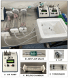
Whole mouse exhaled breath condensate (EBC) collection system version 1.0. For this system, EBC is collected from two unrestrained mice roaming in a sealed glass chamber, which contains a removable metal grate that allows animals to move freely with normal postural movement. Airflow throughout the system is maintained and directed towards a condenser. The components of this mouse EBC collection system include: (A) an air pump that controls airflow (2.0 mL/min) of compressed breathing-grade air that is transported through 1/4 inch plastic tubes; (B) a one-way Balston 0.01 mic 93% airflow filter that maintains air sterility; (C) a glass mouse chamber (containing two mice); that is (D) securely sealed on both ends by caps with gaskets; connected to (E) a glass condenser sealed on both ends by caps, which is placed on ice to allow for the collection of EBC. It is estimated that ~62.5 µL of EBC can be captured from two mice within one h of collection.
Figure 3
Figure 3
MicroRNA analysis of EBC collected from unrestrained animals. (A) Timeline of the weekly EBC collections from animal pairs, separated by sex (circles for females, triangles for males), between healthy control mice (blue) and lung tumor-bearing mice (red), for a period of 16 weeks. Discovery analyses were performed using total small-RNA extracted from EBC collected at weeks 0, 5, 9, and 13 using Next-Generation Sequencing (NGS). Validation analyses were conducted using total small-RNA extracted from EBC at weeks 1, 8, and 15 using quantitative reverse transcription PCR (RT-qPCR). Proteomic analyses were conducted on EBC collected and pooled for weeks 12, 14, and 16 from control and tumor-bearing mice groups; (B) PCA plots for miRNA expression of healthy control mice measured at weeks 0, 5, 9, and 13 for both females and males (top), and lung tumor-bearing mice at the same time points for both females and males (bottom); (C) Heatmap classification of the top 233 miRNAs detected by NGS using small RNAs extracted from EBC of healthy control (blue) and lung tumor-bearing (red) mice at weeks 0 (yellow), 5 (green), 9 (pink), and 13 (purple) for both females (grey) and males (black). The purple box highlights miRNAs commonly identified between condensates from control and lung tumor-bearing animals. The red box highlights miRNAs predominantly identified in condensates obtained from lung tumor-bearing animals. Two miRNAs, namely miR-374a and miR-584, which are bolded in orange text, are identified to be predominantly upregulated in condensates of lung tumor-bearing animals and are also identified to be upregulated in EBC obtained from lung tumor-bearing animals (system v2.0) displayed in Figure 7C and D. Taqman© qPCR analyses of hsa-miR-222 and has-miR-210 using total small-RNA purified from EBC collected at weeks 1 (blue), 8 (light purple), and 15 (dark purple), separately for females and males, with data calculated using the 2ΔΔCt formula between healthy mice (i.e., using week 1 Ct values as the reference), and lung tumor-bearing mice, both normalized to exogenous ath-miR-159a (100 pg) as an internal “housekeeping” control that was spiked in EBC before RNA extractions and qPCR analyses.
Figure 4
Figure 4
Proteomic analysis of EBC from healthy controls and lung tumor-bearing mice. Pie chart distribution of all 286 identified proteins in pooled EBC samples of control (n = 6) and lung tumor-bearing (n = 6) mice, stratified based on the preferential organ/tissue origin of each individual protein as informed by ProteinAtlas, and distributed as either from lung, skin, urine, breast, testis, colon, or undetermined tissue origins (i.e., Other).
Figure 5
Figure 5
Nose and mouth EBC collection system v2.0 for restrained individual animals. The system designed and described here includes additional devices that enable air flow and direct collection of EBC from the nose and mouth of restrained mice. The system is composed of: (A) an air pump controlling airflow (2.0 mL/min) from an air tank transported by 1/4 inch tubes; (B) an airflow valve to maintain air directionality and sterility; (C) an exhaled chamber that is tightly connected with gaskets; to the (D) mouse immobilization chamber where the animal is restrained and through 1/4 inch tubes; towards (E) a glass condenser sealed on both ends by caps and sitting on ice for the accumulation of EBC droplets; which is connected to (E). a second air pump set up in vacuum mode to enhance air circulation through the entire system (i.e., set at 0.2 mL/min). This system allows for individual collection of ~29 µL EBC from restrained mice within 2 h.
Figure 6
Figure 6
Purification and analysis of exhaled extracellular vesicles from mouse EBC. EBC collected directly from the nose and mouth of individual animals was evaluated for the presence of exhaled EVS. (A) EBC from healthy control (n = 3) and lung tumor-bearing (n = 3) mice collected at weeks 2, 6, and 11 was evaluated using the Spectradyne nCS1 nanoparticle analyzer, using C400 cartridges for the detection of nanoparticles between 65 and 400 nm; (B) EBC samples from 6 control (left; 3 mL) and 3 lung tumor-bearing (right; 1 mL) mice, collected over 4 weeks, were subjected to ultracentrifugation and the pellets analyzed by imaging using transmission electron microscopy (TEM); (C) EV pellets from healthy control (top panels) and from lung tumor-bearing mice were analyzed using Super-Resolution Microscopy (ONi instrument) using anti-human anti-CD63, anti-CD9, and anti-CD81 anti-tetraspanin antibodies to evaluate the size and identity exhaled EVs contained in the ultracentrifuged EBC pellets of control (top panels) and lung tumor-bearing mice (bottom panels).
Figure 7
Figure 7
MiRNA analyses of EBC and exhaled EVs collected from control and lung tumor-bearing mice. (A) Western blot evaluation of anti-human and anti-mouse CD63 antibodies using EVs purified from tissue culture media of human breast cancer MDA-MB-231 cells, human kidney cancer HEK293 cells, and normal mouse bone marrow endothelial cells (BMECs); (B) Heatmap analysis of the top 142 most differentially detectable miRNAs between small-RNA extracted from whole EBC (green), and sequentially purified from human exhaled EVs using the anti-human anti-CD63 EV-CATCHER assay from whole EBC (orange), and mouse exhaled EVs using the anti-mouse anti-CD63 EV-CATCHER assay from the same whole EBC samples (purple), collected at weeks 21 (light grey), 22 (brown), and 23 (dark grey) from female control (blue) and lung tumor-bearing (red) mice detectable at study end (week 24). We conducted our analyses in triplicate (i.e., three repeats per RNA purification) on 9 control female mice and 9 lung tumor-bearing female mice. The EBC collected three times a week from the same 3 females was combined (~ 300 µL) to conduct the three different analyses (whole EBC, human exh-EVs, mouse exh-EVs) in triplicate [i.e., 3 sets of 3 EBC collections per control (n = 9) or lung tumor-bearing group (n = 9)]; (C) Venn Diagram displaying the overlap in the identity of the miRNAs detected between whole EBC of lung tumor-bearing mice (yellow), human exh-EVs in lung tumor-bearing mice (orange), mouse exh-EVs in control mice (purple), and human exh-EVs in controls mice (green, non-specific signal). The miRNAs that were selected for these analyses were detected by NGS but had at least 5 reads in 6 of the 9 samples analyzed and were reproducibly detected at least two of the three weeks (weeks 20, 21, and 22). The Venn diagram indicates that 21 miRNAs were non-specifically detected by use of the anti-human anti-CD63 EV-CATCHER assay with EBC of control mice and represented 9% of all selected miRNAs; (D) Small RNAs extracted from whole EBC samples collected at weeks 2, 6, and 11 were evaluated for expression of miR-222, miR-210, miR-374a, and miR-584 by TaqmanTM quantitative PCR analyses using the 2ΔΔCt method to evaluate fold change by comparison to the control sample at week 2. All 4 miRNAs were selected because they were found to be upregulated in the whole EBC of lung tumor-bearing mice compared to control mice by NGS analyses. MiRNA analyses of EBC and exhaled EVs collected from control and lung tumor-bearing mice. (A) Western blot evaluation of anti-human and anti-mouse CD63 antibodies using EVs purified from tissue culture media of human breast cancer MDA-MB-231 cells, human kidney cancer HEK293 cells, and normal mouse bone marrow endothelial cells (BMECs); (B) Heatmap analysis of the top 142 most differentially detectable miRNAs between small-RNA extracted from whole EBC (green), and sequentially purified from human exhaled EVs using the anti-human anti-CD63 EV-CATCHER assay from whole EBC (orange), and mouse exhaled EVs using the anti-mouse anti-CD63 EV-CATCHER assay from the same whole EBC samples (purple), collected at weeks 21 (light grey), 22 (brown), and 23 (dark grey) from female control (blue) and lung tumor-bearing (red) mice detectable at study end (week 24). We conducted our analyses in triplicate (i.e., three repeats per RNA purification) on 9 control female mice and 9 lung tumor-bearing female mice. The EBC collected three times a week from the same 3 females was combined (~ 300 µL) to conduct the three different analyses (whole EBC, human exh-EVs, mouse exh-EVs) in triplicate [i.e., 3 sets of 3 EBC collections per control (n = 9) or lung tumor-bearing group (n = 9)]; (C) Venn Diagram displaying the overlap in the identity of the miRNAs detected between whole EBC of lung tumor-bearing mice (yellow), human exh-EVs in lung tumor-bearing mice (orange), mouse exh-EVs in control mice (purple), and human exh-EVs in controls mice (green, non-specific signal). The miRNAs that were selected for these analyses were detected by NGS but had at least 5 reads in 6 of the 9 samples analyzed and were reproducibly detected at least two of the three weeks (weeks 20, 21, and 22). The Venn diagram indicates that 21 miRNAs were non-specifically detected by use of the anti-human anti-CD63 EV-CATCHER assay with EBC of control mice and represented 9% of all selected miRNAs; (D) Small RNAs extracted from whole EBC samples collected at weeks 2, 6, and 11 were evaluated for expression of miR-222, miR-210, miR-374a, and miR-584 by TaqmanTM quantitative PCR analyses using the 2ΔΔCt method to evaluate fold change by comparison to the control sample at week 2. All 4 miRNAs were selected because they were found to be upregulated in the whole EBC of lung tumor-bearing mice compared to control mice by NGS analyses.
Figure 8
Figure 8
Proteomic analysis of EBC pooled from controls and lung tumor-bearing mice. Pie chart distribution of the 231 identified proteins stratified based on the preferential organ/tissue expression of each individual protein based on ProteinAtlas, and classified as either from lung, skin, urine, breast, testis, upper digestive tract, colon, and other undetermined tissue origins (i.e., Other).

Similar articles

Cited by

References

    1. Sung H, Ferlay J, Siegel RL, et al. Global cancer statistics 2020: GLOBOCAN estimates of incidence and mortality worldwide for 36 cancers in 185 countries. CA Cancer J Clin. 2021;71:209–49. doi: 10.3322/caac.21660. - DOI - PubMed
    1. Siegel RL, Miller KD, Fuchs HE, Jemal A. Cancer statistics, 2021. CA Cancer J Clin. 2021;71:7–33. doi: 10.3322/caac.21654. - DOI - PubMed
    1. Stella GM, Kolling S, Benvenuti S, Bortolotto C. Lung-seeking metastases. Cancers. 2019;11:1010. doi: 10.3390/cancers11071010. - DOI - PMC - PubMed
    1. Caparica R, Mak MP, Rocha CH, et al. Pulmonary nodules in patients with nonpulmonary cancer: not always metastases. J Glob Oncol. 2016;2:138–44. doi: 10.1200/jgo.2015.002089. - DOI - PMC - PubMed
    1. Chen H, Stoltzfus KC, Lehrer EJ, et al. The epidemiology of lung metastases. Front Med. 2021;8:723396. doi: 10.3389/fmed.2021.723396. - DOI - PMC - PubMed

LinkOut - more resources