

Intramuscular Lipoma of the Sternocleidomastoid Muscle: A Rare Entity Revisited
- PMID: 38864025
- PMCID: PMC11165958
- DOI: 10.1177/2632010X241260200
Intramuscular Lipoma of the Sternocleidomastoid Muscle: A Rare Entity Revisited
Abstract
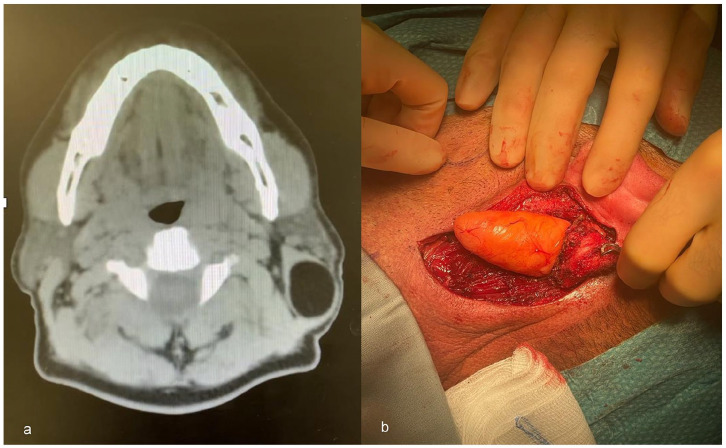
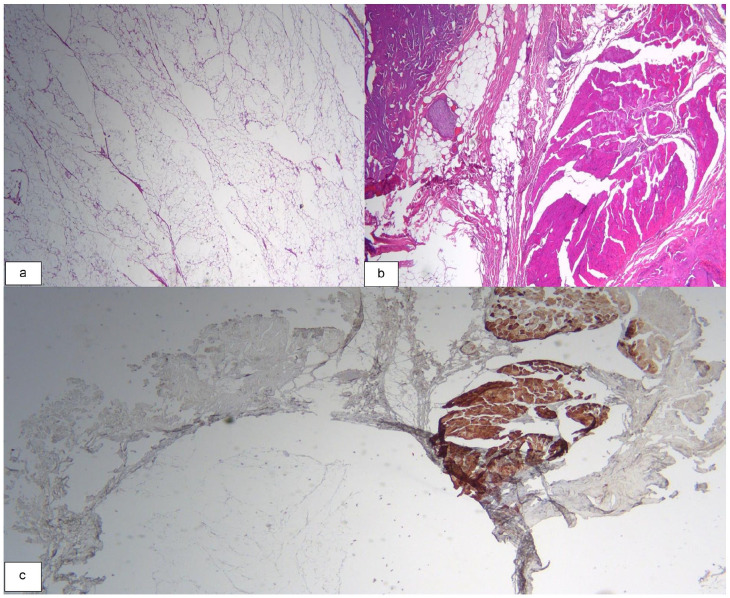
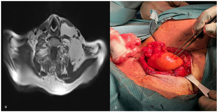
Intramuscular lipomas, typically found in subcutaneous tissue, rarely affect deeper muscular planes, especially those of the head and neck region. The following are 3 cases of intramuscular lipomas involving the sternocleidomastoid muscle. The first 2 patients presented with painless, palpable masses confirmed by diagnostic imaging as well-circumscribed intramuscular lipomas. One was treated surgically, while the other was managed conservatively with monitoring and close follow-up. The third patient reported dysphagia associated with occasional dyspnea and mild pain. The mass was identified as infiltrative lipoma and was resected surgically. Complete tumor removal with no recurrence at 6 months was observed for the first and last cases. The second case was serially followed at 3 and 6 months with no interval changes. We report the largest case series on intramuscular lipomas of the sternocleidomastoid muscle to enhance our understanding of this rare entity.
Keywords: Intramuscular; benign pathology; lipoma; lipomatous tumor; sternocleidomastoid muscle.
© The Author(s) 2024.
Conflict of interest statement
The author(s) declared no potential conflicts of interest with respect to the research, authorship, and/or publication of this article.
Figures
Similar articles
-
Intramuscular lipoma of the sternocleidomastoid muscle.J Craniofac Surg. 2010 Nov;21(6):1976-8. doi: 10.1097/SCS.0b013e3181f502cd. J Craniofac Surg. 2010. PMID: 21119474 Review.
-
Intramuscular benign lipoma of the sternocleidomastoid muscle: a rare cause of neck mass.Eur Arch Otorhinolaryngol. 2005 Feb;262(2):148-50. doi: 10.1007/s00405-003-0732-6. Epub 2004 Jun 10. Eur Arch Otorhinolaryngol. 2005. PMID: 15197561
-
Well-circumscribed intramuscular lipoma of the sternocleidomastoid muscle.Auris Nasus Larynx. 2004 Sep;31(3):283-5. doi: 10.1016/j.anl.2004.03.017. Auris Nasus Larynx. 2004. PMID: 15364365 Review.
-
Giant Lipoma in the Trapezius Muscle: A Rare Case Report.Aesthet Surg J Open Forum. 2024 Sep 26;6:ojae081. doi: 10.1093/asjof/ojae081. eCollection 2024. Aesthet Surg J Open Forum. 2024. PMID: 39430212 Free PMC article.
-
Painful Intramuscular Lipoma of the Infraspinatus: Unusual Location and Presentation.Orthopedics. 2016 Mar-Apr;39(2):e370-3. doi: 10.3928/01477447-20160307-03. Epub 2016 Mar 11. Orthopedics. 2016. PMID: 26966945
References
-
- Johnson CN, Ha AS, Chen E, Davidson D. Lipomatous soft-tissue tumors. J Am Acad Orthop Surg. 2018;26:779-788. - PubMed
-
- Pélissier A, Sawaf MH, Shabana AH. Infiltrating (intramuscular) benign lipoma of the head and neck. J Oral Maxillofac Surg. 1991;49:1231-1236. - PubMed
-
- de Bree E, Karatzanis A, Hunt JL, et al.. Lipomatous tumours of the head and neck: a spectrum of biological behaviour. Eur Arch Otorhinolaryngol. 2015;272:1061-1077. - PubMed
-
- Fletcher CD, Martin-Bates E. Intramuscular and intermuscular lipoma: neglected diagnoses. Histopathology. 1988;12:275-287. - PubMed
Publication types
LinkOut - more resources
Full Text Sources
