. 2024 Oct 3;147(10):3624-3637.
doi: 10.1093/brain/awae187.

The visual cortex in the blind but not the auditory cortex in the deaf becomes multiple-demand regions

Affiliations

The visual cortex in the blind but not the auditory cortex in the deaf becomes multiple-demand regions

Hasan Duymuş et al. Brain. .

Abstract

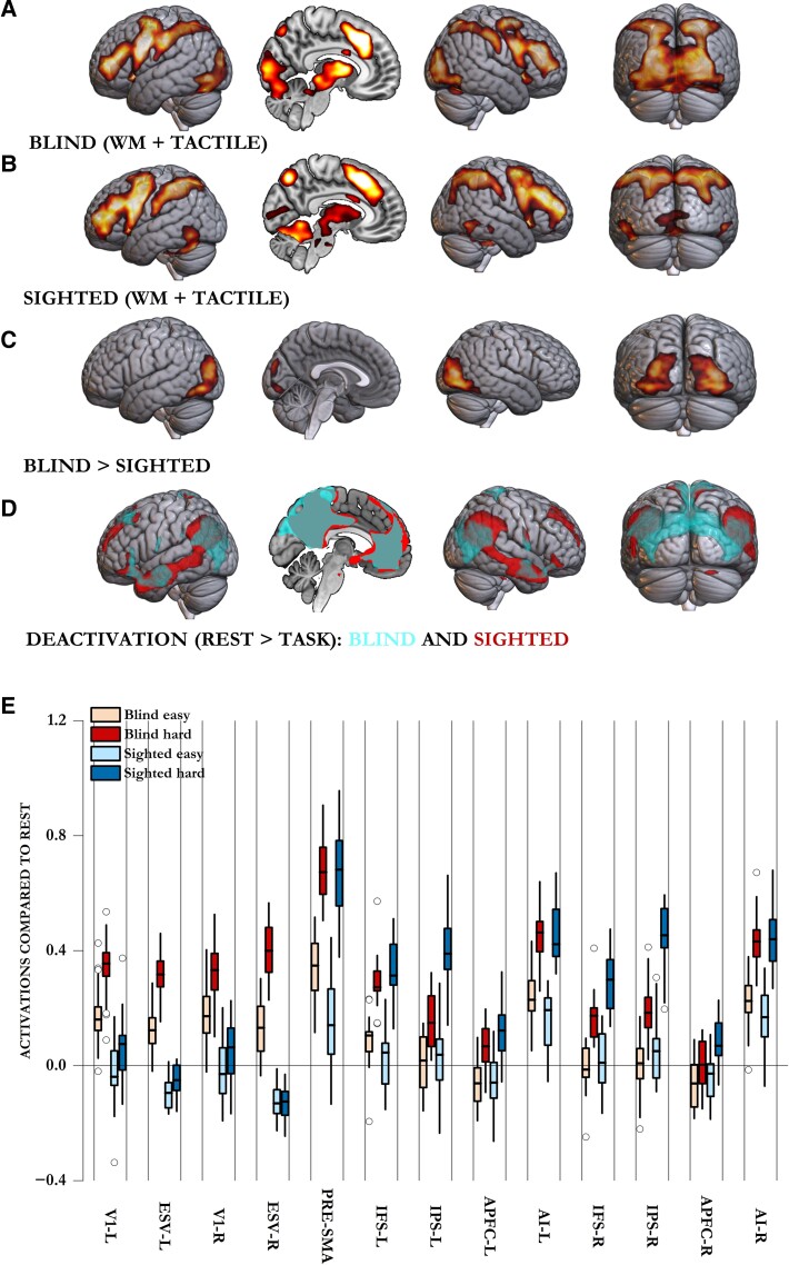
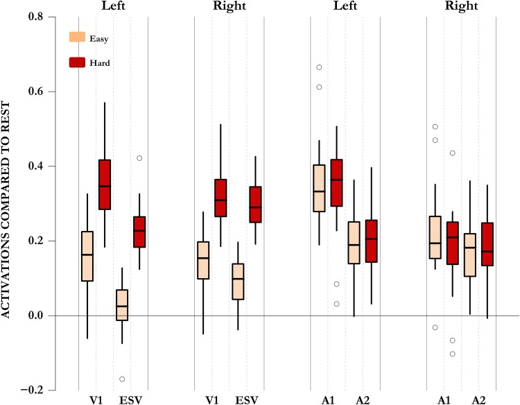
The fate of deprived sensory cortices (visual regions in the blind and auditory regions in the deaf) exemplifies the extent to which experience can change brain regions. These regions are frequently seen to activate during tasks involving other sensory modalities, leading many authors to infer that these regions have started to process sensory information of other modalities. However, such observations can also imply that these regions are now activating in response to any task event, regardless of the sensory modality. Activating in response to task events, irrespective of the sensory modality involved, is a feature of the multiple-demands (MD) network. This is a set of regions within the frontal and parietal cortices that activate in response to any kind of control demand. Thus, demands as diverse as attention, perceptual difficulty, rule-switching, updating working memory, inhibiting responses, decision-making and difficult arithmetic all activate the same set of regions that are thought to instantiate domain-general cognitive control and underpin fluid intelligence. We investigated whether deprived sensory cortices, or foci within them, become part of the MD network. We tested whether the same foci within the visual regions of the blind and auditory regions of the deaf activated in response to different control demands. We found that control demands related to updating auditory working memory, difficult tactile decisions, time-duration judgments and sensorimotor speed all activated the entire bilateral occipital regions in the blind but not in the sighted. These occipital regions in the blind were the only regions outside the canonical frontoparietal MD regions to show such activation in response to multiple control demands. Furthermore, compared with the sighted, these occipital regions in the blind had higher functional connectivity with frontoparietal MD regions. Early deaf, in contrast, did not activate their auditory regions in response to different control demands, showing that auditory regions do not become MD regions in the deaf. We suggest that visual regions in the blind do not take a new sensory role but become part of the MD network, and this is not a response of all deprived sensory cortices but a feature unique to the visual regions.

Keywords: blind; cognitive control; deaf; multiple-demands network; neuroplasticity.


Conflict of interest statement

The authors report no competing interests.

Figures

Figure 1
Figure 1
Overview of the four functional MRI tasks: tactile decision-making, auditory working memory-updating, time-duration judgement and motor speed tasks. All four tasks had an easy block and a hard block, each lasting 15–30 s. (A) Tactile decision-making: participants decided which of the two shapes was larger in size. The task was done using a plexiglass tablet that had an easy part and a hard part, each of which had five trials. Each trial involved a pair of shapes drawn with raised margins. Margins of shapes in easy trials were more raised, making them easier to perceive by touch. Furthermore, the difference in size between shapes of a pair was larger in easy trials. (B) Auditory working memory-updating: each block had 10 trials. Participants decided whether the letter they heard in a given trial was the same as one trial (on easy blocks) or three (on hard blocks) trials prior. (C) Time-duration judgement: each block had 10 trials. In each trial, participants heard two sequential tones of variable durations separated by a silent pause. They had to decide which of the two was longer in duration. Tones in easy blocks had larger differences in their durations, making it easier to discern the longer tone. (D) Motor speed: participants heard one of four numbers (1–4) and had to press the corresponding button. In easy blocks, they had 1.5 s to respond, whereas in hard blocks they had only 0.5 s. Deaf participants did a visual instead of auditory working memory-updating task (see the ‘Materials and methods’ section).
Figure 2
Figure 2
Multiple demand regions, whole-brain activation, global maxima and occipital voxel response of the blind across the four diverse cognitive demands. (A) Multiple demand (MD) regions include prefrontal regions extending from the inferior frontal junction along the inferior frontal sulcus and middle frontal gyrus up to the anterior prefrontal cortex, frontal operculum extending into the anterior insula, frontal eye fields, a blob extending from the pre-supplementary motor area to the anterior cingulate cortex, and parietal regions along the intraparietal sulcus. White circles show the location of spherical regions of interest used in later analyses (shown in Figs 3 and 6). (B) Combined across the four tasks, almost the entire occipital cortices in the blind, along with the frontoparietal MD regions, showed higher activation during hard compared with easy blocks. This was also the case in each of the four tasks individually. (C) Global maxima (or the voxel most intensely activating to control demands) occurred most frequently in these occipital regions. Lateral prefrontal cortex (PFC) in this analysis included the inferior frontal sulcus, inferior frontal junction, middle frontal gyrus, anterior prefrontal cortex, frontal operculum and anterior insula, bilaterally. IPS included the intraparietal sulcus, superior parietal lobule and the upper half of the inferior parietal lobule. The SMA and ACC included the pre-supplementary motor area, anterior cingulate cortex and any global maxima on the medial side of the prefrontal cortices. (D) Response of voxels that activated intensely to one control demand, to the remaining three control demands. BF10 represents the likelihood of these voxels activating across the three remaining control demands. WM = working memory.
Figure 3
Figure 3
Comparison of blind and sighted participants across tactile decision-making and auditory working memory-updating tasks. Although both blind and sighted participants activated their canonical frontoparietal MD regions during the hard blocks of these tasks (A and B), only blind participants activated their occipital regions. Interestingly, sighted participants did activate two visual regions (BA 37 in posterior inferior temporal lobes and small clusters in V1) across the control demands of these non-visual tasks. These, however, were lower in intensity and spread than in blind participants. (C) Whole-brain contrast of blind versus sighted participants showed that the two control demands activated visual occipital regions of the blind significantly more than those of the sighted. (D) Sighted participants, unlike blind participants, showed task-related deactivation of parts of their extrastriate occipital regions. (E) Activations compared with rest across occipital and frontoparietal MD regions. Occipital regions in the blind activated significantly more during tasks than rest and during hard compared with easy blocks. These regions in the sighted either deactivated (ESV) or did not activate more than rest (V1 during easy blocks). Only during the hard blocks did V1 activate slightly more than the rest. In the sighted, hard blocks did not elicit significantly higher activity than easy blocks. Although both blind and sighted participants showed frontoparietal MD activation during hard compared with easy blocks, this activation was lower in the blind. AI = anterior insula; APFC = anterior prefrontal cortex; ESV = extrastriate visual cortex; IFS = inferior frontal sulcus; IPS = intraparietal sulcus; L = left; MD = multiple demand; pre-SMA = pre-supplementary motor area; R = right; V1 = primary visual cortex; WM = working memory.
Figure 4
Figure 4
Functional connectivity in the blind compared with the sighted. Regions that showed higher functional connectivity in the blind compared with the sighted when prefrontal MD regions were seeded (top) and when occipital regions were seeded (bottom). Seeded regions are marked in green. The prefrontal seed region included the inferior frontal sulcus and the middle frontal gyrus extending up to anterior prefrontal regions. Occipital seed included medial and lateral occipital regions (Brodmann area 17, 18 and 19). Seeding prefrontal MD regions showed higher connectivity between them and visual occipital regions in the blind compared with sighted participants. Seeding occipital cortices showed higher connectivity in the blind compared with sighted participants, between them and key MD regions (middle frontal gyrus, inferior frontal sulcus, premotor regions, anterior insula, pre-supplementary motor areas and intraparietal sulcus) in addition to temporoparietal junctions. MD = multiple demand.
Figure 5
Figure 5
Responses of visual and auditory regions of the blind to the auditorily presented tasks. Responses of occipital and auditory regions of blind participants to easy and hard blocks of auditorily presented tasks (auditory WM-updating, time judgement and sensorimotor speed). Note that both V1 and extrastriate visual regions (ESV) activated intensely to control demands (evident in very high BF10 values from Bayesian paired t-tests in Table 1), whereas neither the primary nor the secondary auditory cortices showed any evidence of control-related activation. In these areas, there was significant evidence in favour of the null hypothesis that these regions did not activate to control demands (BF01 > 3; Table 1). A1 = primary auditory cortex; A2 = secondary auditory cortex; BF = Bayes factor; ESV = extrastriate visual cortex; V1 = primary visual cortex; WM = working memory.
Figure 6
Figure 6
Responses of primary and secondary auditory regions in the deaf during tactile decision-making and working memory-updating tasks. Response of anatomically localized auditory regions and frontoparietal MD regions in the deaf across the two control demands (tactile decision-making and visual WM-updating). Note that none of the auditory regions showed any activation above the resting baseline during easy or hard task blocks, nor did any of these regions show higher activation during hard compared with easy blocks. Numbers above auditory ROI plots show the Bayes factor in favour of the null hypothesis (BF01) that hard and easy blocks did not differ in their activation levels. Most auditory regions showed significant evidence in favour of this null hypothesis (BF01 > 3). BF = Bayes factor; L = left; MD = multiple demand; R = right; ROI = region of interest; WM = working memory.

Similar articles

Cited by

References

    1. Burton H, Snyder AZ, Diamond JB, Raichle ME. Adaptive changes in early and late blind: A fMRI study of verb generation to heard nouns. J Neurophysiol. 2002;88:3359–3371. - PMC - PubMed
    1. Ding H, Qin W, Liang M, et al. . Cross-modal activation of auditory regions during visuo-spatial working memory in early deafness. Brain. 2015;138:2750–2765. - PubMed
    1. Finney EM, Fine I, Dobkins KR. Visual stimuli activate auditory cortex in the deaf. Nat Neurosci. 2001;4:1171–1173. - PubMed
    1. Kanjlia S, Loiotile RE, Harhen N, Bedny M. ‘Visual’ cortices of congenitally blind adults are sensitive to response selection demands in a go/no-go task. Neuroimage. 2021;236:118023. - PMC - PubMed
    1. Kupers R, Beaulieu-Lefebvre M, Schneider FC, et al. . Neural correlates of olfactory processing in congenital blindness. Neuropsychologia. 2011;49:2037–2044. - PubMed