Comparative Study
. 2024 Jul 2;26(7):euae158.
doi: 10.1093/europace/euae158.

Electroanatomical adaptations in the guinea pig heart from neonatal to adulthood

Affiliations
Comparative Study

Electroanatomical adaptations in the guinea pig heart from neonatal to adulthood

Kazi T Haq et al. Europace. .

Abstract

Aims: Electroanatomical adaptations during the neonatal to adult phase have not been comprehensively studied in preclinical animal models. To explore the impact of age as a biological variable on cardiac electrophysiology, we employed neonatal and adult guinea pigs, which are a recognized animal model for developmental research.

Methods and results: Electrocardiogram recordings were collected in vivo from anaesthetized animals. A Langendorff-perfusion system was employed for the optical assessment of action potentials and calcium transients. Optical data sets were analysed using Kairosight 3.0 software. The allometric relationship between heart weight and body weight diminishes with age, it is strongest at the neonatal stage (R2 = 0.84) and abolished in older adults (R2 = 1E-06). Neonatal hearts exhibit circular activation, while adults show prototypical elliptical shapes. Neonatal conduction velocity (40.6 ± 4.0 cm/s) is slower than adults (younger: 61.6 ± 9.3 cm/s; older: 53.6 ± 9.2 cm/s). Neonatal hearts have a longer action potential duration (APD) and exhibit regional heterogeneity (left apex; APD30: 68.6 ± 5.6 ms, left basal; APD30: 62.8 ± 3.6), which was absent in adults. With dynamic pacing, neonatal hearts exhibit a flatter APD restitution slope (APD70: 0.29 ± 0.04) compared with older adults (0.49 ± 0.04). Similar restitution characteristics are observed with extrasystolic pacing, with a flatter slope in neonates (APD70: 0.54 ± 0.1) compared with adults (younger: 0.85 ± 0.4; older: 0.95 ± 0.7). Neonatal hearts display unidirectional excitation-contraction coupling, while adults exhibit bidirectionality.

Conclusion: Postnatal development is characterized by transient changes in electroanatomical properties. Age-specific patterns can influence cardiac physiology, pathology, and therapies for cardiovascular diseases. Understanding heart development is crucial to evaluating therapeutic eligibility, safety, and efficacy.

Keywords: Cardiac electrophysiology; Developmental biology; Optical mapping; Pediatric models.


Conflict of interest statement

Conflict of interest: None declared.

Figures

Graphical Abstract
Graphical Abstract
Age-dependent adaptations in the guinea pig heart include adjustments in allometric scaling and cardiac electrophysiology.
Figure 1
Figure 1
Heart weight–body weight relationship. A) The linear regression of normal heart weight vs. body weight in neonatal (n = 10), younger adult (n = 13), and older adult guinea pigs (n = 26). B) The regression slope of heart weight and body weight across different age groups. C) The heart weight-to-body weight ratio in each age group (mean ± SEM). Comparisons by Welch’s ANOVA (unequal standard deviations between groups) with multiple comparisons test, **P < 0.01.
Figure 2
Figure 2
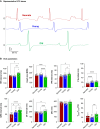
In vivo ECG recordings across different age groups. A) Representative ECG traces (lead I) from neonatal (red), younger adult (blue), and older adult guinea pigs (green). ECGs were recorded in anaesthetized animals using subcutaneous needle electrodes. B) Comparison of ECG metrics between neonates (n = 10), young adults (n = 13), and older adults (n = 9). All comparisons by ordinary one-way ANOVA with multiple comparisons test (with the exception of Welch’s ANOVA for RR interval due to unequal standard deviations). Individual replicates are shown; values are reported as mean ± SEM. *P < 0.05, **P < 0.01, ***P < 0.005, ****P < 0.001. ECG, electrocardiogram.
Figure 3
Figure 3
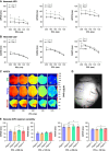
Ventricular activation dynamics differ across guinea pig age groups. A) Illustrative examples of ventricular AP activation maps (time of dV/dtmax) in a neonate, younger adult, and older adult guinea pig heart. Electrical pacing (160 ms PCL) was applied near the geometric centre, revealing a distinct activation pattern in neonatal hearts. B) Illustrative examples of epicardial CV measured from the earliest to the latest activation site, as indicated by the single CV vector (black arrow). Electrical pacing (160 ms PCL) was applied near the apex. C) Epicardial CV restitution curves during S1–S1 pacing (220–120 ms PCL), featuring neonates (red, n = 7), younger adults (blue, n = 11), and older adults (green, n = 21). D) Illustration of connexin-43 (red) localization in ventricular myocardium from neonatal and adult guinea pig; nuclei stained with DAPI (blue). 50 μm scale. E) Measurements of intercalated disc length and connexin-43 gene expression (Gja1). Replicate values are reported as mean ± SEM. Comparisons by two-way ANOVA with multiple comparisons; *P < 0.05 compared to neonate (red), young adult (blue), or old adult (green). AP, action potential; CV, conduction velocity; PCL, pacing cycle length.
Figure 4
Figure 4
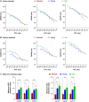
Cardiac electrical restitution properties in response to dynamic stimulation. A) Action potential duration restitution curves at APD30, APD50, and APD70 in neonatal (n = 8), younger adult (n = 6–7), and older adult hearts (n = 18–21). B) Calcium transient duration restitution curves at CaD30, CaD50, and CaD70 in neonatal (n = 9), younger adult (n = 10), and older adult hearts (n = 12). C) Maximum restitution slopes; individual replicates shown. Ventricles were dynamically paced (S1–S1) at the apex; the PCL was decremented from 220 to 120 ms at 20 ms intervals. D) APD50 (top), CaD50 (middle), and heart rate (bottom) measured during NSR in the excised hearts. Values reported as mean ± SEM. Comparisons by two-way ANOVA with multiple comparisons; *P < 0.01, **P < 0.01, ****P < 0.001 compared with neonate (red), young adult (blue), or old adult (green). NSR, normal sinus rhythm; PCL, pacing cycle length.
Figure 5
Figure 5
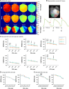
Intragroup and regional variability in neonatal guinea pig hearts (dynamic stimulation). A) Action potential duration and B) CaD restitution curves generated from neonatal hearts isolated on postnatal Day 0 (n = 4–5) and postnatal Day 1 (n = 4). Dynamic pacing (S1–S1) was applied to the apex; the PCL was decremented from 200 to 120 ms. Values reported as mean ± SEM. Comparison by two-way ANOVA with multiple comparisons, *P < 0.05. C) Illustrative APD70 maps of a neonate, younger adult, and older adult heart in response to dynamic S1–S1 apical pacing. Note the regional variability in the neonatal heart. D) Schematic of four epicardial ventricular regions of interest. E) APD30 and APD70 measurements from neonatal hearts, were collected in response to a slow pacing rate (200 ms PCL, n = 7) and faster pacing rate (120 ms PCL, n = 6). Values reported as mean ± SEM. Comparison by one-way (matched) ANOVA with multiple comparisons; *P < 0.05, **P < 0.01 between different regions. APD, action potential duration; CaD, calcium transient duration; PCL, pacing cycle length.
Figure 6
Figure 6
Cardiac electrical restitution properties in response to extrasystolic stimulation. A) Action potential duration restitution curves at APD30, APD50, and APD70 in neonatal (n = 5–6), younger adult (n = 5), and older adult hearts (n = 6–11). B) Calcium transient duration restitution curves at CaD30, CaD50, and CaD70 in neonatal (n = 5–6), younger adult (n = 5), and older adult hearts (n = 6). C) Maximum restitution slopes; individual replicates shown. Extrasystolic stimulation was applied to the ventricle (S1–S2) at the apex. Note the different PCL ranges that were achievable in neonatal vs. adult hearts. Values reported as mean ± SEM. Statistical comparisons between slope measurements, as determined by one-way ANOVA with multiple comparisons; *P < 0.05, **P < 0.01. APD, action potential duration; CaD, calcium transient duration; PCL, pacing cycle length.
Figure 7
Figure 7
Regional variability in neonatal guinea pig hearts (extrasystolic stimulation). A) Illustrative APD50 maps of a neonate, younger adult, and older adult heart in response to extrasystolic S1–S2 apical pacing (maps highlight the ‘S2’ beat). B) Representative AP traces from two regions of the neonatal heart (Region 2: left basal in orange vs. Region 3: right apex in green). C) Regional heterogeneity in (APD and CaD restitution curves generated from neonatal hearts (n = 6–7). D) and E) APD70 and CaD70 restitution curves generated from younger (n = 6–9) and older adult hearts (n = 6–8). Values reported as mean ± SEM. Comparison by two-way ANOVA with multiple comparisons, *P < 0.05 between denoted specific regions. AP, action potential; APD, action potential duration; CaD, calcium transient duration.
Figure 8
Figure 8
Action potential and CaT coupling characteristics across guinea pig age groups. A) Top: Representative traces of AP and CaT were recorded simultaneously from a neonate, younger adult, and older adult heart. Bottom: Representative AP–CaT latency maps (CaD–APD) measured at 30 and 70% duration. B) Quantitative comparison of AP–CaT latency at 30, 50, and 70% duration in neonatal (n = 4), younger adult (n = 6), and older adult (n = 8) guinea pigs. Positive values indicate that the CaD is longer, while negative values indicate that the APD is longer for a given measurement. All measurements were recorded in response to dynamic (S1–S1) ventricular epicardial pacing at the apex. Values reported as mean ± SEM. AP, action potential; APD, action potential duration; CaD, calcium transient duration; CaT, calcium transient.

Update of

Similar articles

Cited by

References

    1. Abman SH, Baker C, Gien J, Mourani P, Galambos C. The Robyn Barst memorial lecture: differences between the fetal, newborn, and adult pulmonary circulations: relevance for age-specific therapies (2013 Grover Conference series). Pulm Circ 2014;4:424–40. - PMC - PubMed
    1. Packer M, Carver JR, Rodeheffer RJ, Ivanhoe RJ, DiBianco R, Zeldis SM et al. Effect of oral milrinone on mortality in severe chronic heart failure. The PROMISE study research group. N Engl J Med 1991;325:1468–75. - PubMed
    1. Shipley JB, Tolman D, Hastillo A, Hess ML. Milrinone: basic and clinical pharmacology and acute and chronic management. Am J Med Sci Elsevier 1996;311:286–91. - PubMed
    1. Tan CMJ, Lewandowski AJ. The transitional heart: from early embryonic and fetal development to neonatal life. Fetal Diagn Ther 2020;47:373–86. - PMC - PubMed
    1. Lue HC, Wu MH, Wang JK, Lin MT, Lu CW, Chiu SN et al. Normal ECG standards and percentile charts for infants, children and adolescents. Pediatr Neonatol 2023;64:256–73. - PubMed

Publication types