Review
. 2024 Jun 6;13(1):2351789.
doi: 10.1080/20450907.2024.2351789. Epub 2024 Jun 12.

Metastatic glioblastoma to the lungs: a case report and literature review

Affiliations
Review

Metastatic glioblastoma to the lungs: a case report and literature review

Carlen A Yuen et al. CNS Oncol. .

Abstract

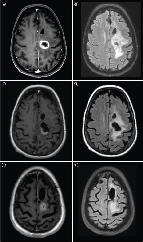
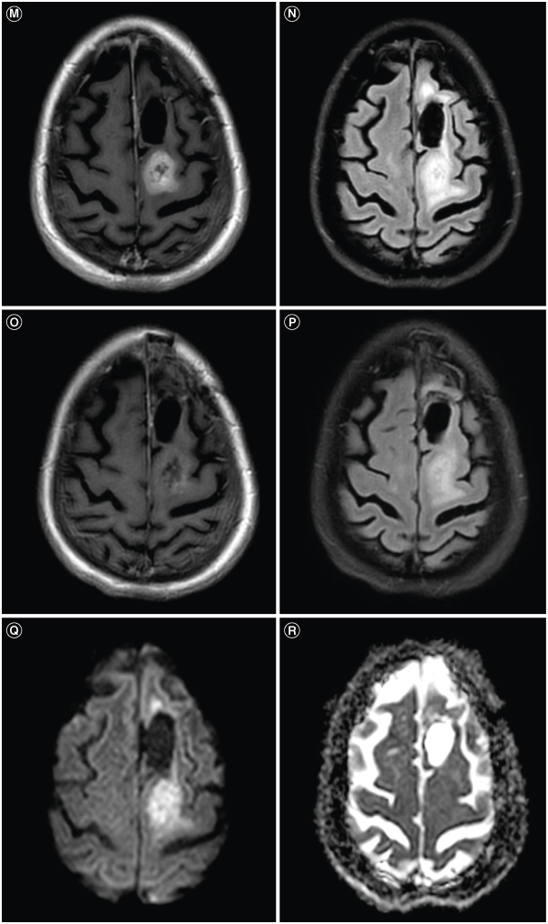
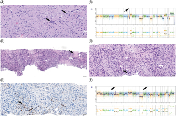
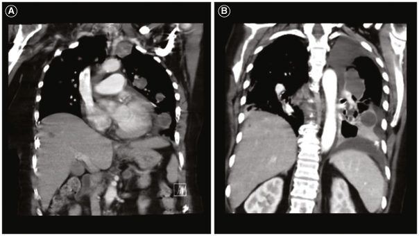
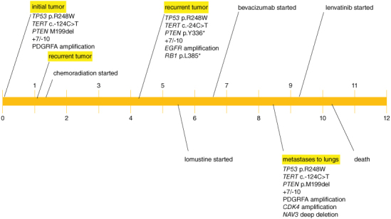
Glioblastoma is the most common malignant primary brain tumor. Despite its infiltrative nature, extra-cranial glioblastoma metastases are rare. We present a case of a 63-year-old woman with metastatic glioblastoma in the lungs. Sarcomatous histology, a reported risk factor for disseminated disease, was found. Genomic alterations of TP53 mutation, TERT mutation, PTEN mutation, and +7/-10 were also uncovered. Early evidence suggests these molecular aberrations are common in metastatic glioblastoma. Treatment with third-line lenvatinib resulted in a mixed response. This case contributes to the growing body of evidence for the role of genomic alterations in predictive risk in metastatic glioblastoma. There remains an unmet need for treatment of metastatic glioblastoma.

Keywords: VEGF; extra-neural glioblastoma; gliosarcoma; lenvatinib; metastatic glioblastoma; vascular endothelial growth factor inhibitor.

Plain language summary

Glioblastoma is the most common malignant primary brain tumor. Glioblastoma can spread into healthy tissue, but metastases beyond the brain are rare. We present a case of a 63-year-old woman with metastatic glioblastoma in the lungs. We identified risk factors associated with spread beyond the brain, including factors related to tissue structure and specific molecular alterations. Treatment with third-line lenvatinib resulted in a mixed response. This case adds to the limited existing data for the use of molecular alterations to serve as risk factors for metastatic glioblastoma. Treatment options are needed for this devastating disease.


Conflict of interest statement

Xiao-Tang Kong, MD received honorarium from Zai Lab for invited speeches for symposiums prior to July of 2021. The authors have no other competing interests or relevant affiliations with any organization or entity with the subject matter or materials discussed in the manuscript apart from those disclosed.

Figures

Figure 1.
Figure 1.
Patient's intracranial glioblastoma throughout the therapy course. (A) Preoperative axial T1 post-contrast brain MRI shows a left frontal heterogeneously enhancing mass. (B) Preoperative axial T2/FLAIR brain MRI shows surrounding vasogenic edema. (C) Postoperative axial T1 postcontrast brain MRI shows near total resection of the previously seen enhancing tumor. (D) Post-operative axial T2/FLAIR brain MRI shows persistent hyperintensity. (E) Radiation planning axial T1 post-contrast brain MRI shows interval development of contrast-enhancing lesions at the posterior margin of the resection cavity and posterior to the resection cavity (yellow arrows). (F) Radiation planning axial T2/FLAIR brain MRI shows hyperintensity associated with the new enhancing lesions. (G) Post-radiation axial T1 post-contrast brain MRI shows interval enlargement of the prior lesion posterior to the resection cavity. (H) Post-radiation T2/FLAIR axial brain MRI shows surrounding vasogenic edema. (I) Re-resection postoperative T1 postcontrast axial brain MRI shows near total resection of the prior enhancing mass. (J) Re-resection postoperative T2/FLAIR axial brain MRI shows improvement in the surrounding vasogenic edema. (K) Post 2 cycles of lomustine and 3 cycles of bevacizumab, T1 post-contrast axial brain MRI shows interval development of a contrast-enhancing mass. (L) Post 2 cycles of lomustine and 3 cycles of bevacizumab, T2/FLAIR axial brain MRI shows surrounding vasogenic edema. (M) Post 2 cycles of lomustine and 4 cycles of bevacizumab, T1 post-contrast axial brain MRI shows interval enlargement of the contrast-enhancing mass. (N) Post 2 cycles of lomustine and 4 cycles of bevacizumab, T2/FLAIR axial brain MRI shows surrounding vasogenic edema. (O) Post 10 days of lenvatinib, T1 post-contrast axial brain MRI shows an interval decrease in the previously seen contrast-enhancing mass. (P) Post 10 days of lenvatinib, T2/FLAIR axial brain MRI shows an interval decrease in the previously seen T2/FLAIR hyperintensity. (Q) Post 10 days of lenvatinib, DWI axial brain MRI shows associated abnormal signals with the aforementioned mass. (R) Post 10 days of lenvatinib, ADC axial brain MRI shows minimal ADC correlate along the lateral margin of the mass.
Figure 1.
Figure 1.
Patient's intracranial glioblastoma throughout the therapy course. (A) Preoperative axial T1 post-contrast brain MRI shows a left frontal heterogeneously enhancing mass. (B) Preoperative axial T2/FLAIR brain MRI shows surrounding vasogenic edema. (C) Postoperative axial T1 postcontrast brain MRI shows near total resection of the previously seen enhancing tumor. (D) Post-operative axial T2/FLAIR brain MRI shows persistent hyperintensity. (E) Radiation planning axial T1 post-contrast brain MRI shows interval development of contrast-enhancing lesions at the posterior margin of the resection cavity and posterior to the resection cavity (yellow arrows). (F) Radiation planning axial T2/FLAIR brain MRI shows hyperintensity associated with the new enhancing lesions. (G) Post-radiation axial T1 post-contrast brain MRI shows interval enlargement of the prior lesion posterior to the resection cavity. (H) Post-radiation T2/FLAIR axial brain MRI shows surrounding vasogenic edema. (I) Re-resection postoperative T1 postcontrast axial brain MRI shows near total resection of the prior enhancing mass. (J) Re-resection postoperative T2/FLAIR axial brain MRI shows improvement in the surrounding vasogenic edema. (K) Post 2 cycles of lomustine and 3 cycles of bevacizumab, T1 post-contrast axial brain MRI shows interval development of a contrast-enhancing mass. (L) Post 2 cycles of lomustine and 3 cycles of bevacizumab, T2/FLAIR axial brain MRI shows surrounding vasogenic edema. (M) Post 2 cycles of lomustine and 4 cycles of bevacizumab, T1 post-contrast axial brain MRI shows interval enlargement of the contrast-enhancing mass. (N) Post 2 cycles of lomustine and 4 cycles of bevacizumab, T2/FLAIR axial brain MRI shows surrounding vasogenic edema. (O) Post 10 days of lenvatinib, T1 post-contrast axial brain MRI shows an interval decrease in the previously seen contrast-enhancing mass. (P) Post 10 days of lenvatinib, T2/FLAIR axial brain MRI shows an interval decrease in the previously seen T2/FLAIR hyperintensity. (Q) Post 10 days of lenvatinib, DWI axial brain MRI shows associated abnormal signals with the aforementioned mass. (R) Post 10 days of lenvatinib, ADC axial brain MRI shows minimal ADC correlate along the lateral margin of the mass.
Figure 2.
Figure 2.
Patient's residual/recurrent glioblastoma patholog. (A) Histologic section of the residual/recurrent glioblastoma with giant cell features (black arrows), Hematoxylin and Eosin, scale bar 50 microns. (B) Whole genome copy number profile of residual/recurrent glioblastoma showing gains of chromosomes 7, 19 and 20, losses of chromosomes 10, 13, 18, 21 and 22 and amplification of EGFR on chromosome 7 (black arrow). (C) Histologic section of the lung mass demonstrating predominantly spindle cell neoplasm with areas of necrosis (black arrow) morphologically consistent with residual/recurrent glioblastoma with sarcomatous features. Hematoxylin and Eosin, scale bar 100 microns. (D) Histologic section of the lung mass demonstrating predominantly spindle cell neoplasm consistent with residual/recurrent glioblastoma with sarcomatous features, scattered multinucleated giant cells (black arrow) are present similar to the patient's intracranial tumor. Hematoxylin and Eosin, scale bar 50 microns. (E) Immunohistochemical stain for GFAP performed on lung mass showed scattered positive cells (black arrow), confirming the glial nature of the neoplasm. Scale bar 50 microns. (F) Whole genome copy number profile of lung mass showing gains of chromosomes 1p, 7 and 20, losses of chromosomes 4, 10, 13, 16, 18, 21 and 22 and amplifications of PDGFRA and CDK4 on chromosomes 4 and 12, respectively (black arrows).
Figure 3.
Figure 3.
Patient's metastatic glioblastoma to the lungs. (A) Coronal computed tomography (CT) chest shows multiple enhancing pulmonary metastases. (B) Coronal CT chest shows further progression of pulmonary metastases and new hepatic metastases.
Figure 4.
Figure 4.
Patient disease course (timeline in months).

Similar articles

Cited by

References

    1. Ostrom QT, Price M, Neff C, et al. . CBTRUS Statistical Report: Primary Brain and Other Central Nervous System Tumors Diagnosed in the United States in 2015–2019. Neuro Oncol. 2022;24(Suppl. 5):v1–v95. doi:10.1093/neuonc/noac202 - DOI - PMC - PubMed
    1. Liu J, Shen L, Tang G, et al. . Multiple extracranial metastases from glioblastoma multiforme: a case report and literature review. J Int Med Res. 2020;48(6):300060520930459. doi:10.1177/0300060520930459 - DOI - PMC - PubMed
    1. Carvalho J, Barbosa CCL, Feher O, et al. . Systemic dissemination of glioblastoma: literature review. Rev Assoc Med Bras (1992). 2019;65(3):460–468. doi:10.1590/1806-9282.65.3.460 - DOI - PubMed
    1. Robert M, Wastie M. Glioblastoma multiforme: a rare manifestation of extensive liver and bone metastases. Biomed Imaging Interv J. 2008;4(1):e3. doi:10.2349/biij.4.1.e3 - DOI - PMC - PubMed
    1. Beauchesne P. Extra-neural metastases of malignant gliomas: myth or reality? Cancers (Basel). 2011;3(1):461–477. doi:10.3390/cancers3010461 - DOI - PMC - PubMed