

Streamlining Acute Abdominal Aortic Dissection Management-An AI-based CT Imaging Workflow
- PMID: 38864947
- PMCID: PMC11612133
- DOI: 10.1007/s10278-024-01164-0
Streamlining Acute Abdominal Aortic Dissection Management-An AI-based CT Imaging Workflow
Abstract
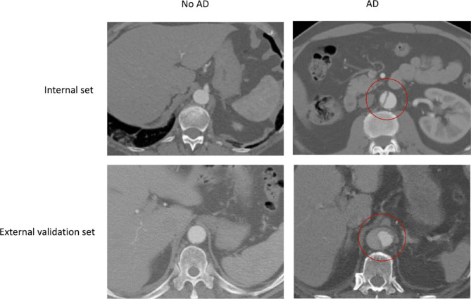
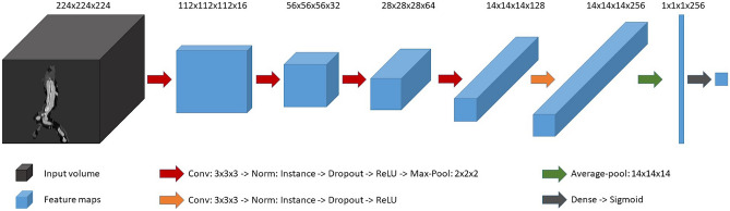
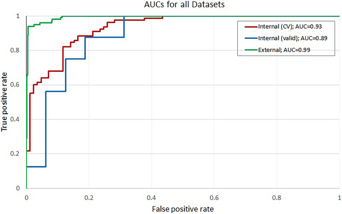
Life-threatening acute aortic dissection (AD) demands timely diagnosis for effective intervention. To streamline intrahospital workflows, automated detection of AD in abdominal computed tomography (CT) scans seems useful to assist humans. We aimed at creating a robust convolutional neural network (CNN)-based pipeline capable of real-time screening for signs of abdominal AD in CT. In this retrospective study, abdominal CT data from AD patients presenting with AD and from non-AD patients were collected (n 195, AD cases 94, mean age 65.9 years, female ratio 35.8%). A CNN-based algorithm was developed with the goal of enabling a robust, automated, and highly sensitive detection of abdominal AD. Two sets from internal (n = 32, AD cases 16) and external sources (n = 1189, AD cases 100) were procured for validation. The abdominal region was extracted, followed by the automatic isolation of the aorta region of interest (ROI) and highlighting of the membrane via edge extraction, followed by classification of the aortic ROI as dissected/healthy. A fivefold cross-validation was employed on the internal set, and an ensemble of the 5 trained models was used to predict the internal and external validation set. Evaluation metrics included receiver operating characteristic curve (AUC) and balanced accuracy. The AUC, balanced accuracy, and sensitivity scores of the internal dataset were 0.932 (CI 0.891-0.963), 0.860, and 0.885, respectively. For the internal validation dataset, the AUC, balanced accuracy, and sensitivity scores were 0.887 (CI 0.732-0.988), 0.781, and 0.875, respectively. Furthermore, for the external validation dataset, AUC, balanced accuracy, and sensitivity scores were 0.993 (CI 0.918-0.994), 0.933, and 1.000, respectively. The proposed automated pipeline could assist humans in expediting acute aortic dissection management when integrated into clinical workflows.
Keywords: Abdomen; Aortic dissection; Computed tomography; Convolutional neural network; Deep learning.
© 2024. The Author(s).
Conflict of interest statement
Declarations. Ethics Approval: This retrospective study was approved by the local institutional review board (2021–635). Written informed consent was not required due to the retrospective nature of the study population enrolled. Competing Interests: SOS: the Department of Radiology and Nuclear Medicine has general research agreements with Siemens Healthineers. FGZ: the Department of Computer Assisted Clinical Medicine has general research agreements with Siemens Healthineers. Others: no conflicts of interest declared.
Figures


Similar articles
-
Deep learning for automatic bowel-obstruction identification on abdominal CT.Eur Radiol. 2024 Sep;34(9):5842-5853. doi: 10.1007/s00330-024-10657-z. Epub 2024 Feb 22. Eur Radiol. 2024. PMID: 38388719
-
Deep learning algorithm for detection of aortic dissection on non-contrast-enhanced CT.Eur Radiol. 2021 Feb;31(2):1151-1159. doi: 10.1007/s00330-020-07213-w. Epub 2020 Aug 28. Eur Radiol. 2021. PMID: 32857203
-
Automatic Segmentation, Detection, and Diagnosis of Abdominal Aortic Aneurysm (AAA) Using Convolutional Neural Networks and Hough Circles Algorithm.Cardiovasc Eng Technol. 2019 Sep;10(3):490-499. doi: 10.1007/s13239-019-00421-6. Epub 2019 Jun 19. Cardiovasc Eng Technol. 2019. PMID: 31218516
-
Role of Artificial Intelligence in Detecting and Classifying Aortic Dissection: Where Are We? A Systematic Review and Meta-Analysis.Radiol Cardiothorac Imaging. 2025 Jun;7(3):e240353. doi: 10.1148/ryct.240353. Radiol Cardiothorac Imaging. 2025. PMID: 40503985
-
Artificial intelligence-powered solutions for automated aortic diameter measurement in computed tomography: a narrative review.Ann Transl Med. 2024 Dec 24;12(6):116. doi: 10.21037/atm-24-171. Epub 2024 Dec 18. Ann Transl Med. 2024. PMID: 39817238 Free PMC article. Review.
Cited by
-
Automated Detection and Differentiation of Stanford Type A and Type B Aortic Dissections in CTA Scans Using Deep Learning.Diagnostics (Basel). 2024 Dec 25;15(1):12. doi: 10.3390/diagnostics15010012. Diagnostics (Basel). 2024. PMID: 39795540 Free PMC article.
-
From promise to practice: a scoping review of AI applications in abdominal radiology.Abdom Radiol (NY). 2025 Jul 28. doi: 10.1007/s00261-025-05144-y. Online ahead of print. Abdom Radiol (NY). 2025. PMID: 40719923 Review.
-
Multi-Stage Cascaded Deep Learning-Based Model for Acute Aortic Syndrome Detection: A Multisite Validation Study.J Clin Med. 2025 Jul 7;14(13):4797. doi: 10.3390/jcm14134797. J Clin Med. 2025. PMID: 40649169 Free PMC article.
References
-
- Vilacosta I, San RJA, di BR, Eagle K, Estrera AL, Ferrera C, Kaji S, Nienaber CA, Riambau V, Sch äfers H-J, Serrano FJ, Song J-K, Maroto L (2021) Acute Aortic Syndrome Revisited. J Am Coll Cardiol 78:2106–2125. 10.1016/j.jacc.2021.09.022 - PubMed
-
- Bossone E, Eagle KA (2021) Epidemiology and management of aortic disease: aortic aneurysms and acute aortic syndromes. Nat Rev Cardiol 18:331–348. 10.1038/s41569-020-00472-6 - PubMed
-
- Benkert AR, Gaca JG (2021) Initial Medical Management of Acute Aortic Syndromes. In: Sellke FW, Coselli JS, Sundt TM, Bavaria JE, Sodha NR (eds) Aortic Dissection and Acute Aortic Syndromes. Springer International Publishing, Cham, pp 119–129
-
- Orabi NA, Quint LE, Watcharotone K, Nan B, Williams DM, Kim KM (2018) Distinguishing acute from chronic aortic dissections using CT imaging features. Int J Cardiovasc Imaging 34:1831–1840. 10.1007/s10554-018-1398-x - PubMed
MeSH terms
LinkOut - more resources
Full Text Sources
Medical