. 2025 Oct;17(5):3416-3428.
doi: 10.1007/s12602-024-10300-9. Epub 2024 Jun 12.

A Lactobacillus Combination Ameliorates Lung Inflammation in an Elastase/LPS-induced Mouse Model of Chronic Obstructive Pulmonary Disease

Affiliations

A Lactobacillus Combination Ameliorates Lung Inflammation in an Elastase/LPS-induced Mouse Model of Chronic Obstructive Pulmonary Disease

Huan-Ting Shen et al. Probiotics Antimicrob Proteins. 2025 Oct.

Abstract

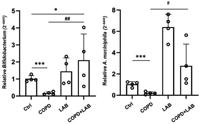
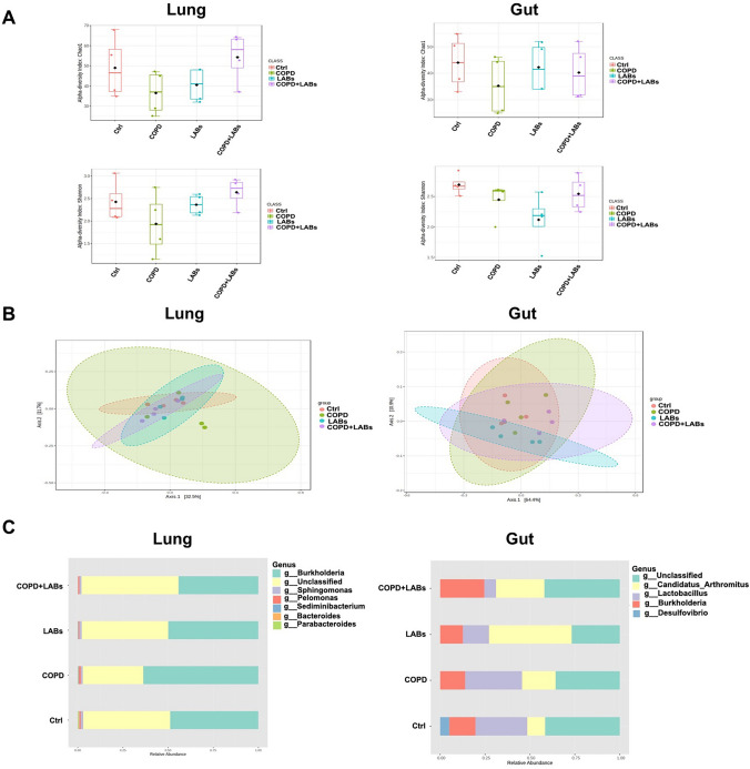
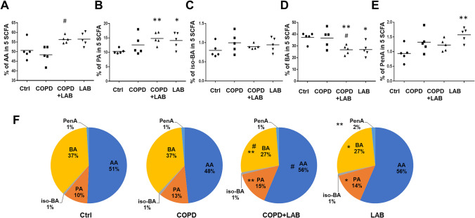
Chronic obstructive pulmonary disease (COPD) is the world's leading lung disease and lacks effective and specific clinical strategies. Probiotics are increasingly used to support the improvement of the course of inflammatory diseases. In this study, we evaluated the potential of a lactic acid bacteria (LAB) combination containing Limosilactobacillus reuteri GMNL-89 and Lacticaseibacillus paracasei GMNL-133 to decrease lung inflammation and emphysema in a COPD mouse model. This model was induced by intranasal stimulation with elastase and LPS for 4 weeks, followed by 2 weeks of oral LAB administration. The results showed that the LAB combination decreased lung emphysema and reduced inflammatory cytokines (IL-1β, IL-6, TNF-α) in the lung tissue of COPD mice. Microbiome analysis revealed that Bifidobacterium and Akkermansia muciniphila, reduced in the gut of COPD mice, could be restored after LAB treatment. Microbial α-diversity in the lungs decreased in COPD mice but was reversed after LAB administration, which also increased the relative abundance of Candidatus arthromitus in the gut and decreased Burkholderia in the lungs. Furthermore, LAB-treated COPD mice exhibited increased levels of short-chain fatty acids, specifically acetic acid and propionic acid, in the cecum. Additionally, pulmonary emphysema and inflammation negatively correlated with C. arthromitus and Adlercreutzia levels. In conclusion, the combination of L. reuteri GMNL-89 and L. paracasei GMNL-133 demonstrates beneficial effects on pulmonary emphysema and inflammation in experimental COPD mice, correlating with changes in gut and lung microbiota, and providing a potential strategy for future adjuvant therapy.

Keywords: Lactobacillus; COPD; Gut microbiota; Gut-lung axis; Short chain fatty acids.


Conflict of interest statement

Declarations. Conflict of Interest: The authors have declared that no competing interest exists. Ethical Statements: There was no involvement of human subjects in this study. Protocols of animal experiments were reviewed and approved by the IACUC Laboratory Animal Center of GenMont Biotech Incorporation with the approval number as 110,009.

Figures

Fig. 1
Fig. 1
Oral administration of LAB containing GMNL-89 and GMNL-133 ameliorates pulmonary emphysema in LPS/elastase-induced COPD mice. (A) The timeline for inducing COPD and inoculating mice (n = 4 per group) with LAB, consisting of GMNL-89 and GMNL-133, was illustrated according to the descriptions in the Methods section. (B) Histological analysis of lung tissues was performed by paraffin sectioning and H&E staining. (C) The mean linear intercept of the lung tissues was used to evaluate the degree of lung emphysema, which was determined using ImageJ software. Ctrl, naïve mice used as control; COPD, intranasal treatment with elastase and LPS; LAB, administration of LAB without induction of COPD; COPD + LAB, induction of COPD followed by oral administration of LAB. *** p < 0.001, compared with Ctrl group. ### p < 0.001, compared with COPD
Fig. 2
Fig. 2
Oral administration of LAB containing GMNL-89 and GMNL-133 reduces macrophage and neutrophil infiltration. BALFs were collected from mice in each group (n = 3 per group), and total cell counts were determined by Giemsa staining (A). Infiltrating macrophages (B) and neutrophils (C) were determined by flow cytometric analysis of CD45 + CD11b + and CD45 + Ly6G + cells, respectively. Ctrl, naïve mice used as control; COPD, intranasal treatment with elastase and LPS; LAB, administration of LAB without induction of COPD; COPD + LAB, induction of COPD followed by oral administration of LAB
Fig.3
Fig.3
Oral feeding of LAB combination reduces the levels of inflammatory cytokines in lung tissues. The expressions of IL-6 and TNF-α in elastase-LPS-induced COPD mice (n = 4 per group) were determined by immunohistochemistry. (A, C) The images represent IL-6 (A) and TNF-α (C) for each group. (B, D) The quantification results of IL-6 (B) and TNF-α (D) were analyzed using TissueFAX software. Ctrl, naïve mice used as control; COPD, intranasal treatment with elastase and LPS; LAB, administration of LAB without induction of COPD; COPD + LAB, induction of COPD followed by oral administration of LAB. ** p < 0.01 and *** p < 0.001, compared to Ctrl group. ### p < 0.001, compared to COPD group
Fig. 4
Fig. 4
Stool levels of Bifidobacterium and A. muciniphila were increased in experimental COPD mice after consumption of the LAB combination. Mouse stool DNA samples (n = 4 per group) were collected on the last day after a two-week period of probiotics consumption. The relative levels of Bifidobacterium and A. muciniphila in stools were determined by qPCR, normalized against total bacteria. Individual data points for each measurement are shown on the graph. Ctrl, naïve mice used as control; COPD, intranasal treatment with elastase and LPS; LAB, administration of LAB without induction of COPD; COPD + LAB, induction of COPD followed by oral administration of LAB. * p < 0.05 compared with Ctrl group. # p < 0.05 compared with the COPD group
Fig. 5
Fig. 5
Changes in the gut and lung microbiome of experimental COPD mice after oral consumption of LAB combination. DNA was extracted from mouse lung and intestinal samples (n = 4 per group), and microbiome analysis was performed by NGS of 16S rRNA genes. (A) α-diversity was indicated by the Chao1 and Shannon indices. (B) β-diversity was measured by PCoA analysis based on weighted UniFrac. (C) The relative abundance at the genus level in the lung and intestine of mice was analyzed by LEfSe. Ctrl, naïve mice used as control; COPD, intranasal treatment with elastase and LPS; LAB, administration of LAB without induction of COPD; COPD + LAB, induction of COPD followed by oral administration of LAB
Fig. 6
Fig. 6
Oral administration of LAB combination significantly increases the SCFA levels of acetic acid and propionic acid in the cecum of experimental COPD mice. Cecal contents (n = 5 per group) were collected to determine SCFAs using the GC-FID method, including acetic acid (AA) (A), propionic acid (PA) (B), iso-butyric acid (iso-BA) (C), butyric acid (BA) (D), and pentanoic acid (PenA) (E). The pie chart (F) shows the percentage of each SCFA. Ctrl, naïve mice used as control; COPD, intranasal treatment with elastase and LPS; LAB, administration of LAB without induction of COPD; COPD + LAB, induction of COPD followed by oral administration of LAB. * p < 0.05 compared with Ctrl group. ** p < 0.01 compared with Ctrl group. # p < 0.05 compared with COPD group
Fig. 7
Fig. 7
Correlation of emphysema and pro-inflammatory cytokines with beneficial bacteria in the gut or lungs. DNA was extracted from fecal samples (A, B) and lung tissue (C, D) (n = 4 per group, total of 16 mice) and was used to detect beneficial bacteria by NGS of 16S rRNA genes (A, B) or by qPCR with specific primers (C, D). The levels of IL-6 (A) and TNF-α (D) in lung tissues were determined by IHC. The levels of pulmonary emphysema were evaluated by mean linear intercept (MLI) length (B, C). The correlations between C. arthromitus and IL-6 (A), Adlercreutzia and MLI length (B), Lactobacillus spp. and MLI length (C), and Lactobacillus spp. and TNF-α (D) were determined by Pearson correlation coefficient (r) using SPSS software

References

    1. Hogg JC, Chu F, Utokaparch S et al (2004) The nature of small-airway obstruction in chronic obstructive pulmonary disease. N Engl J Med 350(26):2645–2653. 10.1056/NEJMoa032158 - PubMed
    1. Corlateanu A, Covantev S, Mathioudakis AG, Botnaru V, Siafakas N (2016) Prevalence and burden of comorbidities in chronic obstructive pulmonary disease. Respir Investig 54(6):387–396. 10.1016/j.resinv.2016.07.001 - PubMed
    1. Wang C, Xu J, Yang L et al (2018) Prevalence and risk factors of chronic obstructive pulmonary disease in China (the China Pulmonary Health [CPH] study): a national cross-sectional study. Lancet 391(10131):1706–1717. 10.1016/s0140-6736(18)30841-9 - PubMed
    1. Halpin DMG, Criner GJ, Papi A, Singh D, Anzueto A, Martinez FJ, Agusti AA, Vogelmeier CF (2021) Global initiative for the diagnosis, management, and prevention of chronic obstructive lung disease. The, 2020 GOLD science committee report on COVID-19 and chronic obstructive pulmonary disease. Am J Respir Crit Care Med 203(1):24–36. 10.1164/rccm.202009-3533SO - PMC - PubMed
    1. Vij N, Chandramani-Shivalingappa P, Van Westphal C, Hole R, Bodas M (2018) Cigarette smoke-induced autophagy impairment accelerates lung aging, COPD-emphysema exacerbations and pathogenesis. Am J Physiol Cell Physiol 314(1):C73-c87. 10.1152/ajpcell.00110.2016 - PMC - PubMed

MeSH terms

Grants and funding

LinkOut - more resources