. 2024 Aug 1;35(8):ar105.
doi: 10.1091/mbc.E24-02-0067. Epub 2024 Jun 12.

Meiosis-specific functions of kinetochore protein SPC105R required for chromosome segregation in Drosophila oocytes

Affiliations

Meiosis-specific functions of kinetochore protein SPC105R required for chromosome segregation in Drosophila oocytes

Jay N Joshi et al. Mol Biol Cell. .

Abstract

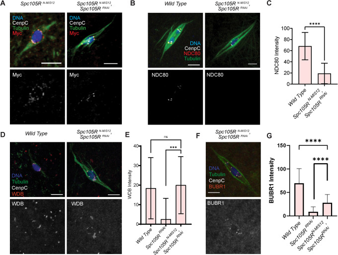
The reductional division of meiosis I requires the separation of chromosome pairs towards opposite poles. We have previously implicated the outer kinetochore protein SPC105R/KNL1 in driving meiosis I chromosome segregation through lateral attachments to microtubules and coorientation of sister centromeres. To identify the domains of SPC105R that are critical for meiotic chromosome segregation, an RNAi-resistant gene expression system was developed. We found that the SPC105R C-terminal domain (aa 1284-1960) is necessary and sufficient for recruiting NDC80 to the kinetochore and building the outer kinetochore. Furthermore, the C-terminal domain recruits BUBR1, which in turn recruits the cohesion protection proteins MEI-S332 and PP2A. Of the remaining 1283 amino acids, we found the first 473 are most important for meiosis. The first 123 amino acids of the N-terminal half of SPC105R contain the conserved SLRK and RISF motifs that are targets of PP1 and Aurora B kinase and are most important for regulating the stability of microtubule attachments and maintaining metaphase I arrest. The region between amino acids 124 and 473 are required for lateral microtubule attachments and biorientation of homologues, which are critical for accurate chromosome segregation in meiosis I.


Figures

FIGURE 1:
FIGURE 1:
Spc105RB rescues the viability and microtubule attachment defects in Spc105RRNAi oocytes. (A) A schematic of the two known Drosophila Spc105R isoforms and mutants used in this study. The first nine amino acids in SPC105RB are changed from MNANKRRSS to MVDLLFLQ in SPC105R A. The coordinates on the schematic represent the first amino acid of each domain. The N-terminal includes the SLRK and RISF motifs and is where the two isoforms differ. Following this is a domain with three MELT-like motifs, a region that contains two KI-like repeats, a central domain containing repeats with the consensus ExxEED, and the C-terminal region containing coiled-coil motifs. An ExxEED-like consensus is found in several Drosophila species (Tromer et al., 2015) although many organisms, including mammals, have repeats of the MELT motif in this domain. The Drosophila ExxEED repeats have been proposed to be phosphomimetic derivatives of the MELT motif (Audett et al., 2022), in part because there is a threonine 5 amino acids upstream of the ExxEED consensus in 14/15 of the repeats, and the 15th is a serine. This threonine is a Aurora B phosphorylation site and is part of the consensus KxRxTLL that is related to the TΩ motif upstream of the MELT motif in other organisms (Tromer et al., 2015). All transgenes included missense mutations to make them resistant to the shRNA GL00392. (B) Viability of Spc105R mutants, all in a Spc105RRNAi background. Data shows the relative amounts of progeny expressing the Spc105RRNAi and mutants (Tub-Gal4) to siblings that did not (no Gal4; n > 200). (C) Confocal images of wild-type, Spc105RRNAi, and Spc105R B Spc105RRNAi oocytes. Merged images show DNA (blue), Tubulin (green), centromeres (white), and SPC105R (red). Centromeres and SPC105R are shown in separate channels. Scale bars are 5 μm.
FIGURE 2:
FIGURE 2:
Homologous chromosome bi-orientation in Spc105R mutants lacking specific domains. (A) Confocal images of Spc105RRNAi oocytes expressing the indicated transgene. Merged images show DNA (blue), Tubulin (green), the X-Chromosome (yellow), the second chromosome (red), and the third chromosome (white). DNA and FISH probes are shown in a separate channel. Scale bars are 5 μm. Examples of monooriented centromeres are circled. (B) Quantification for percent of chromosome monoorientation: Spc105R B Spc105RRNAi oocytes (n = 93), Spc105R ΔN Spc105RRNAi oocytes (n = 117), Spc105R ΔMELT-KI Spc105RRNAi oocytes (n = 123), Spc105R ΔExxEED Spc105RRNAi oocytes (n = 65), Spc105R ΔM Spc105RRNAi oocytes (n = 81), Spc105R C Spc105RRNAi oocytes (n = 63). Significance in frequency of monoorientation in oocytes determined by Fisher’s exact test, with * = p values < 0.01, **** = p value < 0.0001.
FIGURE 3:
FIGURE 3:
The SPC105R C-Terminal Domain recruits NDC80. The N-terminal 1284 amino acids of SPC105R is deleted in Spc105R C, retaining only the last 676 amino acids (Figure 1). (A) Confocal images of Spc105RRNAi oocytes expressing the indicated transgene, with DNA (blue), Tubulin (green), centromeres (white), and the kinetochore protein NDC80 (red). Centromeres and NDC80 are shown in separate channels. Scale bars are 5 μm. (B) Quantification of NDC80 intensity at the centromeres in wild-type (n = 68), Spc105RRNAi (n = 77), Spc105RB Spc105RRNAi (n = 131), and Spc105RC Spc105RRNAi (n = 107). Error bars indicate SD and **** = p value < 0.0001 by an unpaired t test. (C) Confocal images of Spc105RRNAi oocytes expressing the indicated transgene, with DNA (blue), Tubulin (green), and centromeres (white). DNA is shown in separate channels. Scale bars are 5 μm. (D) Quantification of end-to-end chromosome length of wild-type (n = 34), Spc105RRNAi (n = 12), Spc105RC Spc105RRNAi (n = 22), and Spc105RC Spc105RRNAi Ndc80RNAi (n = 30). Error bars indicate SD, and **** = p value < 0.0001 by an unpaired t test.
FIGURE 4:
FIGURE 4:
The SPC105R C-terminal domain recruits proteins required for centromeric cohesion protection in meiosis I. (A) Confocal images of Spc105RRNAi oocytes expressing the indicated transgene with DNA (blue), Tubulin (green), centromeres (white), and MEI-S332 (red) and shown as a separate channel. Scale bars are 5 μm. (B) Quantification of MEI-S332 intensity at the centromeres for wild-type (n = 71), Spc105RRNAi (n = 202), Spc105RB Spc105RRNAi (n = 53) and Spc105RC Spc105RRNAi (n = 114). Error bars indicate SD, and **** = p value < 0.0001by unpaired t test. (C) Confocal images of Spc105RRNAi oocytes expressing the indicated transgene, with DNA (blue), Tubulin (green), centromeres (white), and the PP2A subunit WDB-HA (red), also in a separate channel. Scale bars are 5 μm. (D) Quantification of WDB intensity at the centromeres in wild-type (n = 195), Spc105RB, Spc105RRNAi (n = 134), Spc105RRNAi (n = 189) and Spc105RC Spc105RRNAi (n = 155). Error bars indicate SD and unpaired t test **** = p value < 0.0001. (E) Confocal images of Spc105RRNAi oocytes expressing the indicated transgene, with DNA (blue), Tubulin (green), centromeres (white), and GFP-BUBR1 (red) and in a separate channel. (F) Quantification of BUBR1 intensity at the centromeres, in wild-type (n = 247), Spc105RRNAi (n = 153), Spc105RB Spc105RRNAi (n = 67), Spc105RMELT-KI Spc105RRNAi (n = 89), and Spc105RC Spc105RRNAi (n = 149). Error bars indicate SD and significance is shown by a unpaired t test **** = p value < 0.0001.
FIGURE 5:
FIGURE 5:
The SPC105R N-terminal domain interacts with PP1 to maintain sister centromere coorientation. Mutants with a deletion of the N-terminal domain (Spc105RΔN) or mutations in the SLRK and RISF sequences are shown in Supplemental Figure S4A. (A) Confocal images of PP1-87BRNAi or Spc105RΔN Spc105RRNAi oocytes. Merged images show DNA (blue), Tubulin (green), and centromeres (white). Centromeres are shown in a separate channel along with the average number of foci. Scale bars are 5 μm. (B) Quantification of centromere foci, in the order of the images in A, except wild-type not shown. Sample sizes are: 46, 16, 37, 37, 48, 37, and 20, in order of the graph. (C) Confocal images of oocytes where the RISF site was changed to AAAA, RIAF, or RIDF. Centromeres are shown in a separate channel along with the average number of foci. (D) Quantification of centromere foci, in the order of the images in C, except first two shown in A. Sample sizes are: 37, 46, 17, 29, 19, 20, and 36, in order of the graph. Error bars indicate SD and unpaired t test showed significance in number of foci **** p < 0.0001, ** p = 0.001, ns = not significant.
FIGURE 6:
FIGURE 6:
SPC105R is sufficient for interactions with spindle microtubules. (A) Confocal images of wild-type, Spc105RRNAi or Ndc80RNAi oocytes. Merged images show DNA (blue), Tubulin (green), and centromeres (white). Scale bars are 5 μm. (B) Oocytes in a mei-P22 mutant (mei-P22mut) background. The first image is only Ndc80RNAi mei-P22mut, while the rest express Ndc80RNAi, Spc105RRNAi, and a Spc105R transgene. Due to the absence of crossing over, mei-P22mut mutant oocytes fail to arrest in metaphase, and precociously enter anaphase. (C) Oocytes in mei-P22mut, Spc105R RNAi background. The second image is also expressing a Spc105RN-mis12 transgene. For B and C, merged images show DNA (blue), Tubulin (green), and centromeres (white). Chromosomes are shown in a separate channel. (D) Quantification of the frequency of chromosome separation in oocytes. Sample sizes are: 25, 50, 23, 175, 115, 81, 83, and 21, in order of the graph. Significance in frequency of oocytes entering precocious anaphase determined by Fisher’s exact test, with ** = p value < 0.01, **** = p value < 0.0001, and ns = not significant.
FIGURE 7:
FIGURE 7:
The N-terminal domain of SPC105R fused to MIS12 localizes to the kinetochore and recruits PP2A. (A) Confocal images of stage 14 oocytes expressing Spc105RN-Mis12, either in the presence or absence of Spc105RRNAi. The fusion protein contains the first 1284 amino acids of SPC105R followed by all of MIS12, and was detected using an antibody to the myc tag (red). In all images, CENP-C is in white, and tubulin is in green, and the scale bars are 5 μm. (B) Ndc80 (red) does not localize in Spc105RN-MIS12 Spc105RRNAi oocytes. (C) Measurement of NDC80 intensity in wild-type (n = 137) and Spc105R N-MIS12 Spc105RRNAi (n = 160) oocytes. Error bars indicate SD, and unpaired t test showed significance **** = p < 0.0001. (D) PP2A-B56 subunit WDB localization shown in red and a separate channel in wild-type and Spc105R N-MIS12 Spc105RRNAi oocytes. (E) Measurement of WDB intensity in wild-type (n = 154), Spc105RRNAi (n = 195), and Spc105RN-MIS12 Spc105RRNAi (n = 183) oocytes. Error bars indicate SD and unpaired t test showed significance, *** = p < 0.001 and ns = not significant. (F) GFP-BUBR1 shown in red and in a separate channel in Spc105R N-MIS12 Spc105RRNAi oocytes. (G) Measurement of BUBR1 intensity in wild-type (n = 117), Spc105RRNAi (n = 153), and Spc105RN-MIS12 Spc105RRNAi (n = 141) oocytes. Error bars indicate SD and unpaired t test showed significance, **** = p < 0.0001.
FIGURE 8:
FIGURE 8:
Functional domains of SPC105R based on these and prior studies.

Update of

Similar articles

Cited by

References

    1. Audett MR, Johnson EL, McGory JM, Barcelos DM, Szalai EO, Przewloka MR, Maresca TJ (2022). The microtubule- and PP1-binding activities of Drosophila melanogaster Spc105 control the kinetics of SAC satisfaction. Mol Biol Cell 33, ar1. - PMC - PubMed
    1. Bajaj R, Bollen M, Peti W, Page R (2018). KNL1 Binding to PP1 and Microtubules Is Mutually Exclusive. Structure 26, 1327–1336 e1324. - PMC - PubMed
    1. Barbosa J, Sunkel CE, Conde C (2022). The Role of Mitotic Kinases and the RZZ Complex in Kinetochore-Microtubule Attachments: Doing the Right Link. Front Cell Dev Biol 10, 787294. - PMC - PubMed
    1. Bischof J, Bjorklund M, Furger E, Schertel C, Taipale J, Basler K (2013). A versatile platform for creating a comprehensive UAS-ORFeome library in Drosophila. Development 140, 2434–2442. - PubMed
    1. Bonner AM, Hughes SE, Chisholm JA, Smith SK, Slaughter BD, Unruh JR, Collins KA, Friederichs JM, Florens L, Swanson SK, et al. (2013). Binding of Drosophila Polo kinase to its regulator Matrimony is noncanonical and involves two separate functional domains. Proc Natl Acad Sci USA 110, E1222–E1231. - PMC - PubMed

MeSH terms

LinkOut - more resources