. 2024 Jun 18;121(25):e2320995121.
doi: 10.1073/pnas.2320995121. Epub 2024 Jun 12.

RNF212B E3 ligase is essential for crossover designation and maturation during male and female meiosis in the mouse

Affiliations

RNF212B E3 ligase is essential for crossover designation and maturation during male and female meiosis in the mouse

Yazmine B Condezo et al. Proc Natl Acad Sci U S A. .

Erratum in

Abstract

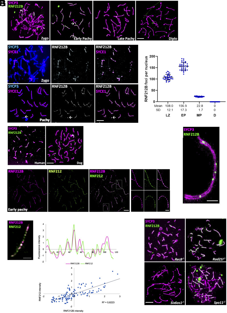
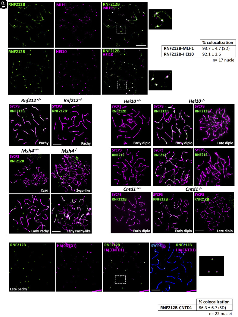
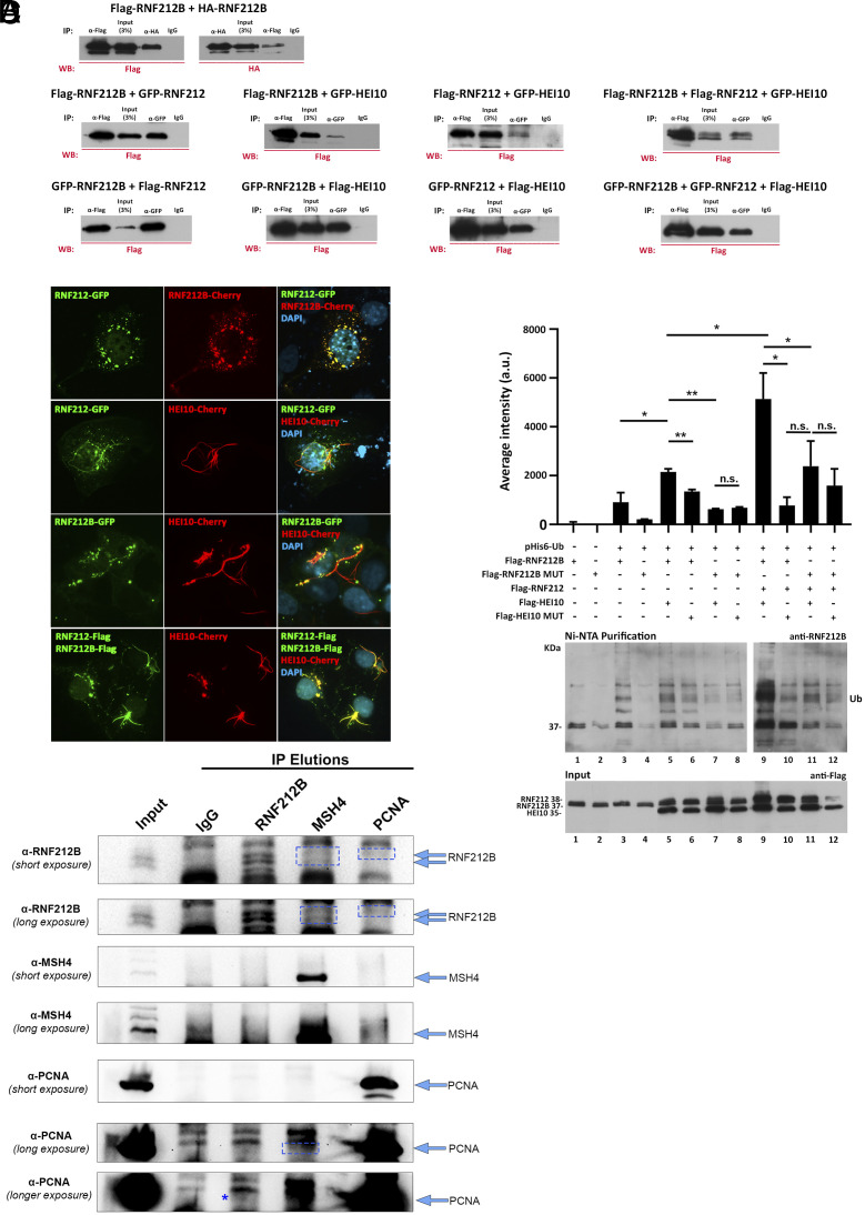
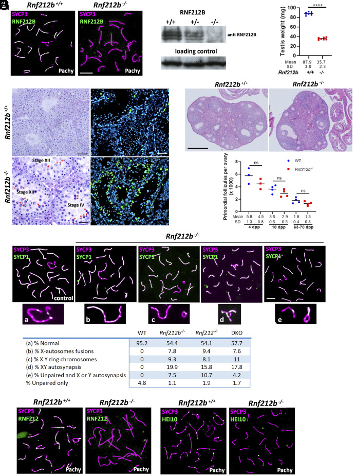
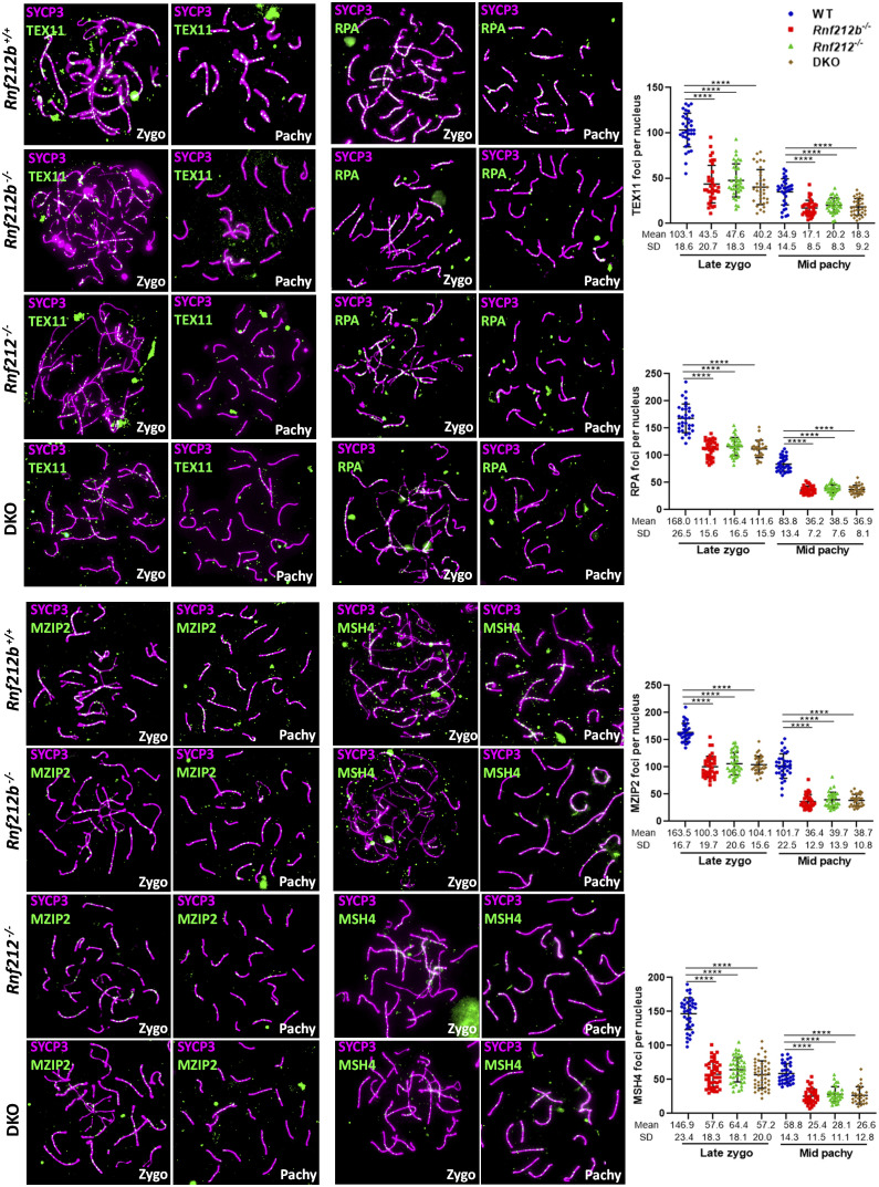
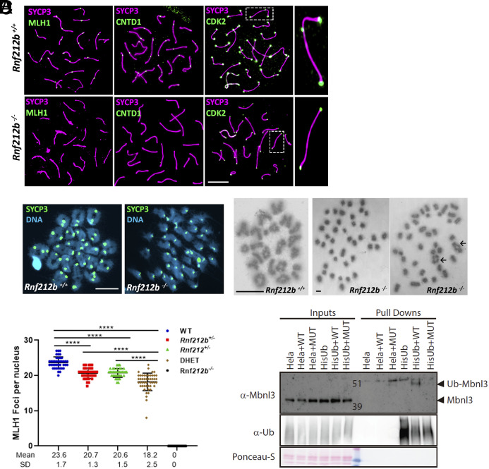
Meiosis, a reductional cell division, relies on precise initiation, maturation, and resolution of crossovers (COs) during prophase I to ensure the accurate segregation of homologous chromosomes during metaphase I. This process is regulated by the interplay of RING-E3 ligases such as RNF212 and HEI10 in mammals. In this study, we functionally characterized a recently identified RING-E3 ligase, RNF212B. RNF212B colocalizes and interacts with RNF212, forming foci along chromosomes from zygonema onward in a synapsis-dependent and DSB-independent manner. These consolidate into larger foci at maturing COs, colocalizing with HEI10, CNTD1, and MLH1 by late pachynema. Genetically, RNF212B foci formation depends on Rnf212 but not on Msh4, Hei10, and Cntd1, while the unloading of RNF212B at the end of pachynema is dependent on Hei10 and Cntd1. Mice lacking RNF212B, or expressing an inactive RNF212B protein, exhibit modest synapsis defects, a reduction in the localization of pro-CO factors (MSH4, TEX11, RPA, MZIP2) and absence of late CO-intermediates (MLH1). This loss of most COs by diakinesis results in mostly univalent chromosomes. Double mutants for Rnf212b and Rnf212 exhibit an identical phenotype to that of Rnf212b single mutants, while double heterozygous demonstrate a dosage-dependent reduction in CO number, indicating a functional interplay between paralogs. SUMOylome analysis of testes from Rnf212b mutants and pull-down analysis of Sumo- and Ubiquitin-tagged HeLa cells, suggest that RNF212B is an E3-ligase with Ubiquitin activity, serving as a crucial factor for CO maturation. Thus, RNF212 and RNF212B play vital, yet overlapping roles, in ensuring CO homeostasis through their distinct E3 ligase activities.

Keywords: crossing over; fertility; meiosis; mouse; recombination.


Conflict of interest statement

Competing interests statement:The authors declare no competing interest.

Figures

Fig. 1.
Fig. 1.
RNF212B colocalizes with RNF212, its loading to the chromosome axis is dependent on synapsis and is independent of Spo11-induced DSB. (A) Colabeling of RNF212B and SYCP3 from Zygonema to Diplonema. (B) Colabeling of spermatocytes spreads with SYCP3, central element protein SYCE1, and RNF212B. Arrow in (A) and (B) indicate the positive labeling of the PAR. (C) Plot quantitation of the number of RNF212B foci. LZ (Late Zygonema), EP (early Pachynema), MP (mid pachynema), and D (Diplonema). (D) Colabeling of RNF212B and SYCP3 in human and dog spermatocytes showing conserved pattern of localization onto the chromosome axes. (E) STED microscopy images of RNF212 and RNF212B colocalization in pachytene spermatocytes. (F) Double immunostaining of RNF212B and RNF212. Immunofluorescence signal levels were measured on synapsed chromosome axes. Upper plot represents normalized signal intensity profiles of RNF212B (magenta) with RNF212 (green). Lower plot shows regression analysis of the correlation between each pair corresponding to chromosome axes. Each of the dots represents the RNF212B fluorescence intensity in axes “x” and RNF212 fluorescence intensity in axe “y” for a given point along the AE. (G) STED images of pachynema chromosome immunostained for SYCP3 and RNF212B. (H) Double immunolabeling of RNF212B and SYCP3 in arrested spermatocytes of Rec8−/−, Rad21l−/−, Six6os1−/−, and Spo11−/−. Bar in panels (A), (B), (D), and (H) 10 μm. Bars in panels (G) and (F) represents 2.5 μm. All experiments have been carried out in at least three mice and 15 cells per mouse.
Fig. 2.
Fig. 2.
RNF212B colocalizes with CO-associated proteins and its loading is dependent on RNF212, and its unloading on HEI10 and CNTD1. (A) Double immunolabeling of RNF212B with MLH1 and HEI10 in pachynema spermatocytes. Right panel show magnification of the indicated region. The quantitation of the colocalization of RNF212B with MLH1 and HEI10 are shown in right table. (B) Double immunolabeling of RNF212B and SYCP3 in Rnf212−/−in pachynema spermatocytes showing absence of foci in comparison with the numerous foci of the wild-type control. (C) Double immunolabeling of RNF212B and SYCP3 in Msh4+/+ and Msh4−/−spermatocytes at zygonema and early pachynema, showing persistence of RNF212B loading in the absence of MSH4. (D) Double immunolabeling of SYCP3 with RNF212B and RNF212 in Hei10−/− early diplonema spermatocytes showing numerous foci at the synapsed LEs in comparison with the wild-type diplonemas which lack foci and in Hei10−/− late diplonemas when foci have completely disappeared. (E) Colabeling of RNF212B, anti-HA (CNTD1), and SYCP3 showing colocalization. The Right panel shows magnification of the indicated region. The quantitation of the colocalization of RNF212B with CNTD1 is shown in the lower table. (F) Double immunolabeling of RNF212B and SYCP3 in Cntd1−/−early diplonema spermatocytes showing numerous foci at the synapsed LEs in comparison with the wild-type diplonema spermatocytes which lack foci and in Cntd1−/− late diplonema when foci have completely disappear. Bar in panels 10 μm. All experiments have been carried out in at least three mice and 15 cells per mouse.
Fig. 3.
Fig. 3.
RNF212B interactions and Ubiquitylation activity. (A) Co immunoprecipitation of RNF212B with RNF212B, RNF212, and HEI10. HEK293T cells were cotransfected with plasmids encoding RNF212, RNF212B, and HEI10 tagged with FLAG or GFP. Protein complexes were immunoprecipitated overnight with an antibody anti-FLAG, anti-GFP, or IgGs (as a negative control) and analyzed by immunoblotting. Positive interaction of RNF212B with RNF212 and HEI10 as well as RNF212B (self-interaction) is shown. Additionally, a positive interaction between RNF212 and HEI10 is observed. (B) COS7 cells immunofluorescence shows colocalization of transfected RNF212B with RNF212 and HEI10. COS7 cells were transfected to express RNF212B (fused to either Cherry, Flag, or GFP) along with RNF212 (fused with Flag or GFP) and/or HEI10 (Cherry-tagged). Nuclear and punctuate cytoplasmic RNF212B signal perfectly colocalizes with RNF212 signal (Top). Transfected HEI10 forms a ring fiber-like pattern that when cotransfected with RNF212 and/or RNF212B recruits them to this ring structures altering their localization pattern. (Scale bars: 10 μm.) (C) RNF212B has autoubiquitynilation activity. HEK293T cells were cotransfected with Flag-tagged plasmids encoding RNF212, RNF212B (WT or mutant), and/or HEI10 (WT or mutant) in the presence or absence of a plasmid encoding 6xHis-Ubiquitin. Protein complexes were purified using Ni-NTA, favoring the purification of proteins containing 6xHis sequence. Then, ubiquitylation was analyzed by immunoblotting using antibodies anti-RNF212B and anti-Flag (Input). The results show RNF212B autoubiquitinylation. As observed in the statistics of the quantification of average intensity for each well, the strongest ubiquitinylation was exhibited when RNF212B was cotransfected along with RNF212 and HEI10 and was abolished when a RNF212B dead mutant was transfected. Quantification of the results of three independent experiments. Two-tailed Welch’s t test analysis: *P < 0.05. **P < 0.005. (D) IPs in isolated germ cells with the antibodies indicated (Top) against RNF212B, MSH4, and PCNA, and western blot analysis of the eluted proteins with the same antibodies (Left) detecting specific bands for RNF212B in the MSH4 and PCNA elution, and PCNA in the RNF212B and MSH4 elution (boxes and asterisk). Arrows indicate sizes for bands of interest in each blot.
Fig. 4.
Fig. 4.
Characterization of Rnf212b-deficient mice. (A) Double immunolabeling of RNF212B and SYCP3 showing absence of fluorescent signal in the Rnf212b−/−and foci labeling in the control spermatocytes. Bar 10 μm. (B) Western blot analysis of whole testis extracts showing two bands in the wild type and heterozygous that are absent in the Rnf212b−/−. (C) Plot representation of the reduction in the weight of the testis from Rnf212b−/−in comparison with the wild-type (WT). (D) Left panel, histological section of a testis from Rf212b+/+ and Rf212b−/−mice showing abnormal accumulation of tubules at stage XII of the seminiferous epithelial cycle with abnormal and apoptotic metaphase I cells (red crosses) and at stage IV of the seminiferous epithelial cycle having apoptotic pachynemas (red asterisks). Bar 50 μm in Rnf212b+/+and 25 μm in Rf212b−/−. Right panel, immunofluorescence detection of apoptotic cells by TUNEL staining showing an increase of apoptotic cells in Rf212b−/−seminiferous tubules. Bar 50 μm. (E) Upper panel, histological staining of representative sections of ovaries from an Rnf212b+/+and an Rnf212b−/−adult mice, showing oocytes and follicles at all stages of follicular development. Bar 200 μm. Lower panel, representative plot of primordial follicles quantitation from Rnf212b−/−and wild-type ovaries showing a reduced trend in primordial follicles numbers in the mutant ovaries, which is not statistically different. (F) Double immunolabeling of SYCP3 and SYCP1 showing aberrant synapsis and structural abnormalities in the XY chromosome pair including X-autosome fusions (b), ring chromosomes (c), XY autosynapsis (d), and unpaired and autosynapsis (e). The quantitation numerical values of these aberrant configurations in the XY bivalent in Rnf212b−/−, Rnf212−/−, Rnf212b−/− Rnf212−/−(dKO), and wild-type spermatocytes (WT) are shown in the lower table. (G) Double immunolabeling of SYCP3 with RNF212 and with HEI10 showing absence of fluorescent foci in Rnf212b−/− in comparison with the wild-type controls. Bar in (EG) 10 μm. All experiments have been carried out in at least three mice and 100 cells per mouse in (F), and 15 cells in (G).
Fig. 5.
Fig. 5.
Localization of pro-CO factors is decreased in wild-type, Rnf212b−/−, Rnf212−/−, and Rnf212b−/− Rnf212−/− (DKO) spermatocytes. Double immunolabeling of SYCP3 and TEX11, RPA, MZIP2, or MSH4. In late zygonema and mid pachynema, foci were significantly reduced in Rnf212b−/−, Rnf212−/−, and Rnf212b−/− Rnf212−/− (DKO) in comparison with the wild-type control. Plots in the Right of the panels represent the quantitation of the values. Two-tailed Welch’s t test analysis: ****P < 0.0001. Bar in all panels, 10 μm. All experiments have been carried out in at least three mice and 15 cells per mouse.
Fig. 6.
Fig. 6.
Major CO pathway disrupted in the absence of RNF212B, and MBNL3 ubiquitination by RNF212B. (A) Double immunolabeling of SYCP3 with MLH1, CNTD1, and CDK2 in Rnf212b−/−and wild-type mouse spermatocytes (Rnf212b+/+). Right magnified panels show the presence (Up) and absence (Low) of an interstitial CDK2 foci at a CO are shown. (B) Metaphase I cells from chromosome spreads (stained with SYCP3 and DAPI, Left panel) and from Methanol/Acetic chromosome preparations stained with Giemsa (Right) show the presence of univalents (Rnf212b−/−) and a few bivalents (black arrows) in comparison with all bivalents (Rnf212b+/+). (C) Plot quantitation of the number of MLH1 foci in pachynema spermatocytes from Rnf212b+/+(WT), Rnf212b+/−, and Rnf212+/−single mutants, Rnf212+/− Rnf212b+/− (DHet) and Rnf212b−/−. Welch’s t test analysis: ****P < 0.0001. (D) Ubiquitination assay performed on Hela cells to verify MBNL3 ubiquitination by RNF212B activity. Hela cells stably expressing 10xHis-Ub were transfected with the indicated expression vectors. 10xHis-ubiquitin conjugated proteins were purified from cell lysates using Ni-NTA beads. Purified proteins (Pull-Downs) and Lysate samples (Inputs) were analyzed by western blotting using specific Ubiquitin antibody and MBNL3 antibody to detect its ubiquitinated form. We observe a 10 KDa higher band corresponding to the ubiquitinated form of MBNL3 that is only detectable when transfecting HELA-Ub with RNF212B-WT expression vector. No band is detected when transfecting RNF212B dead mutant form (MUT). Bar in all panels, 10 μm. All experiments have been carried out in at least three mice and 12 cells per mouse in (A) and (C), and 5 cells in (B).

References

    1. Zickler D., Kleckner N., Meiosis: Dances between homologs. Annu. Rev. Genet. 57, 1–63 (2023). - PubMed
    1. Llano E., Pendas A. M., Synaptonemal complex in human biology and disease. Cells 12, 1718 (2023). - PMC - PubMed
    1. Gray S., Cohen P. E., Control of meiotic crossovers: From double-strand break formation to designation. Annu. Rev. Genet. 50, 175–210 (2016). - PMC - PubMed
    1. Stoop-Myer C., Amon A., Meiosis: Rec8 is the reason for cohesion. Nat. Cell Biol. 1, E125–127 (1999). - PubMed
    1. Kudo N. R., et al. , Role of cleavage by separase of the Rec8 kleisin subunit of cohesin during mammalian meiosis I. J. Cell Sci. 122, 2686–2698 (2009). - PMC - PubMed

LinkOut - more resources