. 2024 Aug 3;45(29):2660-2673.
doi: 10.1093/eurheartj/ehae357.

Non-coding RNA yREX3 from human extracellular vesicles exerts macrophage-mediated cardioprotection via a novel gene-methylating mechanism

Affiliations

Non-coding RNA yREX3 from human extracellular vesicles exerts macrophage-mediated cardioprotection via a novel gene-methylating mechanism

Alessandra Ciullo et al. Eur Heart J. .

Abstract

Background and aims: Extracellular vesicles (EVs) secreted by cardiosphere-derived cells exert immunomodulatory effects through the transmission of small non-coding RNAs.

Methods: The mechanism and role of yREX3, a small Y RNA abundant in EVs in myocardial injury, was investigated.

Results: yREX3 attenuates cardiac ischaemic injury by selective DNA methylation. Synthetic yREX3 encapsulated in lipid nanoparticles triggers broad transcriptomic changes in macrophages, localizes to the nucleus, and mediates epigenetic silencing of protein interacting with C kinase-1 (Pick1) through methylation of upstream CpG sites. Moreover, yREX3 interacts with polypyrimidine tract binding protein 3 (PTBP3) to methylate the Pick1 gene locus in a DNA methyltransferase-dependent manner. Suppression of Pick1 in macrophages potentiates Smad3 signalling and enhances efferocytosis, minimizing heart necrosis in rats with myocardial infarction. Adoptive transfer of Pick1-deficient macrophages recapitulates the cardioprotective effects of yREX3 in vivo.

Conclusions: These findings highlight the role of a small Y RNA mined from EVs with a novel gene-methylating mechanism.

Keywords: Efferocytosis; Inflammation; Macrophages; Myocardial infarction; Pick1; Small non-coding RNA.


Figures

Structured Graphical Abstract
Small non-coding RNA, yREX3, induces methylation-mediated silencing of Pick1. Pick1 silencing in macrophages enhances their capacity to clear dead cells and promote tissue healing after injury.
Figure 1
Figure 1
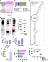
Characterization of extracellular vesicles from therapeutically enhanced cardiac stromal cells (IMEX). (A) Engineering of immortalized cardiosphere-derived cells (imCDCs) from primary cardiosphere-derived cells and preparation of extracellular vesicles from both (cardiosphere-derived cell extracellular vesicles and IMEX, respectively) for RNA sequencing. (B) Nanosight tracking analysis of particle size distribution and concentration of IMEX (1000 kDa), (C) non-extracellular vesicle fraction of 1000 kDa and non-conditioned culture media (used to produce extracellular vesicles: inset). (D) Extracellular vesicle purity is shown as particles (IMEX) vs. micrograms of protein. (E) Western blot for typical exosome markers (CD81, HSP90, CD63, Alix, TSG101). IMEX is negative for Calnexin (n = 3 samples/group, mean ± standard error of the mean). (F) IMEX visualized by cryotransmission electron microscopy. Scale bar: 200 nm
Figure 2
Figure 2
yREX3 is a small RNA enriched in IMEX. (A) Top 10 most expressed small non-coding RNAs in IMEX (expressed as log10 number of reads and fold change compared with primary cardiosphere-derived cell extracellular vesicles). (B) Per cent genomic origins of yREX3 show most sequence hits derive from the human YRNA 4 and its pseudogenes (lavender bar). (C) Alignment of yREX3 (in red) with the human YRNA 4 gene. Previously characterized small Y RNAs EV-YF1 and NT4 (renamed here as yREX1 and yREX2) are indicated in blue and green, respectively. (D) Northern blot analysis of yREX3 RNA expressed by immortalized cardiosphere-derived cells and (E) IMEX. (F) The sizes of individual RNA species are indicated. The band corresponding to the 26-nucleotide transcript is indicated in red. (G, H) Enrichment of yREX3 (in sequencing reads) in immortalized cardiosphere-derived cells to primary cardiosphere-derived cells. This enrichment was more pronounced in IMEX compared with primary cardiosphere-derived cell extracellular vesicles. (I) Quantitative polymerase chain reaction demonstrating the abundance of yREX3 in IMEX compared with primary cardiosphere-derived cell extracellular vesicles (n = 3 replicates per group, mean ± standard error of the mean, significance was determined using Student’s independent t-test, ***P < .001). (J) The abundance of yREX3 in IMEX was further confirmed in quantitative polymerase chain reaction by using 1 × 10¹⁰ and 1 × 10⁷ IMEX as input; data are expressed as Cq cycles in quantitative polymerase chain reaction and no IMEX (phosphate-buffered saline only, indicated as 0) used as a negative control. (K) yREX3 RNA is contained inside IMEX as shown by protection from RNase A degradation and proteinase K treatment (n = 3 biological replicates per group, mean ± standard error of the mean, significance was determined using one-way analysis of variance with Tukey’s post-test, **P < .01)
Figure 3
Figure 3
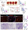
yREX3 is cardioprotective in vivo and enhances macrophage activation. (A) Rats underwent myocardial infarction (induced by 45 min of ischaemia followed by reperfusion) and 20 min after reperfusion they received saline, yREX3, IMEX, or scrambled sequence (Scr) intramyocardially. Forty-eight hours post-myocardial infarction, yREX3 showed cardioprotective activity as shown by reduced scar mass measured by 2,3,5-triphenyl-2H-tetrazolium chloride staining (representative images; B) and infarct mass quantification (C) and lower circulating cardiac Troponin I levels (D) compared with animals injected with vehicle or scramble (n = 6–10 animals/group, mean ± standard error of the mean). Alternatively, rats received a retro-orbital injection of yREX3 (400 ng/animal); pink triangles in C and D (all data presented as mean ± standard error of the mean, comparison between groups were evaluated using one-way analysis of variance with Tukey’s post-test with **P < .01 and ***P < .001). (E) Schematic of cell isolation from rat pups (cardiomyocytes and cardiac fibroblast) and the mother rat (bone marrow-derived macrophages (Mϕ). (F) Transcriptomic data of cardiomyocytes, cardiac fibroblasts, and BMDM (Mϕ) exposed to yREX3 (80 nM for 24 h) show significant differential gene expression compared with vehicle-exposed cells (n = 3 samples/group), expressed as the number of genes up- and down-regulated in the three cell types after in vitro exposure to yREX3. (G, H) In vitro, yREX3 enhances macrophage proliferation as analysed by BrdU incorporation (n = 3–6 replicates from two different experiments) and representative images (scale bar: 200 µm). (I, J) yREX3 potentiates macrophage migration at 24 h post-exposure using a Bowden chamber assay (migration calculated as integrated density/area, n = 5 independent experiments) and representative images (scale bar: 100 µm). All data presented as mean ± standard error of the mean. Comparison between groups was evaluated using one-way analysis of variance with Tukey’s post-test with **P < .01 and ***P < .001
Figure 4
Figure 4
yREX3 methylates pick1 to enhance phagocytosis in macrophages. (A) yREX3 can shuttle between cytoplasm and nucleus in BMDM as shown by the quantitative polymerase chain reaction for yREX3 of cytoplasmic and nuclear RNA in BMDM transfected with yREX3 at different time points (data expressed as log2-fold change using snoU6 as housekeeping (scramble vs. vehicle and yREX3 vs. vehicle). (B) Analysis of global methylation levels in BMDM 48 h post-exposure to vehicle, yREX3, or scramble (Scr) with or without the DNA methyltransferase inhibitor RG108. (C) Methylation peaks for the Pick1 gene in yREX3-transfected BMDM (data presented as the difference in CpG-methylated regions vs. vehicle-exposed cells at 24 h). (D) Percentage of methylation in vehicle- and yREX3-exposed BMDM at 48 h in the different CpG sites was analysed (only relevant regions with different methylation profiles shown; n = 3–4 biological replicates/group). (E) Quantitative polymerase chain reaction for Pick1 expression levels in vehicle-, yREX3-, and scramble-exposed BMDM at 48 h (data presented as fold change compared with vehicle). (F) Quantitative polymerase chain reaction for Pick1 expression levels in vehicle- and yREX3-exposed human peripheral blood mononuclear cell–derived macrophages at 24 and 48 h (data presented as fold change compared with vehicle) (AC, E, F). All data presented as mean ± standard error of the mean, comparison between groups were evaluated using a Student’s independent t-test and one-way analysis of variance with Tukey’s post-test with *P < .05, **P < .01, and ***P < .001. FC, fold change.
Figure 5
Figure 5
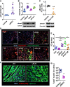
Pick1 suppression activates Smad3 and enhances efferocytosis in macrophages. (A) Phosphorylation levels of Smad3 expressed as phosphorylated/total in vehicle- and yREX3-exposed BMDM (n = 5–6 technical replicates/group). (B) Western blot showing increased phosphorylation of Smad3 (Ser213) in macrophages after exposure to yREX3 or a small interfering RNA targeted against Pick1 (siPick1) compared with scramble (siScr) or vehicle (n = 3–5 biological replicates per group). (C) Western blot showing decreased phosphorylation of Smad3 (Ser213) in macrophages after exposure to a Pick1 overexpressing vector (Pick1 OE) at 24 h compared to vehicle (n = 4 biological replicates per group). (D and E) Efferocytosis assay showing increased uptake of DiO-labelled dead rat cardiomyocytes by yREX3-exposed or small interfering RNA targeted against Pick1–exposed macrophages compared to those exposed to vehicle, Scr, or Pick1 overepxression (representative images taken at 48 h, n = 3–6 biological replicates/group). (F and G) TUNEL assay in rat heart sections 48 h after myocardial infarction in animals receiving vehicle or yREX3 i.v. administration and representative images. All data presented as mean ± standard error of the mean, comparison between groups using Student’s independent t-test or one-way analysis of variance with Tukey’s post-test with *P < .05, **P < .01, and ***P < .001
Figure 6
Figure 6
yREX3 binds PTBP3 through a six-cytosine motif. (A) Mass-spectrometry analysis of RNA-protein pull-down showing peptide hits identified in yREX3- and scrambled-exposed macrophages. (B) Enzyme-linked immunosorbent assay for PTBP3 in RNA-protein pull-down experiments using control (vehicle) or scrambled sequence and yREX3. (C) Quantitative polymerase chain reaction demonstrating yREX3 expression when PTBP3 is immunoprecipitated in macrophages exposed to vehicle, scramble, or yREX3 in vitro. (D) Quantitative polymerase chain reaction demonstrating down-regulation of PTBP3 after transfection of macrophages with a small interfering RNA targeted against PTBP3 (50 nM) for 48 h. (E) Quantitative polymerase chain reaction showing that polypyrimidine tract in yREX3 is essential for suppression of Pick1. (F) Analysis of the abundance of single-nucleotide tracts in the 5′ untranslated region, Intron 1 (where CpG sites are methylated), and in mRNA for Pick1 showing they are enriched in cytosine and guanine tracts compared with adenine and thymine (or uracil in the case of mRNA). (G) First six residues at the 5′ end in the yREX3 sequence (here identified as yREX3WT) are cytosines. Oligonucleotides with mutations in the first six residues at the 5′ end and for the scramble sequence. (H) Quantitative polymerase chain reaction for Pick1 expression levels in vehicle, yREX3, yREX3 with a silent mutation (yREX3s6A), yREX3 with a dysfunctional mutation (yREX3s6G), and scramble-exposed BMDM at 48 h (data presented as fold change compared with vehicle). (I) Enzyme-linked immunosorbent assay of PTBP3 in RNA-Protein pull-down experiments using control or scramble, yREX3, and yREX3 with the dysfunctional mutation (yREX3s6G). (J) Enzyme-linked immunosorbent assay of RAVER1 in immunoprecipitation experiments using scramble or yREX3 macrophage lysates (AD, GI). Pooled data presented as mean ± standard error of the mean, comparison between groups using one-way analysis of variance with Tukey’s post-test with *P < .05, **P < .01, and ***P < .001, or Student’s independent t-test (H)
Figure 7
Figure 7
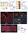
Macrophages mediate the cardioprotective effects of yREX3. (A) Schematic of the experiment design: macrophages exposed for 24 h to yREX3 (MϕyREX3; 80 nM), scramble (MϕScr; 80 nM), vehicle (MϕVeh) or small interfering RNA targeted against Pick1 (MϕsiPick1; 50 nM) were injected via the tail vein of the animals with myocardial infarction, 20 min after reperfusion. (B) At 48 h, rats infused with MϕyREX3 and MϕsiPick1 showed cardioprotection as shown by reduced scar size (2,3,5-triphenyl-2H-tetrazolium chloride (TTC; B, C) and lower cardiac troponin levels at 48 h after I/R (D; n = 5–9 animals/group) compared to rats infused with MϕScr or MϕVeh. (E) In another set of experiments, BMDM-derived macrophages exposed to yREX3 and vehicle were stained for 30 min with DiO prior to tail vein injections in a rat model of I/R, and rats sacrificed after 4 h. Immunofluorescence of heart sections show the localization of DiO-positive cells into the infarct area of the animals. (F) Immunostaining of heart sections show DiO-positive cells are double positive for CD68 (marker of macrophages) and they localize together with other macrophages into the infarct area. (G, H) TUNEL assay in rat heart sections 4 h after myocardial infarction in animals receiving macrophages exposed to vehicle or yREX3 and representative images. (B, D) All data are presented as mean ± standard error of the mean, and comparison between groups was evaluated using a one-way analysis of variance with (B) Tukey’s post-test with *P < .05, or (D) Dunnett’s post-test vs. MϕVeh with *P < .05, **P < .01, and ***P < .001; scale bar: 100 μm

References

    1. Lefer DJ, Marban E.. Is cardioprotection dead? Circulation 2017;136:98–109. 10.1161/CIRCULATIONAHA.116.027039 - DOI - PMC - PubMed
    1. Emmert MY, Burrello J, Wolint P, Hilbe M, Andriolo G, Balbi C, et al. Intracoronary delivery of extracellular vesicles from human cardiac progenitor cells reduces infarct size in porcine acute myocardial infarction. Eur Heart J 2024;45:728–32. 10.1093/eurheartj/ehad636 - DOI - PubMed
    1. Barile L, Marban E. Injury minimization after myocardial infarction: focus on extracellular vesicles. Eur Heart J 2024;15:1602–9. 10.1093/eurheartj/ehae089 - DOI - PMC - PubMed
    1. Marban E. A mechanistic roadmap for the clinical application of cardiac cell therapies. Nat Biomed Eng 2018;2:353–61. 10.1038/s41551-018-0216-z - DOI - PMC - PubMed
    1. Ibrahim AG, Cheng K, Marban E. Exosomes as critical agents of cardiac regeneration triggered by cell therapy. Stem Cell Reports 2014;2:606–19. 10.1016/j.stemcr.2014.04.006 - DOI - PMC - PubMed