

Review
doi: 10.1590/1806-9282.2024S103.
eCollection 2024.
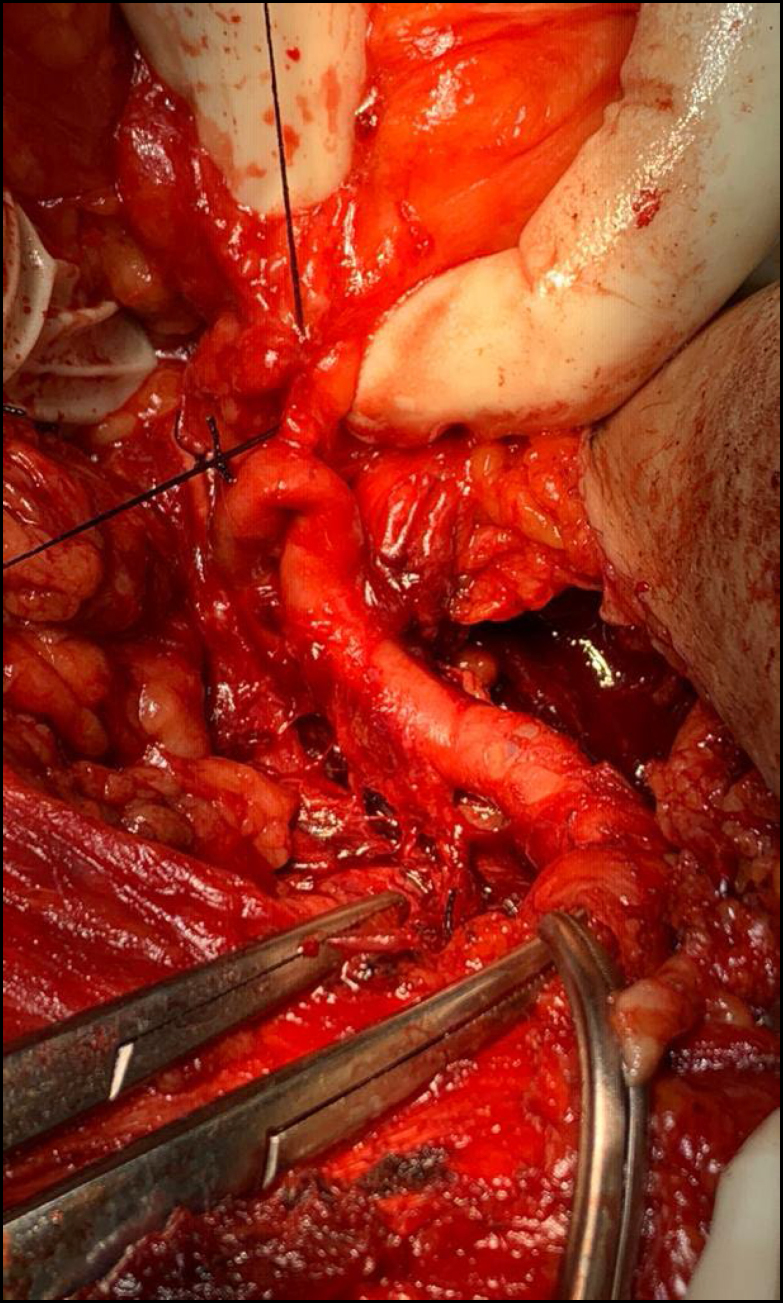
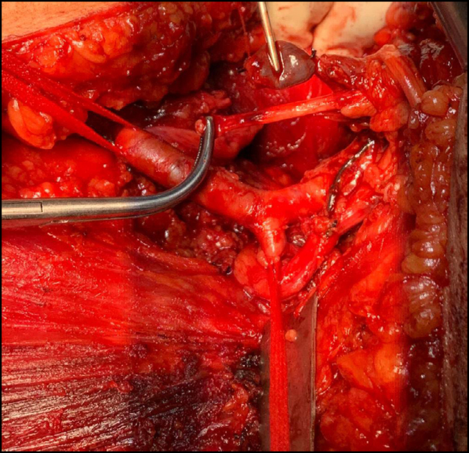
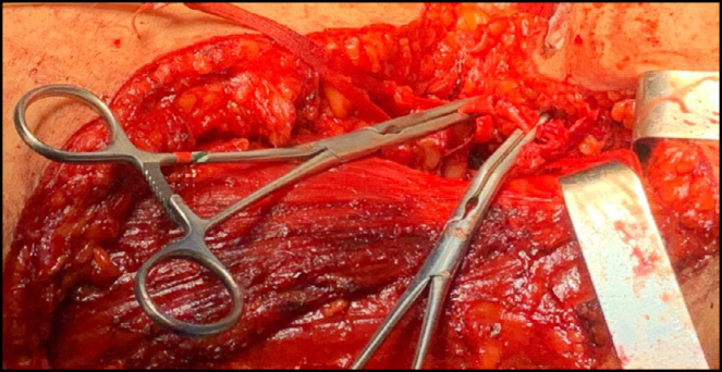
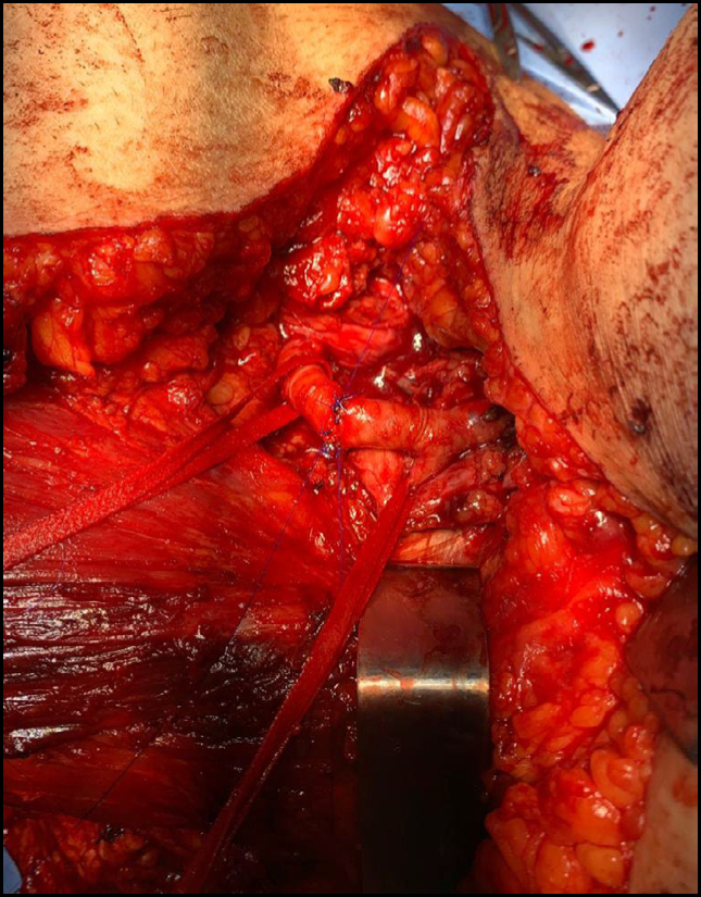
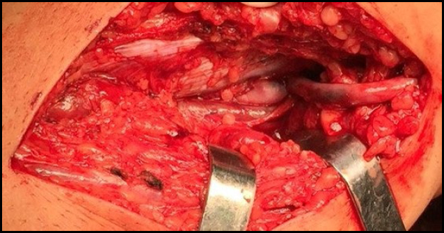
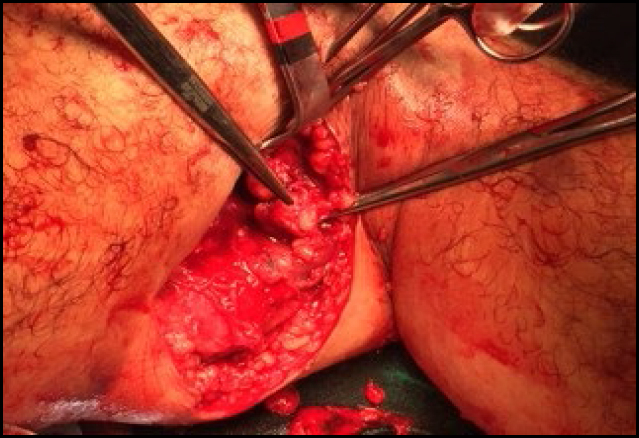
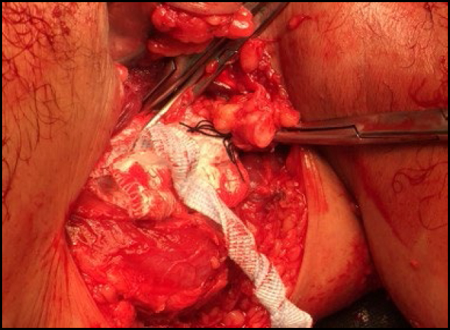
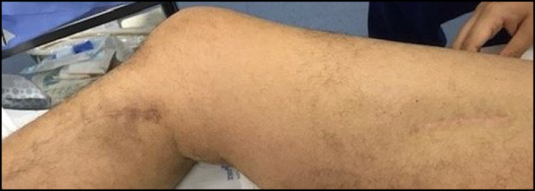
Oncovascular surgery
Affiliations
- PMID: 38865523
- PMCID: PMC11164269
- DOI: 10.1590/1806-9282.2024S103
Item in Clipboard
Review
Oncovascular surgery
Rev Assoc Med Bras (1992).
.
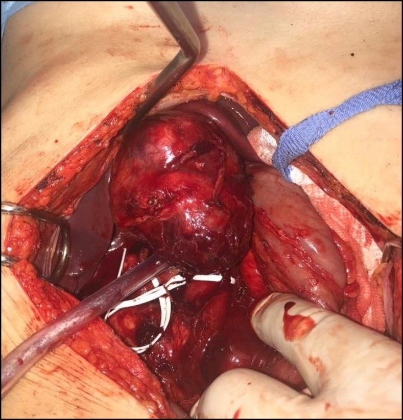
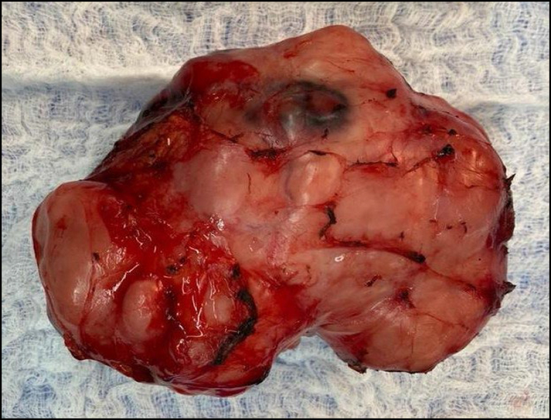
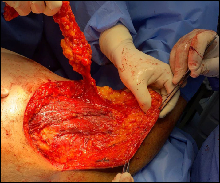
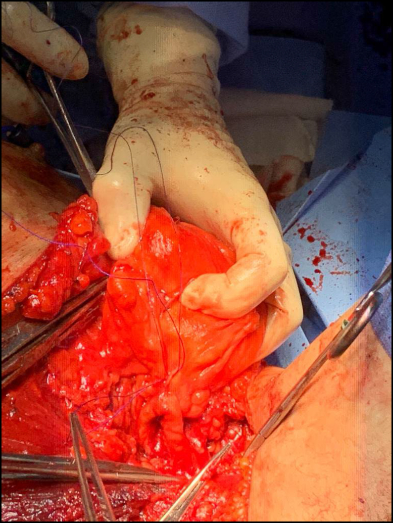
No abstract available
Conflict of interest statement
Conflicts of interest: the authors declare there is no conflicts of interest.
Figures

References
-
- Brandão BL, Silva ACB, Gouvêa MM, Lobão LM. Importância da cirurgia plástica para mulheres mastectomizadas e o papel do Sistema Único de Saúde: revisão integrativa. Rev Bras Cir Plást. 2021;36(4):457–465.
Publication types
MeSH terms
LinkOut - more resources
Full Text Sources
