Practice Guideline
. 2024 Jun 7;70(suppl 1):e2024S108.
doi: 10.1590/1806-9282.2024S108. eCollection 2024.

Hand tumors

Affiliations
Practice Guideline

Hand tumors

Antonio Tufi Neder Filho et al. Rev Assoc Med Bras (1992). .
No abstract available


Conflict of interest statement

Conflicts of interest: the authors declare there is no conflicts of interest.

Figures
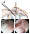
Figure 1
Figure 1. Schematic drawing showing arthroscopic surgical treatment, with the camera positioned in the mid-carpal joint, in its ulnar portal, and the shaver in the radial portal, located transcystic (A). In an initial view, thickening of the dorsal capsule and synovial membrane is observed, typical of a patient with a symptomatic dorsal cyst (B). Similar positioning of the optic after resection of the cyst pedicle and a portion of the dorsal capsule, making the extensor tendons visible (C). S: scaphoid; Si; synovitis; C: capitate; L: lunate; X: dorsal capsule; T: extensor tendon.
Figure 2
Figure 2. Giant cell tumor of the tendon sheath on the palmar aspect of the thumb. (A) Clinical appearance. (B) Surgical appearance of the tumor.
Figure 3
Figure 3. (A) Para-ungual access route. (B) Access with nail resection. (C) Access route through a window in the nail.
Figure 4
Figure 4. (A) T2-weighted magnetic resonance imaging, showing an enchondroma of the proximal phalanx of the left fourth digit, with high signal intensity of the lesion and cortical thinning. (B) X-ray AP view of the left hand showing a lytic, expanding lesion with cortical thinning in the proximal phalanx of the fourth finger, suggestive of enchondroma.
Figure 5
Figure 5. (A) X-ray AP view showing massive osteochondroma on the proximal phalanx of the third finger of the right hand. (B) Clinical image. (C) Exposure of the osteochondroma, with the hyaline cartilage layer being observed.
Figure 6
Figure 6. Computed tomography showing a central nidus with surrounding sclerosis in the trapezoid (arrow). (A) Sagittal. (B) Axial. (C) Coronal. Photos cordially provided by Dr. Fábio Augusto Caporrino from Escola Paulista de Medicina, Federal University of São Paulo.

Similar articles

References

    1. Nahra ME, Bucchieri JS. Ganglion cysts and other tumor related conditions of the hand and wrist. Hand Clin. 2004;20(3):249–260. doi: 10.1016/j.hcl.2004.03.015. - DOI - PubMed
    1. Oliveira RK, Brunelli JPF, Bayer LR, Aita M, Mantovani G, Delgado PJ. Artrhoscopic resection of volar wrist ganglion: surgical technique and case series. Rev Bras Ortop (Sao Paulo) 2019;54(6):721–730. doi: 10.1055/s-0039-1700811. - DOI - PMC - PubMed
    1. Osterman AL, Raphael J. Arthroscopic resection of dorsal ganglion of the wrist. Hand Clin. 1995;11(1):7–12. - PubMed
    1. Mathoulin C, Hoyos A, Pelaez J. Arthroscopic resection of wrist ganglia. Hand Surg. 2004;9(2):159–164. doi: 10.1142/s0218810404002169. - DOI - PubMed
    1. Ho PC, Lo WN, Hung LK. Arthroscopic resection of volar ganglion of the wrist: a new technique. Arthroscopy. 2003;19(2):218–221. doi: 10.1053/jars.2003.50035. - DOI - PubMed

Publication types