

Impact of Topical 0.05% Cyclosporine A Eye Drops on Post-Femtosecond-Assisted Laser In Situ Keratomileusis Ocular Surface Recovery: A Randomized Clinical Trial
- PMID: 38865592
- PMCID: PMC11265643
- DOI: 10.1097/ICL.0000000000001103
Impact of Topical 0.05% Cyclosporine A Eye Drops on Post-Femtosecond-Assisted Laser In Situ Keratomileusis Ocular Surface Recovery: A Randomized Clinical Trial
Abstract
Objectives: To investigate the effect of topical 0.05% cyclosporine A (CsA) eye drops as an adjunct to conventional therapy in maintaining post-femtosecond-assisted laser in situ keratomileusis (FS-LASIK) ocular surface stability.
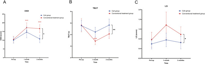
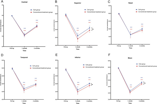
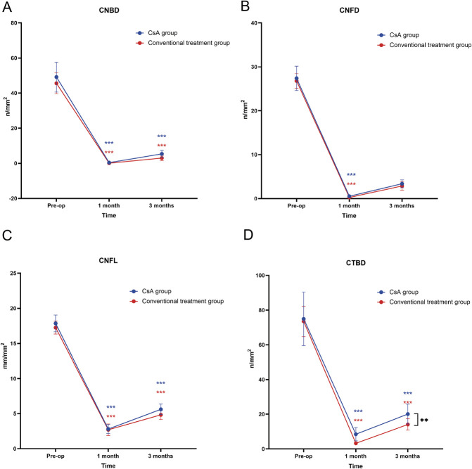
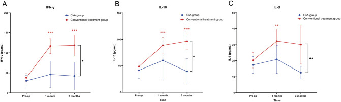
Methods: Sixty-six patients (eyes) undergoing FS-LASIK were randomized into 2 groups: 33 patients (eyes) in group I (conventional treatment group) and 33 patients (eyes) in group II (CsA group). Conventional treatments include topical levofloxacin, fluorometholone, and artificial tears. Group II received topical 0.05% CsA eye drops twice daily for three months in addition to conventional treatment. Ocular Surface Disease Index (OSDI), numerical rating scale (NRS), tear break-up time (TBUT), Schirmer I test (SIt), corneal fluorescein staining (CFS), conjunctival lissamine green (LG) staining, corneal sensitivity, and corneal nerve morphology were measured. In addition, tear inflammatory cytokine levels were measured using the Luminex assay. Follow-up was performed preoperatively and 1 and 3 months postoperatively.
Results: In the CsA group, OSDI, TBUT, LG, corneal sensitivity, and corneal nerve fiber total branch density recovered better than in the conventional treatment group. As for tear inflammatory cytokines, interferon (INF) -γ, interleukin (IL)-10, and IL-6 levels were significantly higher in the conventional treatment group as compared with the CsA group. In addition, no significant differences in NRS, SIt, and CFS scores were observed between the two groups.
Conclusion: In conclusion, 0.05% CsA eye drops is a useful adjunct to conventional treatment for restoring the ocular surface stability after corneal refractive surgery and is more potent in sustaining anti-inflammatory effects.
Copyright © 2024 The Author(s). Published by Wolters Kluwer Health, Inc. on behalf of the Contact Lens Association of Opthalmologists.
Conflict of interest statement
The authors have no funding or conflicts of interest to disclose.
Figures
References
-
- Battat L, Macri A, Dursun D, et al. . Effects of laser in situ keratomileusis on tear production, clearance, and the ocular surface. Ophthalmology 2001;108:1230–1235. - PubMed
-
- Yu EY, Leung A, Rao S, et al. . Effect of laser in situ keratomileusis on tear stability. Ophthalmology 2000;107:2131–2135. - PubMed
-
- Chao C, Stapleton F, Zhou X, et al. . Structural and functional changes in corneal innervation after laser in situ keratomileusis and their relationship with dry eye. Graefes Arch Clin Exp Ophthalmol 2015;253:2029–2039. - PubMed
-
- Denoyer A, Landman E, Trinh L, et al. . Dry eye disease after refractive surgery: Comparative outcomes of small incision lenticule extraction versus LASIK. Ophthalmology 2015;122:669–676. - PubMed
Publication types
MeSH terms
Substances
LinkOut - more resources
Full Text Sources
Medical
Research Materials
