. 2024 Aug 13;8(15):4181-4193.
doi: 10.1182/bloodadvances.2024012874.

CG001, a C3b-targeted complement inhibitor, blocks 3 complement pathways: development and preclinical evaluation

Affiliations

CG001, a C3b-targeted complement inhibitor, blocks 3 complement pathways: development and preclinical evaluation

Ling Li et al. Blood Adv. .

Abstract

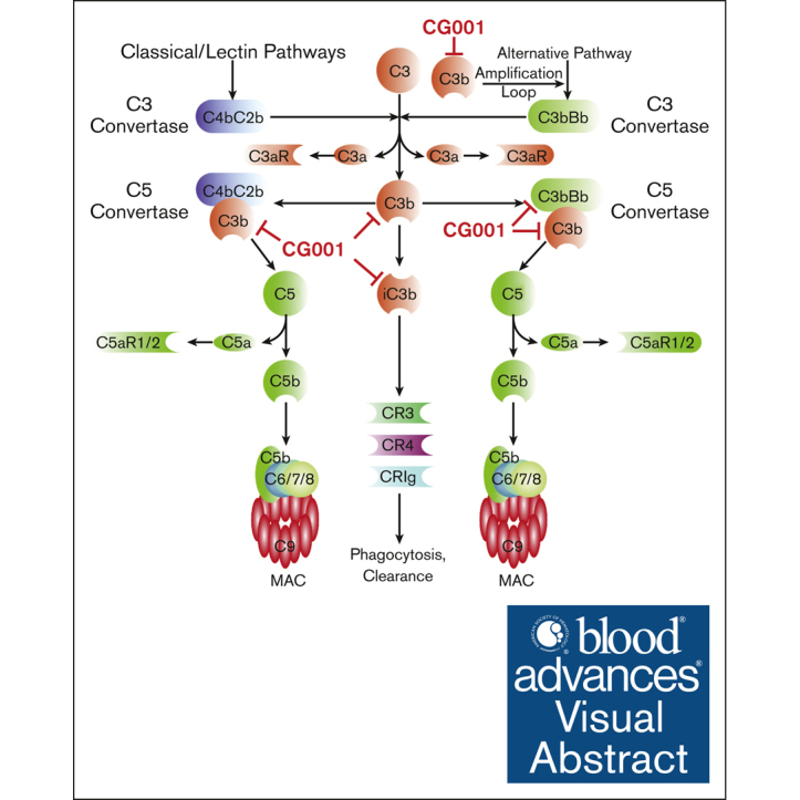
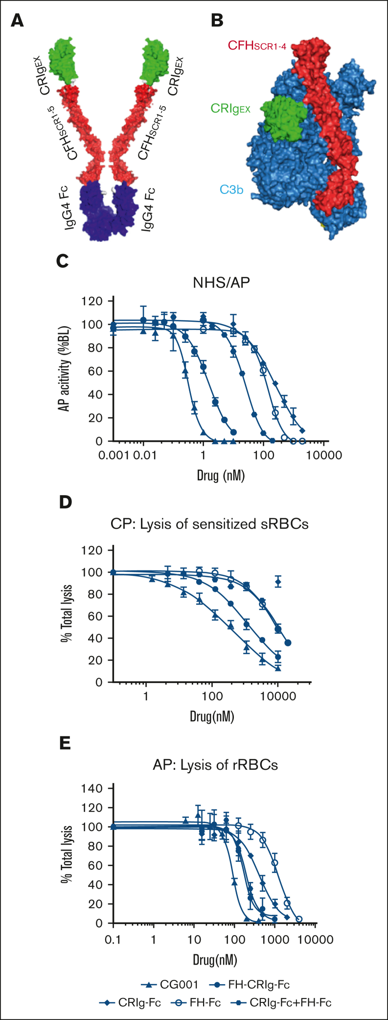
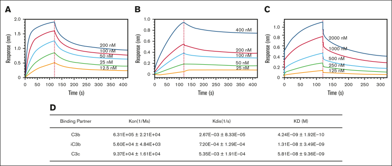
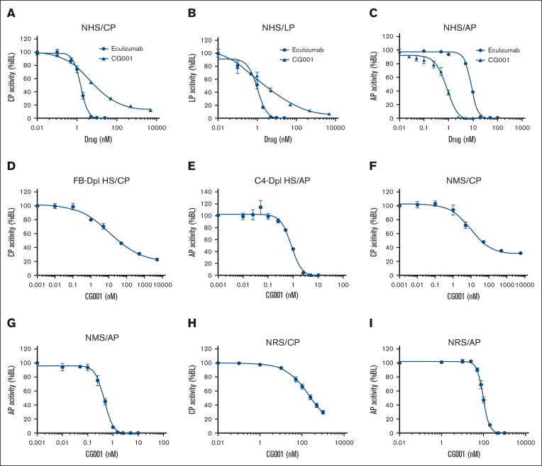
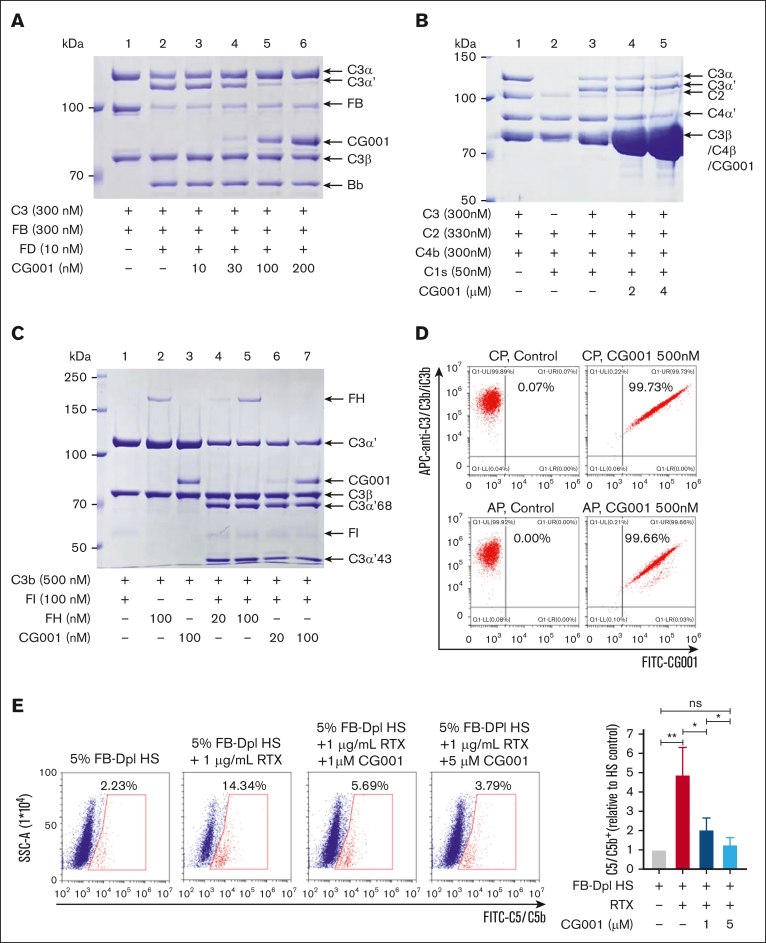
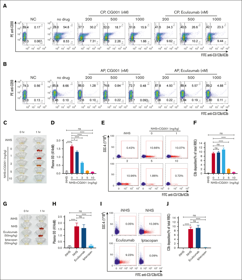
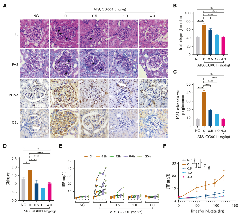
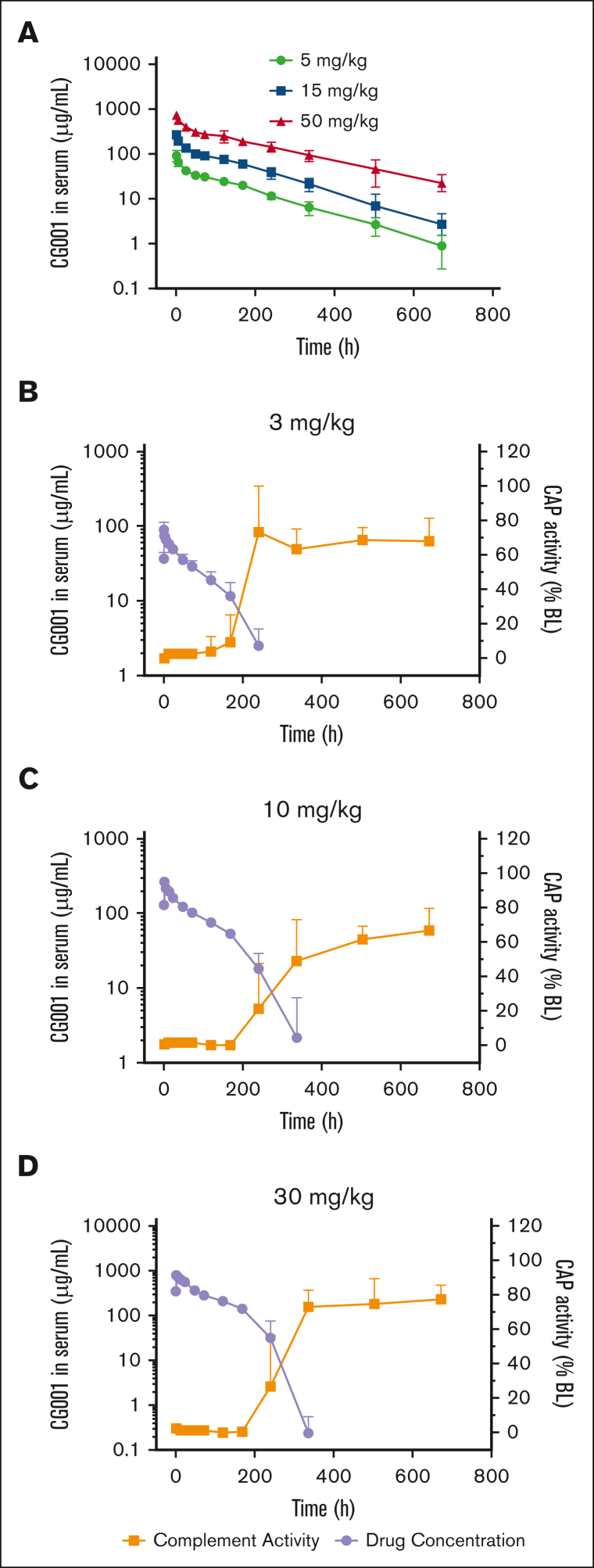
Excessively activated or dysregulated complement activation may contribute to the pathogenesis of a wide range of human diseases, thus leading to a surge in complement inhibitors. Herein, we developed a human-derived and antibody-like C3b-targeted fusion protein (CRIg-FH-Fc) x2, termed CG001, that could potently block all 3 complement pathways. Complement receptor of the immunoglobulin superfamily (CRIg) and factor H (FH) bind to distinct sites in C3b and synergistically inhibit complement activation. CRIg occupancy in C3b prevents the recruitment of C3 and C5 substrates, whereas FH occupancy in C3b accelerates the decay of C3/C5 convertases and promotes the factor I-mediated degradation and inactivation of C3b. CG001 also showed therapeutic effects in alternative pathways-induced hemolytic mouse and classical pathways-induced mesangial proliferative glomerulonephritis rat models. In the pharmacological/toxicological evaluation in rats and cynomolgus monkeys, CG001 displayed an antibody-like pharmacokinetic profile, a convincing complement inhibitory effect, and no observable toxic effects. Therefore, CG001 holds substantial potential for human clinical studies.


Conflict of interest statement

Conflict-of-interest disclosure: W.H. is the inventor of CRIg-FH and CG001, and the founder of ComGen Pharmaceutical. X.L., Luying Lo, and J.Y. are employees of ComGen Pharmaceutical. The remaining authors declare no competing financial interests.

Figures

None
Graphical abstract
Figure 1.
Figure 1.
Design of CG001 and its inhibitory effect on complement pathways. (A) Schematic of CG001. It is composed of the extracellular domain of CRIg (green CRIgEX; PDB, 2ICC13), the short consensus repeat (SCR) 1-5 of FH (red FHSCR1-5; image showing SCR1-4 from PDB, 2WII in panel B), and the Fc fragment of human IgG4 (purple IgG4 Fc; PDB, 4C5415). (B) Composite image of the binding of CRIgEX (green; PDB, 2ICF13) and FHSCR1-4 (red; PDB, 2WII14) to C3b (sky blue). Diagrams were generated with PyMOL molecular visualization software. (C) Comparison of CG001 with other CRIg- or FH-derived fusion proteins to inhibit AP activation using the Wieslab complement system AP kit. The IC50 was 0.83 ± 0.12 nM for CG001; 1.35 ± 0.49 nM for FH-CRIg-Fc; 239.3 ± 123.5 nM for CRIg-Fc; 121.6 ± 36.2 nM for FH-Fc; and 24.55 ± 0.31 for CRIg-Fc + FH-Fc. (D-E) Effect of CG001 on protecting erythrocytes from complement-mediated hemolysis. (D) CP was activated by 0.2% sheep erythrocyte hemolysin and 1% NHS to induce the hemolysis of sheep erythrocytes (sRBCs), whereas (E) AP was directly activated by 12% NHS to induce the hemolysis of rabbit erythrocytes (rRBCs). The IC50 was 446.2 ± 89.06 nM for CG001; 1083 ± 168.18 nM for FH-CRIg-Fc; 5458 ± 1036.81 nM for FH-Fc; and 9269 ± 307.45 nM for CRIg-Fc + FH-Fc for CP. The IC95 was 34.19 ± 3.77 μM for CG001; 68.66 ± 45.85 μM for FH-CRIg-Fc; 111.07 ± 24.13 μM for FH-Fc; and 176.11 ± 7.15 μM for CRIg-Fc + FH-Fc for CP. CRIg showed little inhibitory activity against CP. In addition, the IC50 was 88.88 ± 8.31 nM for CG001; 167 ± 8.66 nM for FH-CRIg-Fc; 416.1 ± 52.83 nM for CRIg-Fc; 1231 ± 146.83 nM for FH-Fc; and 195.4 ± 16.34 nM for CRIg-Fc + FH-Fc for AP. The IC95 was 206.83 ± 7.83 nM for CG001; 419.23 ± 139.03 nM for FH-CRIg-Fc; 2160 ± 345.98 nM for CRIg-Fc; 5040 ± 1846.65 nM for FH-Fc; and 551.05 ± 158.17 nM for CRIg-Fc + FH-Fc for AP (E). Values represent the mean ± standard deviation (SD), and the experiments were performed at least 3 times.
Figure 2.
Figure 2.
Binding affinity of CG001 to human C3 cleavage products. (A-C) Representative profiles of the interactions between CG001 and human C3 products. (A) C3b, (B) iC3b, and (C) C3c. (D) Binding affinity of CG001 to 3 human C3 cleavage products. The interactions were measured by biolayer interferometry, using 3 batches of CG001 drug substances. Values represent the mean ± SD.
Figure 3.
Figure 3.
The in vitro complement inhibitory activity of CG001. (A-C) The complement inhibitory effect of CG001 in NHS. The complement was activated via (A) CP, (B) LP, or (C) AP, and CG001 was compared with eculizumab as a control. (A) IC50 = 4.71 ± 0.28 nM (CG001) or 1.54 ± 0.14 nM (eculizumab) for CP, IC95 = 510.95 ± 91.98 nM (CG001) or 5.5 ± 0.29 nM (eculizumab) for CP; (B) 2.52 ± 0.62 nM (CG001) or 1.02 ± 0.10 nM (eculizumab) for LP, and IC95 was 365.73 ± 114.62 nM (CG001) or 5.71 ± 0.48 nM (eculizumab) for LP; and (C) 0.83 ± 0.12 nM (CG001) or 8.56 ± 0.24 nM (eculizumab) for AP and IC95 was 4.0 ± 0.76 nM (CG001) or 22.0 ± 0.93 nM (eculizumab) for AP. (D-E) The inhibitory effect of CG001 on complement component-depleted human serum. (D) IC50 = 10.932 ± 1.629 nM in FB-depleted human serum selective for CP activity, and (E) IC50 = 0.858 ± 0.034 nM in C4-depleted human serum selective for AP activity. (F-I) The complement inhibitory effect of CG001 in normal monkey serum (NMS) and normal rat serum (NRS). (F) IC50 = 9.335 ± 2.328 nM for CP in NMS, (G) 0.455 ± 0.037 nM for AP in NMS, (H) IC50 = 192.5 ± 36.2 nM for CP in NRS, or (I) 96.0 ± 6.8 nM for AP in NRS. Complement activities in human and monkey sera were measured by the Wieslab complement system kit for panels A-G. To evaluate the complement activities in rat serum, (H) CP was activated by 0.2% sheep erythrocyte hemolysin and 1% NRS to induce hemolysis of sRBCs, whereas (I) AP was directly activated by 18% NHS to induce hemolysis of rRBCs. Values represent mean ± SD, and experiments were performed in triplicate.
Figure 4.
Figure 4.
Molecular mechanism by which CG001 inhibits complement activation. (A) CG001 inhibited the C3 convertase in AP. In the presence of FB and FD, the C3α chain of C3 was cleaved to C3α′ and released C3a, which was inhibited by CG001 in a dose-dependent manner. (B) CG001 failed to inhibit the C3 convertase in CP. In the presence of C2, C4b, and C1s, C3α was cleaved to C3α′ and C3a was released; however, CG001 failed to block this process even at high concentrations. (C) CG001 promoted C3b degradation. The C3α′ chain of C3b was proteolyzed to C3α′68 and C3α′43 by FI in the presence of FH or CG001, in which CG001 displayed a similar potency to FH. (D) CG001 bound to the specific cell surface with C3b/iC3b deposition. CP and AP were activated in Raji cells, and the deposition of C3b/iC3b and CG001 was determined by a flow cytometry assay. (E) CG001 impaired C5 binding to the cell surface upon CP activation. After CP activation by rituximab and FB-depleted human serum in Raji cells, C5 was recruited to the membrane-bound C5 convertase C4bC2aC3b, and the resultant proteolytic C5b fragment was inserted into the cell surface, all of which could be detected by anti-C5/C5b antibody with a flow cytometry assay. CG001 reduced C5/C5b staining by preventing C5 recruitment and C5b production by binding to the C3b subunit of C5 convertase. Values represent mean ± SD, and experiments were performed in triplicate. ∗P < .05 and ∗∗P < .01.
Figure 5.
Figure 5.
CG001 prevented PNH erythrocytes from hemolysis. (A-B) CG001 protected human PNH erythrocytes from CP- and AP-induced hemolysis. Hemolysis and C3b/iC3b deposition (selective for CP) in CD59 PNH erythrocytes were induced by (A) CP or (B) AP activation, which could be effectively prevented by CG001 treatment. However, (A) eculizumab treatment prevented only hemolysis but not C3b/iC3b deposition induced by CP activation and (B) showed little effect on AP-induced hemolysis. (C-J) CG001 blocked both IVH and EVH in the NHS-induced and AP-mediated hemolytic mouse models compared with eculizumab and iptacopan. (C,G) IVH was determined by the plasma images, and (D,H) the quantitative plasma hemoglobin. (E,I) EVH was represented by the deposition of C3b/iC3b in erythrocytes, which is shown as a scatterplot, and (F,J) the related quantitative results. (C-F) CG001, and (G-J) eculizumab and iptacopan. Data are presented as mean ± standard error of the mean (SEM); n = 4; ∗P < .05 and ∗∗∗∗P < .0001.
Figure 6.
Figure 6.
CG001 attenuates complement-mediated glomerular lesions in a rat MsPGN model. (A-D) HE, PAS staining, and immunostaining for proliferating cell nuclear antigen (PCNA) and C3d in kidney tissue sections collected from anti-Thy1 nephritic rats on day 7 after treatment with CG001. (A) Representative photomicrographs of glomeruli and (B) quantitative results of total cell counts, (C) PCNA-positive cell counts in the glomeruli and (D) C3d deposition. Arrows indicate the increased mesangial cellularity, and stars indicate the thickened capillary walls and glomerular basement membrane. (E-F) Effect of CG001 treatment on 24-hour urinary protein collected at the indicated time points. Data are presented as (E) individual and (F) combined results (mean ± SEM); n = 4 (negative control, NC), 8 (rabbit anti-Thy1 serum, anti-rat thymocyte serum [ATS] alone treatment), or 6 (additive CG001 treatment). ∗ P < .05, ∗∗∗P < .001, and ∗∗∗∗P < .0001.
Figure 7.
Figure 7.
PK/PD evaluation of CG001 in rats and cynomolgus monkeys. (A-B) PK was evaluated by measuring the serum CG001 concentration in rats. n = 12 per group. (C-D) PK/PD correlation of CG001 in monkeys. Complement activity was measured by the Wieslab complement system AP kit. n = 6 per group. Values represent mean ± SD.

Similar articles

Cited by

References

    1. Merle NS, Church SE, Fremeaux-Bacchi V, Roumenina LT. Complement system part I - molecular mechanisms of activation and regulation. Front Immunol. 2015;6:262. - PMC - PubMed
    1. Seifert L, Zahner G, Meyer-Schwesinger C, et al. The classical pathway triggers pathogenic complement activation in membranous nephropathy. Nat Commun. 2023;14(1):473. - PMC - PubMed
    1. Ramlall V, Thangaraj PM, Meydan C, et al. Immune complement and coagulation dysfunction in adverse outcomes of SARS-CoV-2 infection. Nat Med. 2020;26(10):1609–1615. - PMC - PubMed
    1. Yamashina M, Ueda E, Kinoshita T, et al. Inherited complete deficiency of 20-kilodalton homologous restriction factor (CD59) as a cause of paroxysmal nocturnal hemoglobinuria. N Engl J Med. 1990;323(17):1184–1189. - PubMed
    1. Risitano AM, Marotta S. Therapeutic complement inhibition in complement-mediated hemolytic anemias: past, present and future. Semin Immunol. 2016;28(3):223–240. - PubMed

Publication types

MeSH terms