. 2024 Jun 25;96(25):10373-10379.
doi: 10.1021/acs.analchem.4c01477. Epub 2024 Jun 12.

Spatially Offset Raman Spectroscopy toward In Vivo Assessment of the Adipose Tissue in Cardiometabolic Pathologies

Affiliations

Spatially Offset Raman Spectroscopy toward In Vivo Assessment of the Adipose Tissue in Cardiometabolic Pathologies

Ewa Stanek et al. Anal Chem. .

Abstract

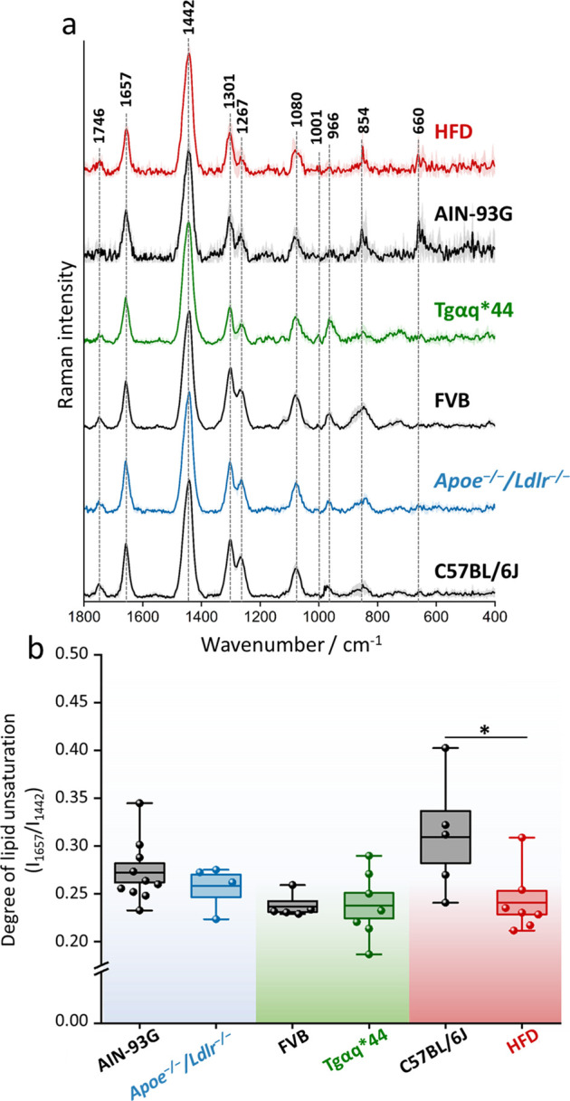
Spatially offset Raman spectroscopy (SORS) enhanced the capabilities of Raman spectroscopy for the depth-resolved analysis of biological and diffusely scattering samples. This technique offers selective probing of subsurface layers, providing molecular insights without invasive procedures. While SORS has found application in biomedical research, up to now, studies have focused mainly on the detection of mineralization of bones and tissues. Herein, for the first time, SORS is used to assess the soft, organic tissue beneath the skin's surface. In this study, we demonstrate the diagnostic utility of a hand-held SORS device for evaluating the chemical composition of the adipose tissue. We compared perigonadal white adipose tissue (gWAT) in a murine model of atherosclerosis, heart failure, and high-fat diet (HFD) induced obesity. Our results reveal distinct chemical differences in gWAT between HFD-fed and control mice, showcasing the potential of SORS for intravital adipose tissue phenotype characterization. Furthermore, our findings underscore the effectiveness of SORS as a valuable tool for noninvasive assessment of the adipose tissue composition, holding potential diagnostic significance for metabolic disorders.


Conflict of interest statement

The authors declare no competing financial interest.

Figures

Figure 1
Figure 1
Representative SORS spectra collected from skin and gWAT. Spectra were acquired at a 0.0 offset and presented as maximally extended in the y-axis. X is the factor that enables comparing intensities of spectra, 20× means that the spectrum is 20 times more intense than the spectrum denoted as 1×.
Figure 2
Figure 2
Spatial offset measurements confirm Raman signals from perigonadal white adipose tissue. Raman spectra collected from different offsets (0 to 5.5) where (a) layer of skin and gWAT were stacked (b) separated by the polypropylene plate and (c) separated by the polypropylene plate with gWAT labeled with β-carotene. All spectra were normalized to the highest band in the red spectrum. X is the factor that enables comparing intensities of spectra, 3× means that the spectrum is three times more intense than the spectrum denoted as 1×, etc.
Figure 3
Figure 3
Chemical composition of the perigonadal white adipose tissue in mice with cardiometabolic diseases. Averaged Raman spectra with the standard deviation on each data point (a) of gWAT acquired post mortem from C57BL/6J, Apoe–/–/Ldlr–/–, FVB, Tgαq*44, and mice fed with a HFD for 4 weeks. The degree of lipid unsaturation (I1657/I1442) was calculated (b) for each studied group. Values shown in box plots: mean (horizontal line), SEM (box), minimal, and maximal values (whiskers). Statistical significance * p < 0.05.
Figure 4
Figure 4
Diet-dependent changes in the lipid profile of perigonadal white adipose tissue. The degree of lipid unsaturation (I1657/I1442) was calculated for each group after 2-week exposure to the AIN-93G and HFD diet with or without additional supplements. Values shown in box plots: mean (horizontal line), SEM (box), minimal, and maximal values (whiskers). Statistical significance * p < 0.05.

Similar articles

Cited by

References

    1. Cordero E. In-Vivo Raman Spectroscopy: From Basics to Applications. J. Biomed. Opt. 2018, 23 (07), 1.10.1117/1.JBO.23.7.071210. - DOI - PubMed
    1. Matousek P.; Clark I. P.; Draper E. R. C.; Morris M. D.; Goodship A. E.; Everall N.; Towrie M.; Finney W. F.; Parker A. W. Subsurface Probing in Diffusely Scattering Media Using Spatially Offset Raman Spectroscopys. Appl. Spectrosc. 2005, 59 (4), 393–400. 10.1366/0003702053641450. - DOI - PubMed
    1. Nicolson F.; Kircher M. F.; Stone N.; Matousek P. Spatially Offset Raman Spectroscopy for Biomedical Applications. Chem. Soc. Rev. 2021, 50 (1), 556–568. 10.1039/D0CS00855A. - DOI - PMC - PubMed
    1. Mosca S.; Dey P.; Salimi M.; Palombo F.; Stone N.; Matousek P. Non-Invasive Depth Determination of Inclusion in Biological Tissues Using Spatially Offset Raman Spectroscopy with External Calibration. Analyst 2020, 145 (23), 7623–7629. 10.1039/D0AN01292K. - DOI - PubMed
    1. Macleod N. A.; Goodship A.; Parker A. W.; Matousek P. Prediction of Sublayer Depth in Turbid Media Using Spatially Offset Raman Spectroscopy. Anal. Chem. 2008, 80 (21), 8146–8152. 10.1021/ac801219a. - DOI - PubMed

Publication types