. 2024 Aug:74:103202.
doi: 10.1016/j.redox.2024.103202. Epub 2024 May 21.

Development of nitroalkene-based inhibitors to target STING-dependent inflammation

Affiliations

Development of nitroalkene-based inhibitors to target STING-dependent inflammation

Fei Chang et al. Redox Biol. 2024 Aug.

Abstract

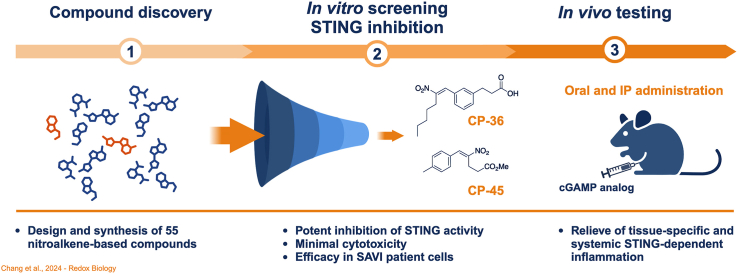
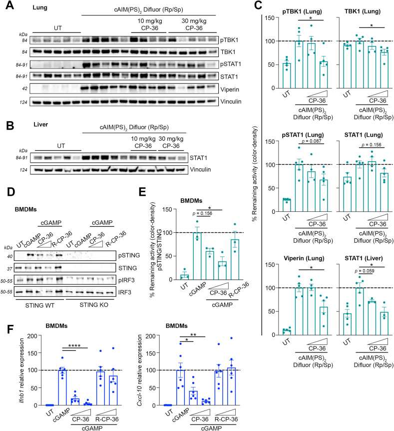
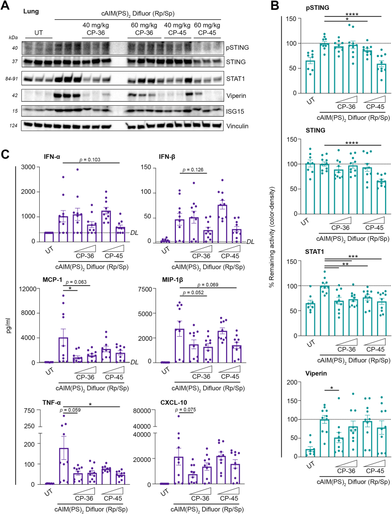
Stimulator of Interferon Genes (STING) is essential for the inflammatory response to cytosolic DNA. Despite that aberrant activation of STING is linked to an increasing number of inflammatory diseases, the development of inhibitors has been challenging, with no compounds in the pipeline beyond the preclinical stage. We previously identified endogenous nitrated fatty acids as novel reversible STING inhibitors. With the aim of improving the specificity and efficacy of these compounds, we developed and tested a library of nitroalkene-based compounds for in vitro and in vivo STING inhibition. The structure-activity relationship study revealed a robustly improved electrophilicity and reduced degrees of freedom of nitroalkenes by conjugation with an aromatic moiety. The lead compounds CP-36 and CP-45, featuring a β-nitrostyrene moiety, potently inhibited STING activity in vitro and relieved STING-dependent inflammation in vivo. This validates the potential for nitroalkene compounds as drug candidates for STING modulation to treat STING-driven inflammatory diseases, providing new robust leads for preclinical development.

Keywords: Drug discovery; Interferon; Nitroalkene-based compounds; STING inhibitors; STING-associated vasculopathy with onset in infancy (SAVI); Stimulator of Interferon Genes (STING).


Conflict of interest statement

Declaration of competing interest The authors declare the following financial interests/personal relationships which may be considered as potential competing interests: Francisco J Schopfer reports a relationship with Creegh Pharmaceuticals Inc that includes: board membership and equity or stocks. Francisco J Schopfer & Fei Chang reports a relationship with Furanica Inc that includes: board membership and equity or stocks. Christian K Holm reports a relationship with UV Medico that includes: consulting or advisory and employment. Francisco J Schopfer, Fei Chang, Christian K Holm, Anne Louise Hansen, Sonia R Salvatore, Luis Villacorta are the inventors of a patent application related to the subject matter of this manuscript. The University of Pittsburgh is the lead institution for this patent application, with joint ownership also held by Aarhus University and Morehouse School of Medicine. Other authors declare that they have no known competing financial interests or personal relationships that could have appeared to influence the work reported in this paper.

Figures

Image 1
Graphical abstract
Fig. 1
Fig. 1
Inhibition of STING by nitroalkene compounds depends on fatty acyl chain length and relative position of the nitroalkene group. The cells were pre-treated with specified concentrations of testing compounds for 2 h, followed by cGAMP stimulation (4 μg/mL) for 24 h (unless otherwise stated below) (A) Levels of NF-κB and IRF activation in THP-1-Dual cells treated with indicated nitroalkene compounds (10 μM) or H-151 (1 μM) and cGAMP stimulation. (B) CXCL-10 release in THP-1 wt cells treated with indicated nitroalkene compounds (10 μM). (C) CXCL-10 release in THP-1 wt cells treated with indicated nitroalkene compounds (2, 5 and 10 μM), CKA (15, 20 and 30 μM), and CP-25 (2, 5, 10 and 15 μM) or H-151 (0.5 and 1 μM). (D) Immunoblot analysis of whole-cell lysate of THP-1 wt cells to evaluate levels of STING, pSTING, TBK1, pTBK1, IRF3, and pIRF3 after treatment with indicated nitroalkene compounds (5 and 10 μM), OA (10 μM), CKA (20 and 30 μM), CP-25 (5 and 10 μM) or H-151 (0.5 and 1 μM), followed by 3 h cGAMP stimulation. (E) Immunoblot analysis of whole-cell lysate of THP-1 wt cells to evaluate levels of IFIT1 and ISG15 after treatment with indicated nitroalkene compounds (5 and 10 μM), 9-NO2OA and OA (10 μM), CKA (20 and 30 μM), CP-25 (5 and 10 μM) or H-151 (1 μM). (F) Type I IFN release in SAVI fibroblasts treated with indicated nitroalkene compounds (1, 2 and 5 μM) or H-151 (2 and 5 μM). (G) Immunoblot analysis of whole-cell lysate of SAVI fibroblasts to evaluate levels of STING, pSTING, TBK1, pTBK1, IRF3 and pIRF3 after treatment with indicated nitroalkene compounds (5 and 10 μM) or H-151 (1 and 2 μM), followed by 3 h cGAMP stimulation. Bars indicate the mean ± SEM of two to three replicates and are normalized to the levels reached with cGAMP, expressed as a percentage. The dotted lines denote 75 % inhibition of NF-kB activation, CXCL-10 response, and type I IFN response, or 50 % inhibition of IRF activation of that induced by cGAMP.
Fig. 2
Fig. 2
Determining thein vitrotherapeutic index of second-generation nitroalkene-based compounds.In vitro therapeutic index based on inhibition of CXCL-10 release plotted against level of cytotoxicity. For CXCL-10 release, THP-1 wt cells were pre-treated with indicated nitroalkene compounds in different concentrations (2, 5, 10 and 15 μM) for 2 h, followed by cGAMP stimulation (4 μg/mL) for 24 h. The cytotoxicity was measured by MTT assay, and THP-1 wt cells were treated with indicated nitroalkene compounds in different concentrations (2, 5, 10, 15, 20, 30, 50, 70 and 100 μM) for 24 h. Data of the therapeutic index represent the mean ± SEM of three replicates. The CXCL-10 release is normalized to the levels reached with cGAMP, expressed as a percentage. The dotted line denotes 75 % inhibition of the CXCL-10 response induced by cGAMP. The cytotoxicity is normalized to untreated cells, expressed as percentage viability. The EC50 value is determined by fitting the curve with nonlinear regression, and compounds with EC50 values above 25 are considered to have low cytotoxicity (bold numbers). The asterisk and bold axes indicate the lead compounds with a high in vitro therapeutical index, due to high potency to inhibit CXCL-10 production with minimal cytotoxicity.
Fig. 3
Fig. 3
Inhibition of STING by second-generation nitroalkene-based compounds and assessment of electrophilic characterization. (A)In vitro therapeutic index based on inhibition of type I IFN release plotted against level of cytotoxicity. For type I IFN release, SAVI fibroblasts were pre-treated with indicated nitroalkene compounds (2, 5, 10 and 15 μM) for 2 h, followed by cGAMP stimulation (4 μg/mL) for 24 h. The cytotoxicity was measured by MTT assay, SAVI fibroblasts were treated with indicated nitroalkene compounds in different concentrations (2, 5, 10, 15, 20, 30, 50, 70 and 100 μM) for 24 h. Data of the therapeutic index represent the mean ± SEM of two (IFN) and three (MTT) replicates. The type I IFN release is normalized to levels reached with cGAMP, expressed as percentage. The cytotoxicity is normalized to untreated cells, expressed as percentage viability. The dotted line denotes 75 % inhibition of the type I IFN response induced by cGAMP. The EC50 value is determined by fitting the curve with nonlinear regression. The asterisk and bold axes indicate the lead compounds with a high in vitro therapeutical index, due to high potency to inhibit type I IFN production with minimal cytotoxicity. (B) Level of NF-κB and IRF activation in THP-1-Dual cells after pre-treatment with indicated nitroalkene compounds (10, 20 and 30 μM) or 9-NO2OA (10 μM), CP-8b (20 μM) and OA (30 μM) for 2 h, followed by cGAMP stimulation (4 μg/mL) for 24 h. Bars indicate the mean ± SEM of three replicates and are normalized to cGAMP levels, expressed as percentage. The dotted lines denote 50 % or 75 % inhibition of the IRF and NF-kB activation induced by cGAMP, respectively. (C) Immunoblot analysis of whole-cell lysate of SAVI fibroblasts to evaluate levels of pSTING, STING, pTBK1, TBK1, pIRF3 and IRF3 after treated with indicated nitroalkene compounds (2, 5 and 10 μM) or CP-8b (5 and 10 μM) for 2 h, followed by cGAMP stimulation (4 μg/mL) for 3 h. (D–F) Reaction kinetics of CP-36 (top panels) and CP-45 (bottom panels) with GSH. (D) Changes in UV–vis absorbance of CP-36 (10 μM) and CP-45 (10 μM) upon reaction with GSH (50 μM) in phosphate buffer (0.1 M, pH 7.4, 0.1 mM DTPA) at 25 °C. Spectra was recorded every 36 s for CP-36 and 12 s for CP-45, capturing approximately 10 half-lives of the reaction between GSH and the nitroalkene. The downward arrows at 323 and 333 nm indicate consumption of CP-36 and CP-45, respectively. The upward arrows at 265/270 nm indicate the formation of a single adduct with GSH (E) Stopped flow kinetic traces of the reaction of CP-36 (20 μM) and CP-45 (μM) with increasing concentrations of GSH, followed at 323 and 333 nm, respectively. (F) Exponential pseudo-first order rate constants as a function of GSH concentration. Kobs values were determined from the best fit of the kinetics traces shown in (E) to a single-phase exponential with slope function and plotted against GSH concentration.
Fig. 4
Fig. 4
Intraperitoneal administration of nitroalkene compound CP-36 dampens STING-dependent inflammationin vivo.(A–C) Mice were administrated with CP-36 i.p. (10 or 30 mg/kg) 2 h before activating STING with i.p. injection of cAIM(PS)2 Difluor (Rp/Sp) (0.2 mg/kg) for 4 h. Immunoblot analysis of homogenized and lysed (A) lung and (B) liver tissue to evaluate the levels of pTBK1, TBK1, pSTAT1, STAT1 and Viperin with (C) densitometry quantification of protein levels. The plots include data from individual mice represented in immunoblots. Bars indicate mean ± SEM and are normalized to the cAIM(PS)2 Difluor (Rp/Sp) group, expressed as a percentage. (D–F) WT or STING-KO BMDMs were pre-treated with CP-36 (5 and 10 μM) or reduced CP-36 (R-CP-36, 5 and 10 μM) for 2 h, followed by cGAMP stimulation (4 μg/mL) for either 3 h or 6 h. (D) Immunoblot analysis of whole-cell lysate (representative blot) to evaluate levels of pSTING, STING, pIRF3 and IRF3 and (E) densitometry quantification of pSTING/STING protein levels after 3 h cGAMP stimulation. Three independent experiments are plotted. (F) qPCR analysis of Ifnb1 and Cxcl-10 expression was quantified relative to 18S mRNA levels after 6 h of cGAMP stimulation. BMDM cultures obtained from 6 individual mice (3 male and 3 female mice) are plotted. Bars indicate mean ± SEM and are normalized to cGAMP levels, expressed as a percentage. Statistical analyses were performed using Welch's t-test, yielding the following p values: For tissues: pTBK1: p = 0.0293, TBK1: p = 0.0283, Viperin: p = 0.0311, STAT1: p = 0.0159. For BMDMs: pSTING/STING: p = 0.0184, Ifnb1: p < 0.0001, Cxcl-10: *, p = 0.0367 and **, p = 0.0076. * Indicates p < 0.05, ** indicates p < 0.01, and **** indicates p < 0.0001.
Fig. 5
Fig. 5
Oral administration of nitroalkene compounds CP-36 and CP-45 dampens STING-dependent inflammationin vivo. Mice were administrated with CP-36 and CP-45 orally (40 or 60 mg/kg) for 2 h before activating STING with i.p. injection of cAIM(PS)2 Difluor (Rp/Sp) (0.2 mg/kg) for 4 h. (A) Immunoblot analysis of homogenized and lyzed lung tissue to evaluate the levels of pSTING, STING, STAT1, Viperin and ISG15 tissue (representative blot) with (B) densitometry quantification of protein levels. (C) Cryo-preserved plasma was analyzed for levels of IFNα, IFNβ, MCP-1, MIP-1β, TNFα, and CXCL-10 using Meso Scale Discovery. DL signifies the Detection Limit for individual assays. Individual mice are plotted. Bars indicate mean ± SEM and are normalized to the cAIM(PS)2 Difluor (Rp/Sp) group, expressed as a percentage. Statistical analyses by Welch's t-test. P values: MCP-1: p = 0.0429, TNF-α: p = 0.0464, pSTING: *, p = 0.0126 and ****, p < 0.0001, STING: p < 0.0001, STAT1: **, p = 0.0024 (CP-36, 40 mg/kg) and p = 0.0042 (CP-45, 40 mg/kg) and ***, p = 0.0006 (CP-36, 60 mg/kg) and p = 0.0008 (CP-45, 60 mg/kg), and Viperin: p = 0.0122. * Indicates p < 0.05, ** indicates p < 0.01, *** indicates p < 0.001, and **** indicates p < 0.0001.

Similar articles

Cited by

References

    1. Zhang X., Shi H., Wu J., Zhang X., Sun L., Chen C., et al. Cyclic GMP-AMP containing mixed phosphodiester linkages is an endogenous high-affinity ligand for STING. Mol Cell. 2013;51(2):226–235. - PMC - PubMed
    1. Diner E.J., Burdette D.L., Wilson S.C., Monroe K.M., Kellenberger C.A., Hyodo M., et al. The innate immune DNA sensor cGAS produces a noncanonical cyclic dinucleotide that activates human STING. Cell Rep. 2013;3(5):1355–1361. - PMC - PubMed
    1. Ablasser A., Goldeck M., Cavlar T., Deimling T., Witte G., Rohl I., et al. cGAS produces a 2'-5'-linked cyclic dinucleotide second messenger that activates STING. Nature. 2013;498(7454):380–384. - PMC - PubMed
    1. Gao P., Ascano M., Wu Y., Barchet W., Gaffney B.L., Zillinger T., et al. Cyclic [G(2',5')pA(3',5')p] is the metazoan second messenger produced by DNA-activated cyclic GMP-AMP synthase. Cell. 2013;153(5):1094–1107. - PMC - PubMed
    1. Sun L., Wu J., Du F., Chen X., Chen Z.J. Cyclic GMP-AMP synthase is a cytosolic DNA sensor that activates the type I interferon pathway. Science. 2013;339:786–791. - PMC - PubMed