. 2024 Aug:74:103224.
doi: 10.1016/j.redox.2024.103224. Epub 2024 Jun 8.

Activation of Sirtuin3 by honokiol ameliorates alveolar epithelial cell senescence in experimental silicosis via the cGAS-STING pathway

Affiliations

Activation of Sirtuin3 by honokiol ameliorates alveolar epithelial cell senescence in experimental silicosis via the cGAS-STING pathway

Qiang Zhou et al. Redox Biol. 2024 Aug.

Abstract

Background: Silicosis, characterized by interstitial lung inflammation and fibrosis, poses a significant health threat. ATII cells play a crucial role in alveolar epithelial repair and structural integrity maintenance. Inhibiting ATII cell senescence has shown promise in silicosis treatment. However, the mechanism behind silica-induced senescence remains elusive.

Methods: The study employed male C57BL/6 N mice and A549 human alveolar epithelial cells to investigate silicosis and its potential treatment. Silicosis was induced in mice via intratracheal instillation of crystalline silica particles, with honokiol administered intraperitoneally for 14 days. Silica-induced senescence in A549 cells was confirmed, and SIRT3 knockout and overexpression cell lines were generated. Various analyses were conducted, including immunoblotting, qRT-PCR, histology, and transmission electron microscopy. Statistical significance was determined using one-way ANOVA with Tukey's post-hoc test.

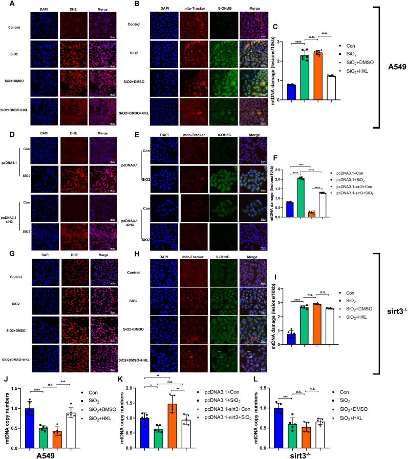
Results: This study elucidates how silica induces ATII cell senescence, emphasizing mtDNA damage. Notably, honokiol (HKL) emerges as a promising anti-senescence and anti-fibrosis agent, acting through sirt3. honokiol effectively attenuated senescence in ATII cells, dependent on sirt3 expression, while mitigating mtDNA damage. Sirt3, a class III histone deacetylase, regulates senescence and mitochondrial stress. HKL activates sirt3, protecting against pulmonary fibrosis and mitochondrial damage. Additionally, HKL downregulated cGAS expression in senescent ATII cells induced by silica, suggesting sirt3's role as an upstream regulator of the cGAS/STING signaling pathway. Moreover, honokiol treatment inhibited the activation of the NF-κB signaling pathway, associated with reduced oxidative stress and mtDNA damage. Notably, HKL enhanced the activity of SOD2, crucial for mitochondrial function, through sirt3-mediated deacetylation. Additionally, HKL promoted the deacetylation activity of sirt3, further safeguarding mtDNA integrity.

Conclusions: This study uncovers a natural compound, HKL, with significant anti-fibrotic properties through activating sirt3, shedding light on silicosis pathogenesis and treatment avenues.

Keywords: Mitochondrial DNA damage; Senescence; Silicosis; Type II alveolar epithelial cell; sirtuin3.


Conflict of interest statement

Declaration of competing interest The authors declare that they have no known competing financial interests or personal relationships that could have appeared to influence the work reported in this paper.

Figures

Image 1
Graphical abstract
Fig. 1
Fig. 1
Honokiol (HKL) restored senescent ATII cells induced by silica via activating the deacetylation activity of sirt3. (A) Cell viability of ATII cells exposed to different concentrations of silica was measured by the cell counting kit 8 (CCK-8) assay (n = 5). (B, C) β-Gal staining was performed to evaluate the senescence in ATII cells to identify the concentration and time of silica stimulation for the construction of senescent ATII cells, Scale bars, 200 μm (n = 5). (D, G) Immunoblot analysis of p-P53, P53, and P21 in ATII cells with different treatments. (E, F) Cell viability of ATII cells was measured by the cell counting kit 8 (CCK-8) assay to identify the time for induction senescence of ATII cells and the intervention time of HKL, respectively (n = 5). (H) β-Gal staining was performed to evaluate the anti-aging effect of HKL. Scale bars, 200 μm (n = 5). (I, K) Immunofluorescence (I) and immunoblot (K) analysis of the abundance of sirt3 protein in HKL-treated senescent ATII cells. Scale bar, 50 μm (n = 5). (J, K) Immunoblot analysis of total acetylated-lysine proteins in A549 cells (J), EMT-related proteins, and senescence-related proteins in HKL-treat senescent ATII cells induced by silica (n = 4 or 5). (L–R) Relative mRNA levels of SASP genes, including IL-1α, IL-1β, IL-6, TNF-α, MCP-1, and CXCL1 in silica-treated ATII cells combined with HKL (n = 5). Bar graphs were presented as mean ± SD (n = 5). Statistical analysis was performed using one-way ANOVA, GraphPad Prism 7.2.0. *P < 0.05, **P < 0.01, ***P < 0.001, ****P < 0.0001; n.s. presents no statistical difference. (For interpretation of the references to color in this figure legend, the reader is referred to the Web version of this article.)
Fig. 2
Fig. 2
HKL reduced mtDNA damage via a sirt3-dependent manner after exposure to silica. (A, D & G) A549 cells (A), sirt3-OE A549 cells (D), and sirt3-KO A549 cells (G) were incubated ex vivo with DHE (red) (5 mM) in DMSO for 30 min before analysis of ROS production by fluorescence microscopy. Scale bar, 100 μm. (B, E & H) Images were taken by confocal microscopy of immunofluorescent staining of the 8-OHdG (green) and MitoTracker™ RED CMXROS (red) and DAPI (blue) in A549 cells (B), sirt3-OE A549 cells (E), and sirt3-KO A549 cells (H). Scale bar, 50 μm. Because permeabilization was not performed, the 8-OHdG in the nucleus was not stained. (C, F & I) Frequency of mtDNA lesions per 10 kb per strand in A549 cells (C), sirt3-OE A549 cells (F), and sirt3-KO A549 cells (I). (J–L) The quantitation of mtDNA copy number in A549 cells (J), sirt3-OE A549 cells (K), and sirt3-KO A549 cells (L). Bar graphs were presented as mean ± SD (n = 5 or 6). Statistical analysis was performed using one-way ANOVA, GraphPad Prism 7.2.0. *P < 0.05, **P < 0.01, ***P < 0.001, ****P < 0.0001; n.s. presents no statistical difference. (For interpretation of the references to color in this figure legend, the reader is referred to the Web version of this article.)
Fig. 3
Fig. 3
HKL attenuates silica-induced acetylation of OGG1 through sirt3. (A & B) Mitochondrial lysates from ATII cells with different treatments were used for immunoprecipitation with anti-acetylated lysine antibody and immune complexes analyzed by Western blots using anti-acetylated lysine antibody. Membranes were stripped and reprobed with anti-OGG1 antibody to determine the level of immunoprecipitated OGG1. The lower panel shows the OGG1 Western blot of input proteins and COX IV as the internal control. (C–F) The bar graph of immunoblot analysis of relative abundance levels of Ace-OGG1 in ATII cells treated as in Fig. 3A&B. Bar graphs were presented as mean ± SD (n = 3). Statistical analysis was performed using one-way ANOVA, GraphPad Prism 7.2.0. *P < 0.05, **P < 0.01, ***P < 0.001, ****P < 0.0001; n.s. presents no statistical difference. (For interpretation of the references to color in this figure legend, the reader is referred to the Web version of this article.)
Fig. 4
Fig. 4
HKL inhibited the cGAS-STING pathway by attenuating mtDNA leakage through activating sirt3. (A, C & E) Images taken by confocal microscopy of immunofluorescent staining of the dsDNA (green) and DAPI (blue) in A549 cells (A), sirt3-OE A549 cells (C), and sirt3-KO A549 cells (E). Scale bar, 50 μm. (B, D & F) The quantitation of cytosolic mtDNA in A549 cells (B), sirt3-OE A549 cells (D), and sirt3-KO A549 cells (F). Bar graphs were presented as mean ± SD (n = 5). Statistical analysis was performed using one-way ANOVA, GraphPad Prism 7.2.0. *P < 0.05, **P < 0.01, ***P < 0.001, ****P < 0.0001; n.s. presents no statistical difference. (G, H & I) Immunoblot analysis of cGAS-STING pathway-related proteins in A549 cells (G), sirt3-OE A549 cells (H), and sirt3-KO A549 cells (I) induced by silica (n = 3). (For interpretation of the references to color in this figure legend, the reader is referred to the Web version of this article.)
Fig. 5
Fig. 5
Honokiol (HKL) attenuated lung pathological injury and inflammation in silicotic mice. (A) Schematic illustration of mouse silicosis model generation. 4 weeks old of C57BL/6 mice were injured by intratracheal instillation of crystalline silica (SiO2), and lung tissues were harvested at d28 and d56 post injury (n = 6). (B) H&E staining, Masson staining, and β-galactosidase staining of lung tissue in each group at 28 days and 56 days after instillation. Scale bars: 100 μm; (C–H) Relative mRNA levels of SASP genes, including IL-1α, IL-1β, IL-6, TNF-α, MCP-1, and CXCL1 in silica-treated mice combined with HKL (n = 6). Bar graphs were presented as mean ± SD (n = 6). Statistical analysis was performed using one-way ANOVA, GraphPad Prism 7.2.0. *P < 0.05, **P < 0.01, ***P < 0.001, ****P < 0.0001; n.s. presents no statistical difference. (For interpretation of the references to color in this figure legend, the reader is referred to the Web version of this article.)
Fig. 6
Fig. 6
Honokiol (HKL) attenuated fibrosis and senescence in silicotic mice. (A) Images taken by confocal microscopy of immunofluorescent staining of the SP-C (green) and P21 (red) and DAPI (blue) in paraffin sections from mice lung tissues. Scale bar, 200 μm. (B) Immunoblot analysis of fibrosis-related proteins and EMT-related proteins in lung tissues from mice of different groups. (C–I) Corresponding densitometry data of fibrosis-related proteins and EMT-related proteins were quantified using Image J Software. Bar graphs were presented as mean ± SD (n = 5). Statistical analysis was performed using one-way ANOVA, GraphPad Prism 7.2.0. *P < 0.05, **P < 0.01, ***P < 0.001, ****P < 0.0001; n.s. presents no statistical difference. (For interpretation of the references to color in this figure legend, the reader is referred to the Web version of this article.)
Fig. 7
Fig. 7
HKL improved the inhibition of sirt3 induced by silica and senescence of ATII cells in silicotic mice. (A) Immunoblot analysis of sirt3 protein in lung tissues from mice of different groups. (B) Representative images of immunohistochemical results of sirt3 in paraffin sections from mice lung tissues. Scale bar:100 μm. (C) Corresponding densitometry data of sirt3 protein were quantified using Image J Software. Bar graphs were presented as mean ± SD (n = 5). (D) Semi-quantification of sirt3 immunohistochemical results using Image J Software (n = 5). Statistical analysis was performed using one-way ANOVA, GraphPad Prism 7.2.0. *P < 0.05, **P < 0.01, ***P < 0.001, ****P < 0.0001; n.s. presents no statistical difference. (E) Representative images of immunofluorescence of SP-C (green) and Acetylated-Lysine (red) and DAPI (blue) in paraffin sections from mice lung tissues. Scale bar, 200 μm. (For interpretation of the references to color in this figure legend, the reader is referred to the Web version of this article.)
Fig. 8
Fig. 8
HKL mitigated mtDNA damage and inhibited ROS accumulation induced by silica in vivo. (A) The paraffin sections from mice lung tissues were incubated ex vivo with DHE (red) (5 mM) in DMSO for 30 min before analysis of ROS production by fluorescence microscopy. Scale bar, 100 μm. (B) Images taken by confocal microscopy of immunofluorescent staining of the dsDNA (green) and DAPI (blue) in paraffin sections from mice lung tissues. Scale bar, 100 μm. (C) Frequency of mtDNA lesions per 10 kb per strand in paraffin sections from mice lung tissues. (D) The quantitation of mtDNA copy number in paraffin sections from mice lung tissues. (E) The quantitation of cytosolic mtDNA in paraffin sections from mice lung tissues. Bar graphs were presented as mean ± SD (n = 5). Statistical analysis was performed using one-way ANOVA, GraphPad Prism 7.2.0. *P < 0.05, **P < 0.01, ***P < 0.001, ****P < 0.0001; n.s. presents no statistical difference. (F) Morphological changes of the mitochondria and LBs in the ATII cells of mice in each group. The red arrows present the mitochondria and the yellow arrows present the LBs. Scale bar, 2 μm. (For interpretation of the references to color in this figure legend, the reader is referred to the Web version of this article.)
Fig. 9
Fig. 9
cGAS-STING pathway was involved in the anti-fibrosis and anti-aging effect of HKL in vivo. (A–H) Representative blots and quantification of cGAS-STING pathway-related proteins in mice lung tissues. Bar graphs were presented as mean ± SD (n = 5). Statistical analysis was performed using one-way ANOVA, GraphPad Prism 7.2.0. *P < 0.05, **P < 0.01, ***P < 0.001, ****P < 0.0001; n.s. presents no statistical difference. (For interpretation of the references to color in this figure legend, the reader is referred to the Web version of this article.)

Similar articles

Cited by

References

    1. Birch J., Gil J. Senescence and the SASP: many therapeutic avenues. Genes & development. 2020;34:1565–1576. doi: 10.1101/gad.343129.120. - DOI - PMC - PubMed
    1. Yao C., Guan X., Carraro G., et al. Senescence of alveolar type 2 cells drives progressive pulmonary fibrosis. Am. J. Respir. Crit. Care Med. 2021;203(6):707–717. doi: 10.1164/rccm.202004-1274OC. - DOI - PMC - PubMed
    1. Calcinotto A., Kohli J., Zagato E., et al. Cellular senescence: aging, cancer, and injury. Physiol. Rev. 2019;99(2):1047–1078. doi: 10.1152/physrev.00020.2018. - DOI - PubMed
    1. Lingampally A., Jones M.R., Bagari S., et al. Use of the reversible Myogenic to Lipogenic Transdifferentiation Switch for the design of preclinical drug Screening in idiopathic pulmonary fibrosis. Front. Bioeng. Biotechnol. 2020;8 doi: 10.3389/fbioe.2020.569865. - DOI - PMC - PubMed
    1. Li T., Yang X.-y., Xu D.-j., et al. OC-STAMP overexpression drives lung alveolar epithelial cell type II senescence in silicosis. Oxid. Med. Cell. Longev. 2021 doi: 10.1155/2021/4158495. - DOI - PMC - PubMed

MeSH terms