. 2024 Jul 11;111(7):1352-1369.
doi: 10.1016/j.ajhg.2024.05.016. Epub 2024 Jun 11.

PSMD11 loss-of-function variants correlate with a neurobehavioral phenotype, obesity, and increased interferon response

Affiliations

PSMD11 loss-of-function variants correlate with a neurobehavioral phenotype, obesity, and increased interferon response

Wallid Deb et al. Am J Hum Genet. .

Abstract

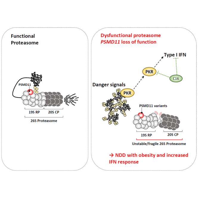
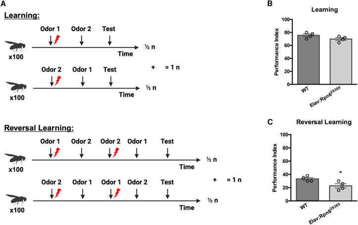
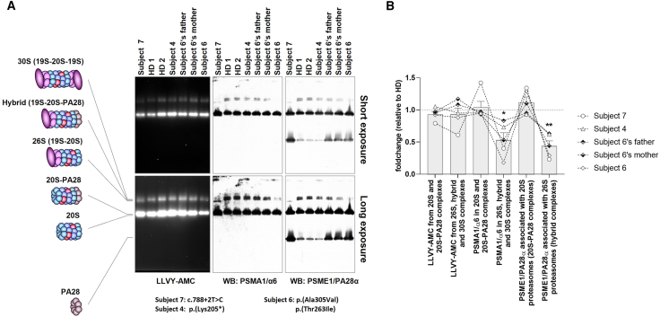
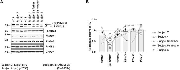
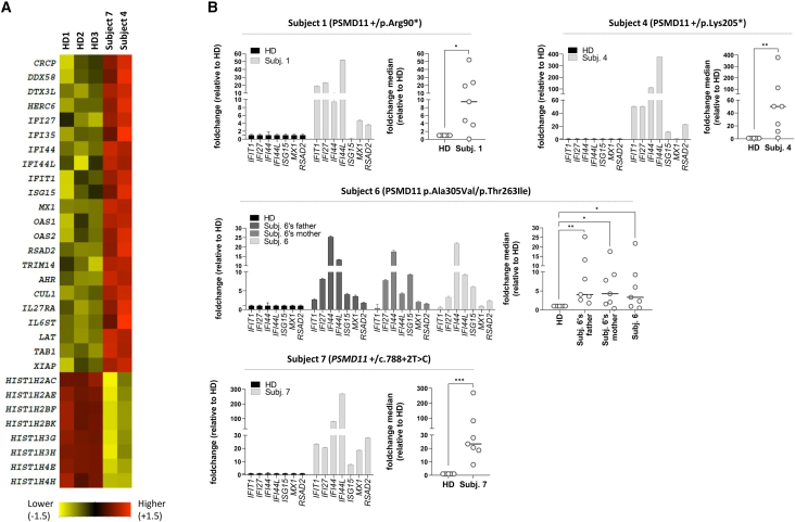
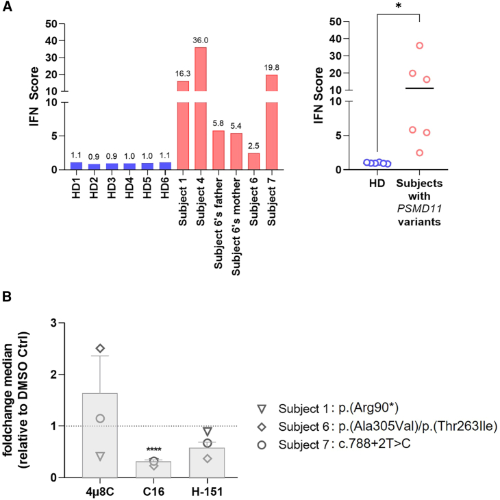
Primary proteasomopathies have recently emerged as a new class of rare early-onset neurodevelopmental disorders (NDDs) caused by pathogenic variants in the PSMB1, PSMC1, PSMC3, or PSMD12 proteasome genes. Proteasomes are large multi-subunit protein complexes that maintain cellular protein homeostasis by clearing ubiquitin-tagged damaged, misfolded, or unnecessary proteins. In this study, we have identified PSMD11 as an additional proteasome gene in which pathogenic variation is associated with an NDD-causing proteasomopathy. PSMD11 loss-of-function variants caused early-onset syndromic intellectual disability and neurodevelopmental delay with recurrent obesity in 10 unrelated children. Our findings demonstrate that the cognitive impairment observed in these individuals could be recapitulated in Drosophila melanogaster with depletion of the PMSD11 ortholog Rpn6, which compromised reversal learning. Our investigations in subject samples further revealed that PSMD11 loss of function resulted in impaired 26S proteasome assembly and the acquisition of a persistent type I interferon (IFN) gene signature, mediated by the integrated stress response (ISR) protein kinase R (PKR). In summary, these data identify PSMD11 as an additional member of the growing family of genes associated with neurodevelopmental proteasomopathies and provide insights into proteasomal biology in human health.


Conflict of interest statement

Declaration of interests A.C. and K.M. are employees of GeneDx, LLC. J.R.L. has stock in 23andMe and is a paid consultant for Genome International.

Figures

None
Graphical abstract
Figure 1
Figure 1
Family pedigrees and cross-species alignment of the regions of the PSMD11 proteasome subunit subjected to missense variants (A) Pedigrees of the 10 affected families carrying variants in PSMD11. Male and females are indicated by squares and circles, respectively. Affected individuals are denoted by filled symbols. Healthy heterozygous carriers are highlighted by a half-filled symbol. (B) Schematic representation of the PSMD11 protein primary structure showing the distribution and localization of the residues subjected to missense (purple) or loss-of-function (red) variants. Exon 7 is predicted to be abnormally spliced in subject 7, marked in red. The proteasome component (PCI) domain is marked in dark green. The lower panel shows multiple sequence alignment of PSMD11 regions prone to amino acid substitutions across species.
Figure 2
Figure 2
In silico protein structure analysis of the PSMD11 subunit affected regions within proteasome complexes (A) Cryo-electron microscopy (EM) structure of the human 26S proteasome without imposed symmetry (PDB ID: 5LN3) containing the PSMD11 subunit marked in orange. Right: the lid and the base of the 19S regulatory particle are shown in ribbon colored in light blue and green. The α- and β-rings of the upper half of the 20S core particle are shown in ribbon colored in pink and brown, respectively. (B–D) Bottom: lateral view of the PSMD11 subunit interacting with the upper α-ring (B), base (C), and lid (D) of the 19S regulatory particle, as indicated.
Figure 3
Figure 3
Rpn6 knockdown (KD) affects reversal learning in Drosophila (A) Schematic of olfactory learning and reversal learning. Classical olfactory conditioning consisted of 100 Drosophila learning the contingency between odor 1 being paired with electric shock and odor 2 with no shock (½ n). Another 100 naive Drosophila were then trained with the reverse odor and shock pairing to counter any odor preference (½ n). Combining these 2 results was considered n = 1. Reversal learning consisted of the same initial training cycle but added a second training cycle immediately after that would reverse the odor and shock pairing from the first training. (B) For learning, the learning performance index (PI) of wild-type and pan-neuronal (Elav) Rpn6-KD Drosophila did not differ significantly (N = 4; t test, p = 0.1136). (C) Conversely, Rpn6-KD Drosophila performed significantly worse than wild-type Drosophila on the reversal learning paradigm (N = 4; t test; p = 0.0314).
Figure 4
Figure 4
T cells carrying PSMD11 variants exhibit reduced amounts of 26S, hybrid, and 30S proteasome complexes (A) Twenty micrograms of T cell lysates from subjects 2, 4, and 6 as well as both parents of subject 6 and 2 unrelated healthy donors (HDs) were separated by 3%–12% native-PAGE. Proteasome chymotrypsin-like activity was assessed in gels using the LLVY-AMC fluorogenic reporter substrate, as indicated. Gels were subsequently subjected to western blotting using antibodies specific for PSMA1 and PSME1, as indicated. The left schematic illustrates the proteasome complexes (30S, hybrid, 26S, 20S-PA28, and 20S) and free regulators (19S and PA28) detected using the 2 antibodies. (B) Quantification of the LLVY-AMC fluorescent signals and PSMA1 and PSME1 immunoreactive bands in 20S, 20S-PA28, 26S, hybrid, and/or 30S proteasome complexes by densitometry, as indicated. Data are presented as activity (LLVY-AMC) and protein (PSMA1 and PSME1) fold changes in subjects 2,4, and 6 as well as both parents of subject 6 vs. healthy donors (HDs) whose densitometric measurements were set to 1 (gridline), as indicated. Columns indicate the fold change mean values ± SEM calculated from the 5 normalizations. Statistical significance was assessed by unpaired Student’s test (p < 0.05, ∗∗p < 0.01).
Figure 5
Figure 5
PSMD11 variants affect the steady-state expression level of unmodified and/or phosphorylated PSMD11 proteins (A) Five to 20 micrograms of RIPA lysates from T cells isolated from subjects 2, 4, and 6 as well as both parents of subject 6 and 3 unrelated healthy donors (HDs) were separated by SDS-PAGE followed by western blotting using antibodies specific for PSMD11, PSMD12, PSMC5, PSMA2, PSME1, and GAPDH (loading control), as indicated. (B) Quantification of the western blots by densitometry. Data are presented as fold changes in subjects 4, 6, and 7 as well as both parents of subject 6 vs. healthy donors (HDs) whose densitometric measurements were set to 1 (gridline), as indicated. Columns indicate the fold change mean values ± SEM calculated from the 5 normalizations.
Figure 6
Figure 6
T cells from subjects with PSMD11 variants develop a type I interferon (IFN) gene signature (A) Non-clustered heatmap visualization of gene expression in T cells isolated from subjects 4 and 7 carrying PSMD11 variants and 3 unrelated healthy donors (HDs). Each column represents 1 individual subject or control and each row represents 1 gene. Color indicates normalized counts of each transcript, with red representing higher expression and yellow relatively lower expression. (B) Gene expression of 7 IFN-stimulated genes (ISG, namely IFIT1, IFI27, IFI44, IFI44L, ISG15, MX1, and RSAD2) was assayed by RT-qPCR on T cells derived from subjects 1, 4, 6, and 7 as well as both parents from subject 6 and healthy donor (HD) controls. Expression levels were normalized to GAPDH and relative quantifications (RQs) are presented as fold change over controls. Shown is also the median fold expression of the 7 ISG over healthy donors (HDs). Statistical significance was assessed by ratio paired t test where indicates p < 0.05, ∗∗ indicates p < 0.01 and ∗∗∗ indicates p < 0.001.
Figure 7
Figure 7
The type I IFN response associated with PSMD11 variants depends on PKR in affected individuals T cells (A) IFN scores for subjects 1, 4, 6, and 7 as well as both parents of subject 6 and 6 unrelated healthy donor (HD) controls were calculated as the median of the relative quantifications of the 7 ISGs over a single calibrator control. Shown are the IFN scores of each sample (left) and the sample groups, namely healthy donors (HDs) and subjects carrying PSMD11 variants, as indicated. Statistical significance was assessed by unpaired t test where indicates p < 0.05. (B) T cells from subjects 6, 7, and 8 were subjected to a 6-h treatment with DMSO (vehicle), 4μ8C (100 μM), C16 (500 nM), or H-151 (2 μM) small-molecule inhibitors before RNA extraction and RT-qPCR for expression analysis of IFIT1, IFI27, IFI44, IFI44L, ISG15, MX1, and RSAD2. Transcript expression was normalized to GAPDH and data are presented as the fold-change median values of the 7 ISGs relative to DMSO (gridline) for each subject in each treatment. Columns indicate the fold-change mean values ± SEM of the subject group (n = 3) for each treatment. Statistical significance was assessed by ratio paired t test where ∗∗∗ indicates p < 0.001.

Similar articles

  • PSMC3 proteasome subunit variants are associated with neurodevelopmental delay and type I interferon production.
    Ebstein F, Küry S, Most V, Rosenfelt C, Scott-Boyer MP, van Woerden GM, Besnard T, Papendorf JJ, Studencka-Turski M, Wang T, Hsieh TC, Golnik R, Baldridge D, Forster C, de Konink C, Teurlings SMW, Vignard V, van Jaarsveld RH, Ades L, Cogné B, Mignot C, Deb W, Jongmans MCJ, Cole FS, van den Boogaard MH, Wambach JA, Wegner DJ, Yang S, Hannig V, Brault JA, Zadeh N, Bennetts B, Keren B, Gélineau AC, Powis Z, Towne M, Bachman K, Seeley A, Beck AE, Morrison J, Westman R, Averill K, Brunet T, Haasters J, Carter MT, Osmond M, Wheeler PG, Forzano F, Mohammed S, Trakadis Y, Accogli A, Harrison R, Guo Y, Hakonarson H, Rondeau S, Baujat G, Barcia G, Feichtinger RG, Mayr JA, Preisel M, Laumonnier F, Kallinich T, Knaus A, Isidor B, Krawitz P, Völker U, Hammer E, Droit A, Eichler EE, Elgersma Y, Hildebrand PW, Bolduc F, Krüger E, Bézieau S. Ebstein F, et al. Sci Transl Med. 2023 May 31;15(698):eabo3189. doi: 10.1126/scitranslmed.abo3189. Epub 2023 May 31. Sci Transl Med. 2023. PMID: 37256937 Free PMC article.
  • De Novo Disruption of the Proteasome Regulatory Subunit PSMD12 Causes a Syndromic Neurodevelopmental Disorder.
    Küry S, Besnard T, Ebstein F, Khan TN, Gambin T, Douglas J, Bacino CA, Craigen WJ, Sanders SJ, Lehmann A, Latypova X, Khan K, Pacault M, Sacharow S, Glaser K, Bieth E, Perrin-Sabourin L, Jacquemont ML, Cho MT, Roeder E, Denommé-Pichon AS, Monaghan KG, Yuan B, Xia F, Simon S, Bonneau D, Parent P, Gilbert-Dussardier B, Odent S, Toutain A, Pasquier L, Barbouth D, Shaw CA, Patel A, Smith JL, Bi W, Schmitt S, Deb W, Nizon M, Mercier S, Vincent M, Rooryck C, Malan V, Briceño I, Gómez A, Nugent KM, Gibson JB, Cogné B, Lupski JR, Stessman HAF, Eichler EE, Retterer K, Yang Y, Redon R, Katsanis N, Rosenfeld JA, Kloetzel PM, Golzio C, Bézieau S, Stankiewicz P, Isidor B. Küry S, et al. Am J Hum Genet. 2017 Feb 2;100(2):352-363. doi: 10.1016/j.ajhg.2017.01.003. Epub 2017 Jan 26. Am J Hum Genet. 2017. PMID: 28132691 Free PMC article.
  • PSMD12 haploinsufficiency in a neurodevelopmental disorder with autistic features.
    Khalil R, Kenny C, Hill RS, Mochida GH, Nasir R, Partlow JN, Barry BJ, Al-Saffar M, Egan C, Stevens CR, Gabriel SB, Barkovich AJ, Ellison JW, Al-Gazali L, Walsh CA, Chahrour MH. Khalil R, et al. Am J Med Genet B Neuropsychiatr Genet. 2018 Dec;177(8):736-745. doi: 10.1002/ajmg.b.32688. Epub 2018 Nov 13. Am J Med Genet B Neuropsychiatr Genet. 2018. PMID: 30421579 Free PMC article.
  • [Neurodevelopmental proteasomopathies: New disorders caused by proteasome dysfunction].
    Cuinat S, Bézieau S, Deb W, Mercier S, Vignard V, Toutain B, Isidor B, Küry S, Ebstein F. Cuinat S, et al. Med Sci (Paris). 2024 Feb;40(2):176-185. doi: 10.1051/medsci/2023221. Epub 2024 Feb 27. Med Sci (Paris). 2024. PMID: 38411426 Review. French.
  • Understanding neurodevelopmental proteasomopathies as new rare disease entities: A review of current concepts, molecular biomarkers, and perspectives.
    Cuinat S, Bézieau S, Deb W, Mercier S, Vignard V, Isidor B, Küry S, Ebstein F. Cuinat S, et al. Genes Dis. 2023 Sep 26;11(6):101130. doi: 10.1016/j.gendis.2023.101130. eCollection 2024 Nov. Genes Dis. 2023. PMID: 39220754 Free PMC article. Review.

Cited by

References

    1. Schmid H.P., Akhayat O., Martins De Sa C., Puvion F., Koehler K., Scherrer K. The prosome: an ubiquitous morphologically distinct RNP particle associated with repressed mRNPs and containing specific ScRNA and a characteristic set of proteins. EMBO J. 1984;3:29–34. doi: 10.1002/j.1460-2075.1984.tb01757.x. - DOI - PMC - PubMed
    1. Arrigo A.P., Tanaka K., Goldberg A.L., Welch W.J. Identity of the 19S “prosome” particle with the large multifunctional protease complex of mammalian cells (the proteasome) Nature. 1988;331:192–194. doi: 10.1038/331192a0. - DOI - PubMed
    1. Falkenburg P.E., Haass C., Kloetzel P.M., Niedel B., Kopp F., Kuehn L., Dahlmann B. Drosophila small cytoplasmic 19S ribonucleoprotein is homologous to the rat multicatalytic proteinase. Nature. 1988;331:190–192. doi: 10.1038/331190a0. - DOI - PubMed
    1. Collins G.A., Goldberg A.L. The Logic of the 26S Proteasome. Cell. 2017;169:792–806. doi: 10.1016/j.cell.2017.04.023. - DOI - PMC - PubMed
    1. Ebstein F., Kloetzel P.-M., Krüger E., Seifert U. Emerging roles of immunoproteasomes beyond MHC class i antigen processing. Cell. Mol. Life Sci. 2012;69:2543–2558. doi: 10.1007/s00018-012-0938-0. - DOI - PMC - PubMed

Publication types