. 2024 Aug 16;30(9):1184-1198.
doi: 10.1261/rna.080013.124.

Modeling the structure and DAP5-binding site of the FGF-9 5'-UTR RNA utilized in cap-independent translation

Affiliations

Modeling the structure and DAP5-binding site of the FGF-9 5'-UTR RNA utilized in cap-independent translation

Amanda Whittaker et al. RNA. .

Abstract

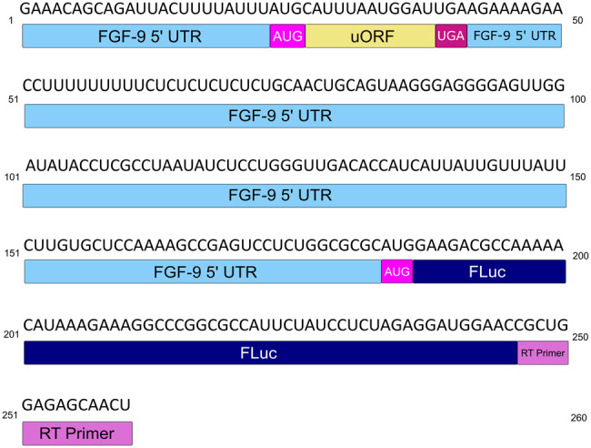
Cap-independent or eukaryotic initiation factor (eIF) 4E-independent, translation initiation in eukaryotes requires scaffolding protein eIF4G or its homolog, death-associated protein 5 (DAP5). eIF4G associates with the 40S ribosomal subunit, recruiting the ribosome to the RNA transcript. A subset of RNA transcripts, such as fibroblast growth factor 9 (FGF-9), contain 5' untranslated regions (5' UTRs) that directly bind DAP5 or eIF4GI. For viral mRNA, eIF recruitment usually utilizes RNA structure, such as a pseudoknot or stem-loops, and the RNA-helicase eIF4A is required for DAP5- or 4G-mediated translation, suggesting these 5' UTRs are structured. However, for cellular IRES-like translation, no consensus RNA structures or sequences have yet been identified for eIF binding. However, the DAP5-binding site within the FGF-9 5' UTR is unknown. Moreover, DAP5 binds to other, dissimilar 5' UTRs, some of which require an unpaired, accessible 5' end to stimulate cap-independent translation. Using SHAPE-seq, we modeled the 186 nt FGF-9 5'-UTR RNA's complex secondary structure in vitro. Further, DAP5 footprinting, toeprinting, and UV cross-linking experiments identify DAP5-RNA interactions. Modeling of FGF-9 5'-UTR tertiary structure aligns DAP5-interacting nucleotides on one face of the predicted structure. We propose that RNA structure involving tertiary folding, rather than a conserved sequence or secondary structure, acts as a DAP5-binding site. DAP5 appears to contact nucleotides near the start codon. Our findings offer a new perspective in the hunt for cap-independent translational enhancers. Structural, rather than sequence-specific, eIF-binding sites may act as attractive chemotherapeutic targets or as dosage tools for mRNA-based therapies.

Keywords: DAP5; DAP5-binding site; FGF-9 RNA; biophysics; cap-independent translation.


Figures

FIGURE 1.
FIGURE 1.
wt FGF-9 5′-UTR construct. The FGF-9 5′ UTR (sky blue) and partial firefly luciferase (FLuc) reporter sequence (navy) RNA used for this study. The FGF-9 uORF (nt [nucleotides] 27–38) is annotated in goldenrod, uAUG in magenta, and uUGA in maroon. A cy5-tagged reverse transcript primer (orchid), corresponding to part of the FLuc reporter, was used to create cDNA from treated RNA.
FIGURE 2.
FIGURE 2.
SHAPE-derived 2′ structure of the FGF-9 5′-UTR mRNA. Normalized SHAPE data from three SHAPE reactions were pooled and input into RNAStructure software as a binding constraint. The minimum-free-energy predicted structure (ΔG = −73.1 kJ) was selected. Nucleotides were annotated in RNA2Drawer on a scale from 0 (ivory, unreactive to 1M7) to 1.0 and above (medium slate blue, always reactive to 1M7, seven SL were predicted to form). The AUG start codon is visible at the base of SL7.
FIGURE 3.
FIGURE 3.
FGF-9 5′ UTR–DAP5 toeprinting data. FGF-9 5′-UTR cDNA was reverse-transcribed from DAP5-bound mRNA. n = 4. Higher peak area corresponds to a higher proportion of RT stops in the presence of DAP5. Size standard peaks visible at 20, 40, 50, 60, 70, 80, 100, 120, 140, 160, 170, and 180 nt. Strong RT stop at nt 93 (black arrow). Start codon: nts184–186 (magenta arrow).
FIGURE 4.
FIGURE 4.
FGF-9 5′ UTR–DAP5 footprinting data. 1M7 reactions were performed on DAP5-bound FGF-9 5′-UTR mRNA. n = 2. (A) Changes in 1M7 reactivity in the presence of DAP5. Normalized 1M7 reactivity in the presence of DAP5 was subtracted from normalized 1M7 reactivity in the absence of DAP5. For clarity, reactivity changes >1 (from a reactive site to a nonreactive site) are shown here. A decrease in 1M7 reactivity > 1 (green) indicates possible contact with DAP5. An increase in 1M7 reactivity (magenta) indicates increased flexibility in the presence of DAP5. Start codon: nt 184–186. Full length of mRNA = 209 nt. Size standard peaks (20, 40, 50, 60, 70, 80, 100, 120, 140, 160, 170, and 180 nt) are not shown here. (B) DAP5-induced protection from 1M7 annotated across the predicted secondary structure of the FGF-9 5′-UTR mRNA. Decreased 1M7 reactivity is annotated in green, with darker green = sharper decrease in reactivity. Toeprinting site (nt 93) from Figure 3 is annotated in aqua blue. AUG (nt 184–186) in magenta.
FIGURE 5.
FIGURE 5.
DAP5–FGF-9 5′-UTR UV cross-linking. DAP5 was cross-linked to the wt FGF-9 5′-UTR RNA construct using UV light. For clarity, DAP5 footprinting data are shown alongside UV cross-linking stops. (A) Difference in peak area between RT stops occurring with and without DAP5. (Orange) More frequent stops with DAP5. (Blue) More frequent stops found without DAP5. Size standard peaks (20, 40, 50, 60, 70, 80, 100, 120, 140, 160, 170, and 180 nt) are not shown here. (B) DAP5 footprinting, toeprinting, and UV cross-linking sites mapped on our putative 2D model. Ivory with red outline = RT stops shared between footprinting stops from Figure 4 (green) and UV cross-linking data (orange). Toeprinting site (nt 93) shown in aqua blue. Start codon (nt 184–186) shown in magenta.
FIGURE 6.
FIGURE 6.
Predicting the FGF-9 5′-UTR tertiary structure. The minimum-free-energy tertiary conformation was predicted by RNAComposer using Figure 2 as a folding constraint. USCF ChimeraX was used to annotate 1M7 reactivity. ΔG = −3060 kcal/mol. SL1–SL5 aggregate closely around the 5′ end of the mRNA, whereas SL6 and SL7 project away. The AUG start codon, tucked between SL5 and SL6, is visualized in an insert (bottom left). SL1 = nt 5−12 and 117–124, SL2 = 20–38, SL3 = 40–62, SL4 = 63–92, SL5 = 94–112, SL6 = 128–138, and SL7 = 140–186. Start codon: 184–186.
FIGURE 7.
FIGURE 7.
DAP5 contacts mapped on our tertiary model of the FGF-9 5′-UTR mRNA. Tertiary prediction from Figure 6 annotated with DAP5 toeprinting, footprinting, and cross-linking data. Ivory with red outline = RT stops shared between footprinting data (green) and UV cross-linking data (orange). Toeprinting site (nt 93) shown in aqua blue. Start codon (nt 184–186) shown in magenta. All other nucleotides are annotated in black.
Amanda Whittaker
Amanda Whittaker

Update of

References

    1. Akirtava C, May GE, McManus CJ. 2022. False-positive IRESes from Hoxa9 and other genes resulting from errors in mammalian 5′ UTR annotations. Proc Natl Acad Sci 119: e2122170119. 10.1073/pnas.2122170119 - DOI - PMC - PubMed
    1. Alard A, Marboeuf C, Fabre B, Jean C, Martineau Y, Lopez F, Vende P, Poncet D, Schneider RJ, Bousquet C, et al. 2019. Differential regulation of the three eukaryotic mRNA translation initiation factor (eIF) 4Gs by the proteasome. Front Genet 10: 254. 10.3389/fgene.2019.00254 - DOI - PMC - PubMed
    1. Antczak M, Popenda M, Zok T, Sarzynska J, Ratajczak T, Tomczyk K, Adamiak RW, Szachniuk M. 2016. New functionality of RNAComposer: application to shape the axis of miR160 precursor structure. Acta Biochim Pol 63: 737–744. 10.18388/abp.2016_1329 - DOI - PubMed
    1. Balvay L, Rifo RS, Ricci EP, Decimo D, Ohlmann T. 2009. Structural and functional diversity of viral IRESes. Biochim Biophys Acta 1789: 542–557. 10.1016/j.bbagrm.2009.07.005 - DOI - PubMed
    1. Braunstein S, Karpisheva K, Pola C, Goldberg J, Hochman T, Yee H, Cangiarella J, Arju R, Formenti SC, Schneider RJ. 2007. A hypoxia-controlled cap-dependent to cap-independent translation switch in breast cancer. Mol Cell 28: 501–512. 10.1016/j.molcel.2007.10.019 - DOI - PubMed

MeSH terms

LinkOut - more resources