. 2024 Jun 25;38(9-10):415-435.
doi: 10.1101/gad.351424.123.

A nuclear architecture screen in Drosophila identifies Stonewall as a link between chromatin position at the nuclear periphery and germline stem cell fate

Affiliations

A nuclear architecture screen in Drosophila identifies Stonewall as a link between chromatin position at the nuclear periphery and germline stem cell fate

Ankita Chavan et al. Genes Dev. .

Abstract

The association of genomic loci to the nuclear periphery is proposed to facilitate cell type-specific gene repression and influence cell fate decisions. However, the interplay between gene position and expression remains incompletely understood, in part because the proteins that position genomic loci at the nuclear periphery remain unidentified. Here, we used an Oligopaint-based HiDRO screen targeting ∼1000 genes to discover novel regulators of nuclear architecture in Drosophila cells. We identified the heterochromatin-associated protein Stonewall (Stwl) as a factor promoting perinuclear chromatin positioning. In female germline stem cells (GSCs), Stwl binds and positions chromatin loci, including GSC differentiation genes, at the nuclear periphery. Strikingly, Stwl-dependent perinuclear positioning is associated with transcriptional repression, highlighting a likely mechanism for Stwl's known role in GSC maintenance and ovary homeostasis. Thus, our study identifies perinuclear anchors in Drosophila and demonstrates the importance of gene repression at the nuclear periphery for cell fate.

Keywords: genome organization; germline stem cell; heterochromatin; nuclear architecture; nuclear periphery.


Figures

Figure 1.
Figure 1.
Discovery of novel regulators of chromatin positioning at the nuclear periphery in Drosophila. (A) Cartoon schematic of HiDRO screening pipeline and the 1 Mb probe regions along chromosome 2R. (B) Percentage of each chromosome 2R region occupied by LADs. (C) Normalized distance from periphery for each chromosome 2R region. (D) Z-score plot for genes affecting peripheral localization of chromosome 2R-C. Genes above the red dashed line represent hits that increase the distance between chromosome 2R-C and the nuclear periphery. These are shown larger in the overlay box. Genes below the blue dashed line represent hits that decrease the distance between chromosome 2R-C and the periphery. (Green) Lamin B, (red) Stwl. (E) Venn diagram indicating the overlap between the peripheral localization and compaction hits. Eleven genes were hits for both metrics, including stwl. (F) Comparison of z-scores for the normalized distance from the periphery across the categories of hits that only affect peripheral distance (Peri. only), all hits that affect compaction (All Comp. Hits), and hits that affect both peripheral distance and compaction (Both). (G) STRING analysis of peripheral hits. (H) Individual Kc167 cell nuclei labeled with probes against chromosome 2R-A (magenta), chromosome 2R-B (yellow), and chromosome 2R-C (blue) from LacZ RNAi (control), stwl RNAi, and Lamin B RNAi. Outlines show the nuclear boundary. (I) Radar plot indicating the screen metrics following Stwl knockdown (left) or Lamin B knockdown (right). Red and blue wedges represent screen metrics in which the knockdown significantly increased or decreased the metric, respectively. (J) Example nucleus showing Stwl immunofluorescence. Scale bar, 5 µm. (K) Cartoon schematic of shell analysis of immunofluorescence. The first four shells were combined to define the periphery, and the fifth shell defines the center. (L) Shell analysis of the indicated nuclear components. The median signal in the periphery and the center was calculated from two replicates of >300 nuclei each.
Figure 2.
Figure 2.
Stwl is a regulator of perinuclear chromatin positioning in female GSCs. (A) Schematic of Drosophila ovary and germarium. The germarium resides at the anterior tip of the ovariole (red box) and is further subdivided into region 1, containing germline stem cells (GSCs; green) and cystoblasts (CBs; purple), and regions 2a/2b, containing differentiated germ cell cysts (yellow). (B) Ovaries from control TM3/StwlRNAi, nos>StwlRNAi, and bam>StwlRNAi imaged 3 days after eclosion. Scale bars, 100 μm. (C) Germaria from nos>mCherryRNAi (control) and nos>StwlRNAi ovaries stained for Vasa (green) and DAPI (magenta). Scale bar, 5 μm. (D) Ovaries from nos>mCherryRNAi (control) and nos>StwlRNAi following 4, 8, and 20 day shifts to 29°C in a Gal80ts background. Scale bars, 100 μm. (E) Germaria from nos>mCherryRNAi (control) and nos>StwlRNAi ovaries stained for Stwl (green), Vasa (blue), and Hts (magenta) following a 4 day shift to 29°C. White arrowheads indicate the GSCs. Scale bars, 5 μm. (F) Oligopaint FISH against chromosome 2R-A (magenta) and IF staining of Vasa (green) in GSCs from nos>mCherryRNAi (control) and nos>StwlRNAi ovaries following a 4 day shift to 29°C. Yellow arrowheads indicate the chromosome 2R-A locus within the nucleus. Yellow dotted lines indicate the nuclear boundary. Scale bars, 5 μm. (G) Quantification of the NE–chromosome 2R-A distance (in micrometers) in GSCs from F. n = 43 GSCs from nos>mCherryRNAi; n = 44 GSCs from nos>StwlRNAi. (**) P < 0.01 from Student's t-test. (H) Histogram of the NE–chromosome 2R-A distance (in micrometers) in GSCs from G. (I) Oligopaint FISH against chromosome 2R-C (magenta) and IF staining of Vasa (green) in GSCs from nos>mCherryRNAi (control) and nos>StwlRNAi ovaries following a 4 day shift to 29°C. Yellow arrowheads indicate the chromosome 2R-C locus within the nucleus. Yellow dotted lines indicate the nuclear boundary. Scale bars, 5 μm. (J) Quantification of the NE–chromosome 2R-C distance (in micrometers) in GSCs from I. n = 45 GSCs from nos>mCherryRNAi (control); n = 49 GSCs from nos>StwlRNAi. (ns) P > 0.05 from Student's t-test. (K) Histogram of the NE–chromosome 2R-C distance (in micrometers) in GSCs from J.
Figure 3.
Figure 3.
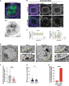
Loss of Stwl leads to defects in perinuclear chromatin organization. (A) IF staining of Stwl (green) and Vasa (blue) in GSCs from young ovaries in a Nup58-GFP (magenta) strain. Scale bar, 5 μm. (B) IF staining of Lamin B (green), FG Nups (magenta), and Vasa (blue) in GSCs from nos>mCherryRNAi (control) and nos>StwlRNAi ovaries following a 4 day shift to 29°C. Yellow arrowheads indicate NPC clusters in the regions lacking Lamin B. Scale bars, 5 μm. (C) Relative fluorescence intensity of Lamin B (green) and FG Nups (magenta) along the nuclear envelope from B. Shaded gray regions highlight NPC clustering in regions lacking Lamin B. (D) TEM image of GSC-like cells from nos>mCherryRNAi (control) ovaries in a bamΔ86/bam1 background following a 4 day shift to 29°C. The top inset shows NPCs (yellow arrowheads), while the bottom inset shows an electron-dense chromatin focus associated with the nuclear envelope. (E) TEM image of GSC-like cells from nos>StwlRNAi ovaries in a bamΔ86/bam1 background following a 4 day shift to 29°C. The top inset shows NPC clusters, while the bottom inset shows the absence of electron-dense chromatin foci in regions containing NPC clusters. (F) Quantification of perinuclear electron-dense chromatin foci in GSC-like cells from D and E. Each dot represents the number of perinuclear chromatin foci per nucleus per micrometer of the nuclear envelope. n = 67 GSCs from nos>mCherryRNAi; n = 60 GSCs from nos>StwlRNAi. (****) P < 0.0001 from Student's t‐‐test. (G) The number of NPCs per micrometer of the NE was quantified from TEM images of GSC-like cells from nos>mCherryRNAi (control; n = 66) and nos>StwlRNAi (n = 59) ovaries in a bamΔ86/bam1 background following a 4 day shift to 29°C. (ns) P > 0.05 from Student's t-test. (H) The percentage of perinuclear electron-dense chromatin foci at NPC clusters versus other regions on the nuclear envelope in GSC-like cells from E. n = 42. (****) P < 0.0001 from Fisher's exact test.
Figure 4.
Figure 4.
Stwl binds and represses the bgcn differentiation gene. (A, left panels) nos; TM3 (control) and nos>StwlEY00146 (StwlOE) ovaries in a bam1/+ background. (Middle and right panels) IF staining of Bam (green), Vasa (magenta), and DAPI (blue) in germaria. Scale bars, 5 μm. (B) Quantification of undifferentiated Bam-negative germ cells from A. n = 15 germaria from the control; n = 45 germaria from stwlOE. (*) P < 0.05 from a Student's t-test. (C) Volcano plot of −log10(P-value) versus log2FC from nos-Gal4/+ (control) and nos>stwlEY00146 (StwlOE) GSC-enriched ovaries (bamΔ86/bam1 background). Differentially expressed genes (log2FC > |0.6|, Padj < 0.01) are indicated as blue dots. Genes upregulated in Stwl-depleted ovaries from Zinshteyn and Barbash (2022) or Kotb et al. (2024) are indicated as magenta dots, while genes upregulated in both studies are indicated as yellow dots. Adjusted P-values following multiple testing correction are shown. (D) Transcripts per million (log2TPM) for the indicated genes from nos-Gal4/+ (control) and nos>stwlEY00146 (StwlOE) GSC-enriched ovaries in a bamΔ86/bam1 background. Adjusted P-values following multiple testing correction are shown. (E) Heat maps of CUT&RUN reads for IgG from young WT ovaries and for Stwl from ovaries enriched for GSC-like cells (nos>bamRNAi). Data are centered on ±3 kb window around 12,888 Stwl peaks (merged within 1 kb) and are shown for two replicates each. (F) Capture of the IGV genome browser (v2.11.4) showing an ∼10 kb region on Drosophila chromosome 3 (y-axis indicates reads per kilobase per million reads). (Blue) Ensembl genes. Shaded areas correspond to Stwl binding peaks.
Figure 5.
Figure 5.
Stwl positions bgcn at the nuclear periphery in female GSCs. (A) Oligopaint FISH against the bgcn locus (magenta) and IF staining of Vasa (green) in GSCs from nos>mCherryRNAi (control) and nos>StwlRNAi ovaries following a 6 day shift to 29°C. Red arrowheads indicate the bgcn locus within the nucleus. Black dotted lines indicate the nuclear boundary. Scale bars, 5 μm. (B) Quantification of the NE–bgcn distance (in micrometers) in GSCs from A. n = 201 GSCs from nos>mCherryRNAi (control); n = 200 GSCs from nos>StwlRNAi. (****) P < 0.0001 from Student's t-test. (C) Histogram of the NE–bgcn distance (in micrometers) in GSCs from B. (D) Oligopaint FISH against the bgcn locus (magenta) and IF staining of Vasa (blue) in region 2a/2b differentiated germline cysts from nos>mCherryRNAi (control) and nos>StwlRNAi ovaries following a 6 day shift to 29°C. Black dotted lines indicate the nuclear boundary. Scale bars, 5 μm. (E) Quantification of the NE–bgcn distance (in micrometers) in region 2a/2b differentiated germ cells from D. n = 106 GSCs from nos>mCherryRNAi (control); n = 65 GSCs from nos>StwlRNAi. (****) P < 0.0001 from Student's t-test. (F) Histogram of the NE–bgcn distance (in micrometers) in region 2a/2b differentiated germ cells from E. (G) Oligopaint FISH against the bgcn locus (magenta) and Smr locus (green) and IF staining of Vasa (blue) in GSCs from nos>mCherryRNAi (control) and nos>StwlRNAi ovaries following a 6 day shift to 29°C. Red arrowheads indicate the bgcn locus within the nucleus. Black dotted lines indicate the nuclear boundary. Scale bars, 5 μm. (H) Quantification of the percentage of GSCs with the indicated categories that relate to the peripheral and internal positions of bgcn and Smr loci. n = 35 GSCs from nos>mCherryRNAi (control); n = 54 GSCs from nos>StwlRNAi. (I) smFISH against bgcn mRNA (green) and poly-A mRNA (magenta) in GSCs from nos>mCherryRNAi (control) following a 6 day shift to 29°C. In the bottom left panel, black dotted lines demarcate region 1 and the germarium boundary. In the bottom right panel, black dotted lines indicate the nuclear boundary. Scale bars, 5 μm. (J) smFISH against bgcn mRNA (green) and poly-A mRNA (magenta) in GSCs from nos>StwlRNAi following a 6 day shift to 29°C. In the bottom left panel, black dotted lines demarcate the germarium boundary. In the bottom right panel, black dotted lines indicate the nuclear boundary. Scale bars, 5 μm. (K) Quantification of the percentage of GSCs and cystoblasts (CBs) with nascent bgcn expression from nos>mCherryRNAi (control) and nos>StwlRNAi ovaries following a 6 day shift to 29°C. For the control, n = 90 GSCs and n = 174 CBs. For nos>StwlRNAi, n = 71 GSCs and n = 146 CBs. (ns) P > 0.05, (***) P < 0.001 from a Fisher's exact test. (L) Quantification of the NE–bgcn nascent focus distance (in micrometers) in GSCs from I and J. n = 51 GSCs from nos>mCherryRNAi (control); n = 37 GSCs from nos>StwlRNAi. (ns) P > 0.05 from Student's t-test. (M,N) Schematic of data from A–C and I–L showing the percentage of GSCs with the bgcn DNA locus and the nascent bgcn RNA focus positioned at the nuclear periphery or in the nuclear interior in GSCs from nos>mCherryRNAi (control) and nos>StwlRNAi ovaries following a 6 day shift to 29°C.
Figure 6.
Figure 6.
Model of Stwl function in female germline stem cells. Stwl-mediated positioning and repression of differentiation genes at the nuclear periphery promote the maintenance of germline stem cell fate.

Update of

References

    1. Ables ET, Drummond-Barbosa D. 2010. The steroid hormone ecdysone functions with intrinsic chromatin remodeling factors to control female germline stem cells in Drosophila. Cell Stem Cell 7: 581–592. 10.1016/j.stem.2010.10.001 - DOI - PMC - PubMed
    1. Akhtar W, de Jong J, Pindyurin AV, Pagie L, Meuleman W, de Ridder J, Berns A, Wessels LFA, van Lohuizen M, van Steensel B. 2013. Chromatin position effects assayed by thousands of reporters integrated in parallel. Cell 154: 914–927. 10.1016/j.cell.2013.07.018 - DOI - PubMed
    1. Akiyama T. 2002. Mutations of stonewall disrupt the maintenance of female germline stem cells in Drosophila melanogaster. Dev Growth Differ 44: 97–102. 10.1046/j.1440-169x.2002.00625.x - DOI - PubMed
    1. Barton LJ, Pinto BS, Wallrath LL, Geyer PK. 2013. The Drosophila nuclear lamina protein otefin is required for germline stem cell survival. Dev Cell 25: 645–654. 10.1016/j.devcel.2013.05.023 - DOI - PMC - PubMed
    1. Bhaskar V, Courey AJ. 2002. The MADF-BESS domain factor Dip3 potentiates synergistic activation by Dorsal and Twist. Gene 299: 173–184. 10.1016/S0378-1119(02)01058-2 - DOI - PubMed