Case Reports
. 2024 Jun 12;17(6):e259272.
doi: 10.1136/bcr-2023-259272.

Challenges of managing anomalous mitral arcade with severe mitral regurgitation and hydrops fetalis in infants

Affiliations
Case Reports

Challenges of managing anomalous mitral arcade with severe mitral regurgitation and hydrops fetalis in infants

Tienake Trisauvapak et al. BMJ Case Rep. .

Abstract

Anomalous mitral arcade (MA) is a rare congenital anomaly. We report a case of MA in a newborn who presented with hydrops fetalis due to severe mitral regurgitation. After birth, he developed severe respiratory failure, congestive heart failure and airway obstruction because an enlarged left atrium from severe mitral regurgitation compressed the distal left main bronchus. There is limited experience in surgical management of this condition in Thailand, and the patient's mitral valve was too small for replacement. Therefore, he was treated with medication to control heart failure and supported with positive pressure ventilation to promote growth. We have followed the patient until the current time of writing this report at the age of 2 years, and his outcome is favourable regarding heart failure symptoms, airway obstruction, growth and development. This case describes a challenging experience in the non-surgical management of MA with severe regurgitation, which presented at birth.

Keywords: Cardiothoracic surgery; Congenital disorders; Heart failure; Nutritional support; Valvar diseases.


Conflict of interest statement

Competing interests: None declared.

Figures

Figure 1
Figure 1
(A) chest X-ray shows considerable cardiomegaly after birth.
Figure 2
Figure 2
(A, B, C, D, E and F) Echo imaging shows absent or short thickened cords (red asterisk), enlarged and elongated papillary muscles (PM) in direct continuity with the anterior mitral valve leaflet (AML) and posterior mitral valve leaflet (PML) (yellow dotted arrows). *Readers can compare the described findings with the illustrated picture (figure 9).
Figure 3
Figure 3
Echo imaging shows a column of fibrous tissue forming an arcade extending between the papillary muscles (PM) (red arrow) in (A, B) the apical four-chamber view and (C, D) the parasternal short-axis view. Abbreviation: MV, mitral valve. *Readers can compare the described findings with the illustrated picture (figure 9).
Figure 4
Figure 4
(A) The apical four-chamber view shows a central coaptation gap (red arrow) and severe mitral regurgitation (MR). (B) The parasternal short-axis view shows a posterior large MR jet from the coaptation gap between the AML and PML (red arrow). (C) The subcostal view shows a large MR jet area in the left atrium, directing towards the pulmonary vein. Abbreviations: LA, left atrium; LV, left ventricle; LVOT, left ventricular outflow tract; AML, anterior mitral valve leaflets; PML, posterior mitral valve leaflets; PV, pulmonary vein.
Figure 5
Figure 5
One week after CHF treatment, a chest X-ray shows reduced cardiac size and left atrial enlargement was still observed, as indicated by the double contour sign (red dashed line). CHF, congestive heart failure.
Figure 6
Figure 6
CT of the airway shows segmental luminal narrowing of the distal left main bronchus.
Figure 7
Figure 7
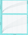
Pattern of growth in this patient from birth to 18 months of age (the figure was created by Nitiroj Bongkotwilawan).
Figure 8
Figure 8
Left atrial volume at (A) 3 days of age compared with (B) 18 months of age. (C) MR is reduced to a mild degree at 18 months of age. MR, mitral regurgitation.
Figure 9
Figure 9
Illustrates the cardinal morphological features of the mitral arcade: absent or short chordae tendineae, interconnecting band of fibrous tissue between two papillary muscles (PM), and elongated PM. It also shows the direct attachment of PM to the mitral leaflet (white arrow) and a view from the left atrium (inset). (Reproduced with permission from: Hakim FA, Krishnaswamy C, Mookadam F. Mitral arcade in adults—a systematic overview. Echocardiography 2013;30:354–9).

Similar articles

References

    1. Ong C, Brown R-M, Gershon A, et al. . Mitral arcade with anomalous papillary muscles: rare cause of mitral stenosis in a young adult. JACC Case Rep 2021;3:1303–9. 10.1016/j.jaccas.2021.06.013 - DOI - PMC - PubMed
    1. Singh R, Wang M. A rare encounter of mitral arcade with anomalous papillary muscles. Cureus 2022;14:e21253. 10.7759/cureus.21253 - DOI - PMC - PubMed
    1. Hakim FA, Krishnaswamy C, Mookadam F. Mitral arcade in adults -- a systematic overview. Echocardiography 2013;30:354–9. 10.1111/echo.12126 - DOI - PubMed
    1. Kim S-J, Shin E-S, Park M-K, et al. . Congenital mitral insufficiency caused by anomalous mitral arcade in an elderly patient: use of echocardiography and Multidetector computed tomography for diagnosis. Circ J 2005;69:1560–3. 10.1253/circj.69.1560 - DOI - PubMed
    1. Su LS, Burkhart HM, O’Leary PW, et al. . Mitral valve arcade with concomitant anomalous left coronary artery from the pulmonary artery. Ann Thorac Surg 2011;92:e121–3. 10.1016/j.athoracsur.2011.06.010 - DOI - PubMed

Publication types

MeSH terms