. 2024 Jun 12;15(1):5032.
doi: 10.1038/s41467-024-49431-6.

UBE2D3 facilitates NHEJ by orchestrating ATM signalling through multi-level control of RNF168

Affiliations

UBE2D3 facilitates NHEJ by orchestrating ATM signalling through multi-level control of RNF168

Zeliha Yalçin et al. Nat Commun. .

Abstract

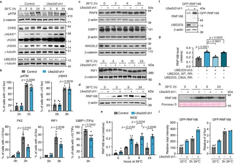
Maintenance of genome integrity requires tight control of DNA damage response (DDR) signalling and repair, with phosphorylation and ubiquitination representing key elements. How these events are coordinated to achieve productive DNA repair remains elusive. Here we identify the ubiquitin-conjugating enzyme UBE2D3 as a regulator of ATM kinase-induced DDR that promotes non-homologous end-joining (NHEJ) at telomeres. UBE2D3 contributes to DDR-induced chromatin ubiquitination and recruitment of the NHEJ-promoting factor 53BP1, both mediated by RNF168 upon ATM activation. Additionally, UBE2D3 promotes NHEJ by limiting RNF168 accumulation and facilitating ATM-mediated phosphorylation of KAP1-S824. Mechanistically, defective KAP1-S824 phosphorylation and telomeric NHEJ upon UBE2D3-deficiency are linked to RNF168 hyperaccumulation and aberrant PP2A phosphatase activity. Together, our results identify UBE2D3 as a multi-level regulator of NHEJ that orchestrates ATM and RNF168 activities. Moreover, they reveal a negative regulatory circuit in the DDR that is constrained by UBE2D3 and consists of RNF168- and phosphatase-mediated restriction of KAP1 phosphorylation.


Conflict of interest statement

The authors declare no competing interests.

Figures

Fig. 1
Fig. 1. UBE2D3 is a regulator of telomere-driven genomic instability.
a Outline of the screen performed to identify factors controlling telomere-driven genomic instability. b Survival assays after 2 weeks of telomere uncapping for 2 independent Ube2d3 shRNAs and 1 Nbs1 shRNA. Representative plates from 8 independent experiments for control and Ube2d3 sh1 and 2 independent experiments for Ube2d3 sh2 and Nbs1 sh are shown. Representative immunoblots for Ube2d3 depletion from 2 independent experiments are shown. c Left: Growth curves of control and Ube2d3 knockdown TRF2ts MEFs (clone C15) upon induction of telomere uncapping (n = 3 independent experiments; mean ± SEM; two-tailed Student’s t-test). Immunoblot for UBE2D3 in cells used for growth assays are shown below (R1, R2, R3 = replicate 1, 2, 3). Right: Representative photographs of arrested and dying control TRF2ts MEFs and proliferating Ube2d3 knockdown TRF2ts MEFs after 12 days of telomere uncapping (TRF2ts clone B17). Scale bar represents 10 μm. Growth curves from these cells can be found in Supplementary Fig. 1c. d Survival assays upon Ube2d1, Ube2d2 and Ube2d3 depletion, after 12 days of telomere uncapping and at 32 °C. Representative plates from 2 independent experiments are shown. Source data are provided as a Source Data file.
Fig. 2
Fig. 2. UBE2D3 promotes NHEJ at uncapped telomeres and in a wider genomic context.
a Quantification of chromosome fusions in TRF2ts cells transduced as indicated upon 24 h of telomere uncapping at 39 °C (n = 4 independent experiments; mean ± SEM; two-tailed Student’s t-test). b Quantification of chromosome fusions in control and UBE2D3-depleted SV40-immortalised wild-type (WT) MEFs, at 6 days after Trf2 shRNA expression (n = 3 independent experiments; mean ± SEM; two-tailed Student’s t-test). c Top: Quantification of chromosome fusions in control and UBE2D3-depleted HeLa cells expressing a doxycycline-inducible TRF2 shRNA (n = 3 independent experiments; mean ± SEM; two-tailed Student’s t-test). Bottom: Immunoblotting for TRF2 and UBE2D3 in cells used for chromosome fusion analysis. The asterisk indicates TRF2. Representative blots from 3 independent experiments. d Top: Quantification of aneuploidy (>4N DNA content) based on DNA content analysis by flow cytometry of TRF2ts MEFs, transduced as indicated, subjected to telomere uncapping and stained with propidium iodide (n = 2 independent experiments). Bottom: Immunoblotting for UBE2D3 in cells used for aneuploidy assays. Representative blots from 2 independent experiments. e Telomeric G-overhang signal in control and UBE2D3-depleted TRF2ts MEFs at 32 °C and after 48 h of telomere uncapping. Left: Native conditions to detect single-strand (ss) TTAGGG repeats. Right: Denaturing conditions for total telomeric DNA. f Quantification of relative telomeric G-overhang signal (n = 2 independent experiments). g NHEJ-mediated repair in U2OS cells transduced as indicated and analysed for random plasmid integration. ShRNAs against the NHEJ promoting factors 53BP1 and RIF1 serve as positive controls (mean ± SEM of n = 3 independent experiments for 53BP1 sh and UBE2D3 sh2 and n = 2 independent experiments for RIF1 sh are shown). Statistical significance was calculated using one-way analysis of variance (ANOVA) with Tukey’s multiple comparisons test. h Top: Survival assays of TRF2ts cells depleted for UBE2D3 with Ube2d3 shRNA1 and complemented with RNAi-resistant (RR) wild-type UBE2D3 (LZRS_UBE2D3_WT_RR) and UBE2D3 C85A (LZRS_UBE2D3_C85A_RR). Representative plates from 3 independent experiments. Bottom: Western blot analysis of Ube2d3 depletion, and UBE2D3 RR and UBE2D3 C85A RR expression in TRF2ts MEFs used in (h). Representative blots from 3 independent experiments. Ref reference. Source data are provided as a Source Data file.
Fig. 3
Fig. 3. UBE2D3 contributes to DDR-induced chromatin ubiquitination and limits RNF168 protein accumulation and recruitment to uncapped telomeres.
a Immunoblotting for pATM, γH2AX and CHK2 in TRF2ts MEFs transduced as indicated and upon telomere uncapping at 39 °C. Asterisk indicates ATM phosphorylated at Ser1981. Representative blots from 4 independent experiments. b Quantification of pATM, γH2AX, FK2 and RIF1 foci in TRF2ts MEFs, transduced as indicated, at 32 °C (0 h) or upon telomere uncapping for 3 h at 39 °C (n = 3 independent experiments for pATM and RIF1, n = 6 independent experiments for γH2AX and n = 4 independent experiments for FK2; mean ± SEM), and quantification of 53BP1 foci at telomeres (telomere dysfunction-induced foci (TIFs)) in TRF2ts MEFs upon telomere uncapping for 3 h at 39 °C (n = 5 independent experiments; mean ± SEM). Statistical significance was calculated using the two-tailed Student’s t-test. c Immunoblotting for RNF8, 53BP1, RIF1 and MAD2L2 in TRF2ts MEFs subjected to telomere uncapping at 39 °C. Representative blots from 4 independent experiments (RNF8 and 53BP1) or 2 independent experiments (MAD2L2 and RIF1). The asterisk indicates 53BP1; below is a non-specific band. d RNF168 levels in TRF2ts MEFs upon telomere uncapping. Representative blots from 5 independent experiments. e Quantification of RNF168 levels in whole cell extracts (WCE) (n = 5 independent experiments; mean ± SEM; one-way analysis of variance (ANOVA) with Tukey’s multiple comparisons test). f Immunoblots for GFP-RNF168 in control and UBE2D3-depleted TRF2ts MEFs. Asterisk indicates endogenous RNF168. Representative blots from 4 independent experiments (quantifications in Supplementary Fig. 5c). g Quantification of RNF168 protein levels in HEK 293T cells with complementation of UBE2D3 depletion by RNAi-resistant (RR) wild-type UBE2D3 (pCDH_UBE2D3_WT_RR) or UBE2D3 C85A (pCDH_UBE2D3_C85A_RR) (n = 4 independent experiments; mean ± SEM; one-way analysis of variance (ANOVA) with Tukey’s multiple comparisons test). Representative immunoblots in Supplementary Fig. 5d. h Immunoblotting of chromatin fractions to assess RNF168 recruitment to chromatin upon telomere uncapping at 39 °C. Representative blots from 3 independent experiments. i Quantification of GFP-RNF168 foci in TRF2ts MEFs at 32 °C and upon 3 h of telomere uncapping at 39 °C. Ref reference. Source data are provided as a Source Data file.
Fig. 4
Fig. 4. UBE2D3 prevents RNF168 hyperaccumulation through ubiquitination and degradation of RNF168 and thereby facilitates telomere NHEJ.
a Immunoblots to evaluate RNF168 protein stability in control and Ube2d3 shRNA2 transduced cells, cultured in the presence of 50 μg/ml cycloheximide (CHX). Representative blots from 5 independent experiments. b Quantification of cycloheximide experiments as in a (n = 5 independent experiments; mean ± SEM; two-tailed Student’s t-test). Significance is shown for Control vs. UBE2D3 sh2: *p = 0.0488 (8 h), ns p = 0.0524 (16 h), *p = 0.0307 (24 h). c Immunoblots for RNF168 in control and UBE2D3-depleted TRF2ts cells at 32 °C or upon telomere uncapping and treated for 4 h with 10 μM MG132. Representative blots of two independent experiments. d Immunoblots for GFP-RNF168 in control and UBE2D3-depleted TRF2ts cells at 32 °C or upon telomere uncapping and treated for 4 h with 10 μM MG132. Representative blots of two independent experiments. The asterisk annotates the band for GFP-RNF168. e HeLa cells with or without expression of His-Ubiquitin were transduced with control or two independent lentiviral shRNAs targeting UBE2D3. His-ubiquitin conjugates were purified and analysed by immunoblotting. Representative of 3 independent experiments. See Supplementary Fig. 6b for UBE2D3 levels in input samples. f Quantification of polyubiquitinated GFP-RNF168 (Ubn) immunoprecipitated from control or UBE2D3-depleted 293T cells, transfected with HA-Ubiquitin and GFP or GFP-RNF168 (n = 2 independent experiments). The eluted poly-HA-Ub signal was corrected over the eluted GFP-RNF168 signal. Corresponding blots in Supplementary Fig. 6e, f. g Quantification of chromosome fusions in control, Ube2d3 shRNA1 or GFP-RNF168 transduced TRF2ts cells upon 24 h of telomere uncapping (n = 3 independent experiments; mean ± SEM; two-tailed Student’s t-test). h Immunoblotting for GFP-RNF168 expression and Ube2d3 depletion in TRF2ts cells used in (g). Asterisk indicates GFP-RNF168. i Immunoblotting for RNF168 in TRF2ts cells transduced as indicated upon 24 h of telomere uncapping at 39 °C. Representative blots from 3 independent experiments. j Chromosome fusions in TRF2ts MEFs transduced with indicated shRNAs upon 24 h of telomere uncapping (n = 3 independent experiments; mean ± SEM; one-way analysis of variance (ANOVA) with Tukey’s multiple comparisons test). Ref reference. Source data are provided as a Source Data file.
Fig. 5
Fig. 5. UBE2D3 promotes KAP1-S824 phosphorylation via regulation of RNF168 stability, thereby facilitating NHEJ at telomeres.
a Immunoblotting for pKAP1 (S824), KAP1 and RNF168 in control and UBE2D3-depleted TRF2ts MEFs upon telomere uncapping at 37 °C. Representative blots from 3 independent experiments. b Quantification of pKAP1 levels in TRF2ts MEFs transduced as indicated. The mean ± SEM from n = 3 independent experiments is shown, except for the 6 h timepoint that is n = 2. Statistical significance was calculated using one-way analysis of variance (ANOVA) with Tukey’s multiple comparisons test. c Quantification of pKAP1 levels in TRF2ts MEFs transduced with Ube2d1 shRNA, Ube2d2 shRNA and/or Ube2d3 sgRNA upon 3 h of telomere uncapping at 37 °C (n = 3 independent experiments; mean ± SEM; one-way analysis of variance (ANOVA) with Tukey’s multiple comparisons test). d Quantification of pKAP1 levels in TRF2ts MEFs transduced with Ube2d3 sh1 and complemented with exogenously expressed control, 3xFLAG-UBE2D1 or 3x-FLAG-UBE2D2 constructs (n = 3 independent experiments; mean ± SEM; one-way analysis of variance (ANOVA) with Tukey’s multiple comparisons test). e Immunoblotting for pKAP1 (S824) in HeLa cells transduced with control or UBE2D3 shRNAs at 30 min post IR (5 Gy). Representative blots from 3 independent experiments. f Quantification of pKAP1 levels in HeLa cells upon IR (representative immunoblots in (e)). The mean ± SEM from 3 independent experiments is shown. Statistical significance was calculated using the two-tailed Student’s t-test. g Chromosome fusions in TRF2ts MEFs transduced with control or Ube2d3 shRNA3, complemented with exogenously expressed KAP1 WT or KAP1 S824D mutant, upon 36 h of telomere uncapping at 37 °C (n = 3 independent experiments; mean ± SEM; two-tailed Student’s t-test). h Chromosome fusions in TRF2ts MEFs transduced with Ube2d3 sh1 and/or an sgRNA targeting Cbx5 (encoding the HP1α protein), along with non-targeting control shRNA/sgRNA (-) where appropriate (n = 3 independent experiments; mean ± SEM two-tailed Student’s t-test). i Immunoblotting for pKAP1 (S824) in TRF2ts MEFs transduced with control, Ube2d3 and/or Rnf168 shRNAs at 32 °C or after 3 h at 37 °C. Representative blots from 2 independent experiments. Ref reference. Source data are provided as a Source Data file.
Fig. 6
Fig. 6. UBE2D3 promotes KAP1 phosphorylation and telomere NHEJ in a PP2A-dependent manner.
a Immunoblotting for pKAP1 (S824) in TRF2ts MEFs transduced with control, Ube2d3 and/or two independent Ppp2ca shRNAs at 32 °C or after 3 h at 37 °C. Representative blots from 2 independent experiments. b Quantification of chromosome fusions in TRF2ts MEFs transduced with control, Ube2d3 and two independent Ppp2ca shRNAs, upon 24 h of telomere uncapping. Two independent experiments are shown. c PP2A activity assays with immunoprecipitated PP2A from TRF2ts MEFs transduced as indicated and cultured at 32 °C or for 3 h at 37 °C to induce telomere uncapping (n = 3 independent experiments; mean ± SEM; two-tailed Student’s t-test). Immunoblots of input and immunoprecipitates are shown in Supplementary Fig. 8e. d PP2A phosphatase activity assay with immunoprecipitated PP2A-alpha (PP2Ac) from RNF168 mutant human cells (RIDDLE) with and without expression of ectopic HA-RNF168. Corrected for the amount of immunoprecipitated PP2A-alpha. Cells were untreated or harvested 30 min after irradiation with 3 Gy (n = 3 independent experiments; mean ± SEM; two-tailed Student’s t-test). The different symbols (dot, square and triangle) represent 3 biologically independent experiments. Immunoblots of input and immunoprecipitates are shown in Supplementary Fig. 8f. e Assessment of in vivo PP2A ubiquitination by immunoprecipitation of endogenous PP2A catalytic C subunit from HEK 293T cell lysates transfected with the indicated plasmids. See Supplementary Fig. 8i for input samples. Representative blots from 3 independent experiments. f Venn diagram showing the overlap in significantly altered phospho-substrates in 3 independent replicate phosphoproteomics analyses of UBE2D3-depleted (Ube2d3 sh1) or PP2A inhibitor-treated (5 μM LB100, 15 h) TRF2ts MEFs upon 3 h of telomere uncapping at 37 °C. Numbers indicate the phospho-substrates that were significantly reduced in UBE2D3-depleted cells or significantly increased in LB100-treated cells. Ube2d3 sh1 and LB100 were compared to control (shScramble) TRF2ts MEFs. g Quantification of pDNA-PKcs (S2056) foci in TRF2ts MEFs, transduced as indicated, at 32 °C (0 h) or upon telomere uncapping for 3 h at 39 °C (n = 3 independent experiments; mean ± SEM; two-tailed Student’s t-test). Ref reference. Source data are provided as a Source Data file.
Fig. 7
Fig. 7. Model for how UBE2D3 facilitates NHEJ by promoting ATM kinase-dependent DDR activities.
On the one hand UBE2D3 promotes DDR-induced chromatin ubiquitination and 53BP1 recruitment, that are known to be mediated by the RNF8 and RNF168 E3 ligases following ATM kinase activation. 53BP1 is known to promote NHEJ by recruiting the shieldin complex and promoting chromatin mobility (not depicted). Thus, UBE2D3 appears to support the role of RNF168 in promoting NHEJ via chromatin ubiquitination and 53BP1 recruitment. On the other hand, UBE2D3 promotes the ubiquitination and proteasomal degradation of RNF168, thereby preventing RNF168 hyperaccumulation. UBE2D3-deficiency and RNF168 hyperaccumulation are associated with impaired KAP1-S824 phosphorylation, that appears to be in part caused by enhanced phosphatase activity by PP2A, but may also involve other mechanisms. By limiting RNF168 accumulation, UBE2D3 prevents disproportionate dephosphorylation of KAP1 that counteracts the ability of ATM to promote efficient NHEJ through phosphorylation of S824 of KAP1. Thus, this implies the existence of a negative feed-back circuit within ubiquitin and kinase signalling in the DDR that is constrained by UBE2D3 and consists of RNF168- and phosphatase-mediated restriction of ATM-dependent KAP1 phosphorylation.

Similar articles

Cited by

References

    1. Hanahan D, Weinberg RA. Hallmarks of cancer: the next generation. Cell. 2011;144:646–674. doi: 10.1016/j.cell.2011.02.013. - DOI - PubMed
    1. Maciejowski J, de Lange T. Telomeres in cancer: tumour suppression and genome instability. Nat. Rev. Mol. Cell Biol. 2017;18:175–186. doi: 10.1038/nrm.2016.171. - DOI - PMC - PubMed
    1. Boersma V, et al. MAD2L2 controls DNA repair at telomeres and DNA breaks by inhibiting 5’ end resection. Nature. 2015;521:537–540. doi: 10.1038/nature14216. - DOI - PMC - PubMed
    1. Konishi A, de Lange T. Cell cycle control of telomere protection and NHEJ revealed by a ts mutation in the DNA-binding domain of TRF2. Genes Dev. 2008;22:1221–1230. doi: 10.1101/gad.1634008. - DOI - PMC - PubMed
    1. Peuscher MH, Jacobs JJ. DNA-damage response and repair activities at uncapped telomeres depend on RNF8. Nat. Cell Biol. 2011;13:1139–1145. doi: 10.1038/ncb2326. - DOI - PubMed

MeSH terms