. 2024 Jun 12;15(1):4941.
doi: 10.1038/s41467-024-49131-1.

descSPIM: an affordable and easy-to-build light-sheet microscope optimized for tissue clearing techniques

Affiliations

descSPIM: an affordable and easy-to-build light-sheet microscope optimized for tissue clearing techniques

Kohei Otomo et al. Nat Commun. .

Abstract

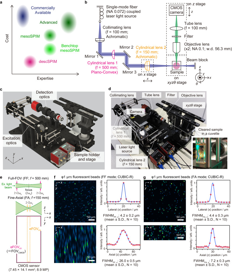
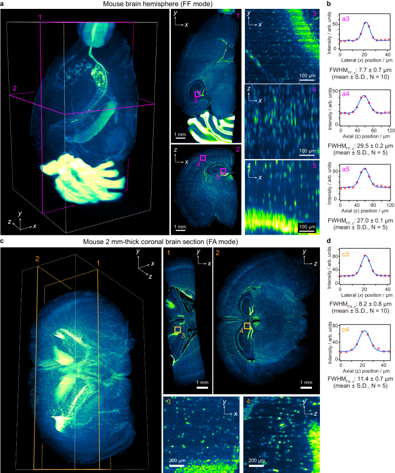
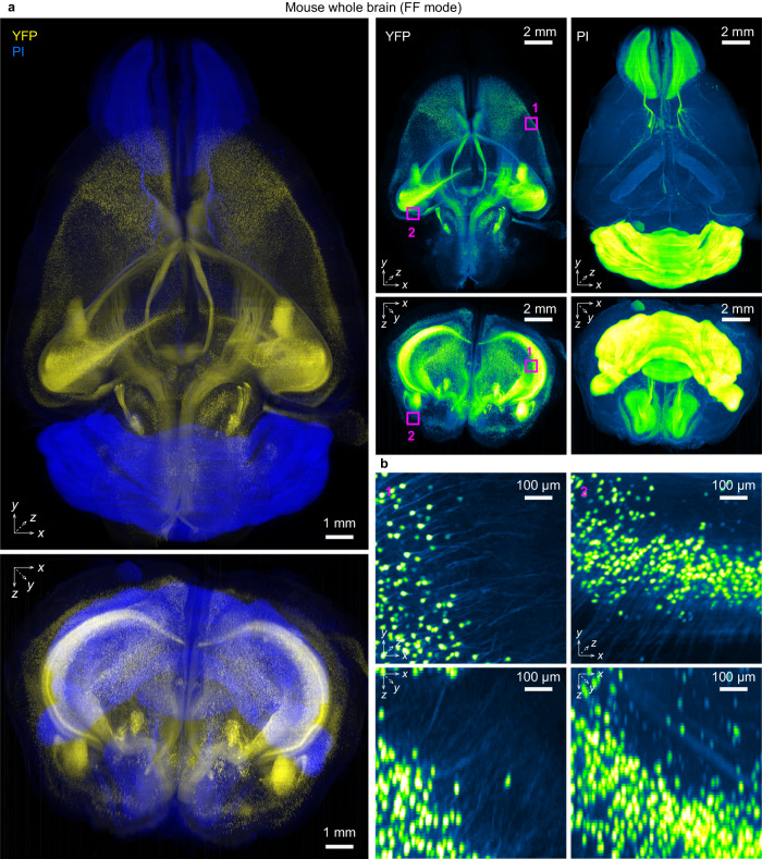
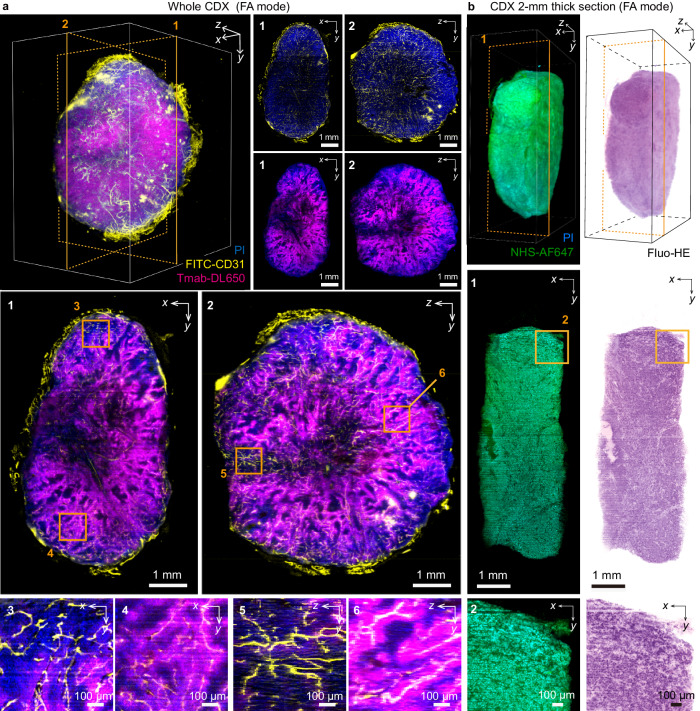
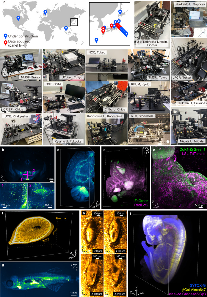
Despite widespread adoption of tissue clearing techniques in recent years, poor access to suitable light-sheet fluorescence microscopes remains a major obstacle for biomedical end-users. Here, we present descSPIM (desktop-equipped SPIM for cleared specimens), a low-cost ($20,000-50,000), low-expertise (one-day installation by a non-expert), yet practical do-it-yourself light-sheet microscope as a solution for this bottleneck. Even the most fundamental configuration of descSPIM enables multi-color imaging of whole mouse brains and a cancer cell line-derived xenograft tumor mass for the visualization of neurocircuitry, assessment of drug distribution, and pathological examination by false-colored hematoxylin and eosin staining in a three-dimensional manner. Academically open-sourced ( https://github.com/dbsb-juntendo/descSPIM ), descSPIM allows routine three-dimensional imaging of cleared samples in minutes. Thus, the dissemination of descSPIM will accelerate biomedical discoveries driven by tissue clearing technologies.


Conflict of interest statement

E.A.S. and K.T. are co-inventors on patents and patent applications owned by RIKEN covering the CUBIC reagents, and E.A.S. is employed by CUBICStars Inc. that offers services based on CUBIC technology. The remaining authors declare no competing interests.

Figures

Fig. 1
Fig. 1. descSPIM concept, components, and optical specifications.
a The target of descSPIM on the expertise and cost axes, where the simultaneous achievement of thorough simplification and practical quality is required. b descSPIM system overview. A one-sided light-sheet illumination formed by a collimated lens and a cylindrical lens comes from the right side of the sample. Either a cylindrical lens (f = 500 mm or f = 150 mm) is used for each illumination mode (Full FOV; FF and Fine axial; FA), respectively. A manual linear translation stage is attached to the latter cylindrical lens for the tilling light-sheet (TLS) imaging. The detection path is composed of a 2× objective lens, tube lens, and CMOS camera, with an effective integration magnification of 1× (3.45 µm × 3.45 µm of the pixel size). c Schematic overview of the descSPIM. d Overview of the actual descSPIM instrument. (inset) The cleared sample can be placed in a four-sided transmission cuvette and directly illuminated for imaging. No oil chamber was adopted. e Two illumination modes adopted to the descSPIM by switching the cylindrical lenses. FF covers the whole CMOS sensor area (7.45 mm in width). FA provides finer axial resolution while covering ~11% (830 µm of the Rayleigh length in width) of the sensor area. eFOV: effective field of view. f, g Estimation of lateral and axial resolutions by point spread functions (PSFs; evaluated with images 1 µm beads images). Comparable xy resolution was achieved by the two illumination modes. FA provides approximately 7 µm of axial full width of half maximum (FWHM) value, over three-times finer than FF mode. The original grayscale 8-bit maps were pseudo-colored with a Green Fire Blue look-up table. The imaging experiments were performed twice with nearly identical results.
Fig. 2
Fig. 2. Optical specifications and imaging results.
a Full FOV (FF)-mode imaging of a propidium iodide (PI)-stained mouse brain hemisphere. Voxel size: 3.45 × 3.45 × 10 µm3. Imaging speed: 180 s. per 900 slices. Data size: 8.02 GB per 8-bit image stack. Flat-field correction (FFC) was applied. The xy images illustrate a homogeneous image contrast across the FOV. The yz images indicate the elongation of PI-stained cell nuclei across the z direction due to the lower axial resolution. Note that the basic system did not include a device for eliminating the stripe shadow artifact (e.g., a galvanometer mirror) for simplification. Despite these technical limitations, the reconstituted 3D image is qualitatively sufficient for evaluating the whole sample structure. The original grayscale 8-bit maps were pseudo-colored with a Green Fire Blue look-up table (LUT). The imaging experiment was performed at least 5 times with nearly identical results. b Lateral and axial spatial resolutions of PI-stained nuclei obtained with FF mode, estimated by the full width of half maximum (FWHM) values of their intensity profiles. The axial FWHM values were consistent with the value measured by beads (Fig. 1f) and about quadrupled by the lateral FWHM of the PI signals. c Fine axial (FA) mode imaging of a PI-stained, 2 mm-thick mouse brain section. Voxel size: 3.45 × 3.45 × 5 µm3. Imaging speed: 372 s per 1860 slices per stack. Data size: 16.54 GB per 8-bit image stack. A total of four stacks with different light-sheet focus positions were collected, then tilling light-sheet (TLS) was applied. The image intensity was corrected by FFC. The original grayscale 8-bit maps were pseudo-colored with a Green Fire Blue LUT. The imaging experiment was performed twice with nearly identical results. d Lateral and axial spatial resolutions of PI-stained nuclei obtained by FA mode, estimated as in (b). The yz images indicate that the FA mode provides a quality of image close to the isotropic resolution for the object with cell nuclei size across the z direction.
Fig. 3
Fig. 3. Practical use of descSPIM in neuroscience.
a An example of propidium iodide (PI)-stained whole Thy1-YFP-H mouse brain imaging with full FOV (FF) mode and flat-field correction (FFC) adoption. PI and YFP were excited with 561 nm and 488 nm laser beams, respectively. Four image-stack tiles were collected from a single direction (angle 0°). Using the θ stage, a second set of four image tiles from the opposite direction (angle 180°) were then obtained. All the image tiles were stitched, aligned, and fused with BigStither and ANTs software. See Supplementary Fig. 13 for the procedure. Voxel size: 3.45 × 3.45 × 10 µm3. b Magnified images of YFP-expressing neurons within the dataset. Signal elongation along the axial direction was more modest in somas (10–20 µm in diameter) than in neurites (approximately 1 µm in diameter), consistent with the light sheet thickness (approximately 25 µm). Quantitative data is shown in Supplementary Fig. 14. The original grayscale 8-bit maps were pseudo-colored with a Blue, Yellow or Green Fire Blue look-up table. The imaging experiment was performed twice with nearly identical results.
Fig. 4
Fig. 4. Practical use of descSPIM in the drug discovery and 3D pathology.
a An example of Trastuzumab-administered whole cell-line derived xenograft (CDX; BT-474 human breast cancer cell line) imaging with fine axial (FA) mode, tilling light-sheet (TLS) method, flat-field correction (FFC) adoption, and multi-directional stack registration and fusing. Anti-CD31 antibody was also perfused before sampling. PI was stained during clearing. DL650 (Trastuzumab), PI, and FITC (CD31) were excited with 647 nm, 515 nm, and 488 nm laser beams, respectively. Using the θ stage, image tiles from the angles 0° and 180° were obtained. The two-sided stacks were then aligned and merged with ANTs and BigStitcher. Orthogonal cross-sections near the center of the specimen are shown as max intensity projection (MIP) images of a thickness of approximately 70 µm. Although validation of the imaging results by other methods is required, the descSPIM’s potential to acquire apparent drug distributions is anticipated to be beneficial for drug discovery and development. Voxel size: 3.45 × 3.45 × 10 µm3. The original grayscale 8-bit maps were pseudo-colored with a Blue, Yellow or Magenta look-up table (LUT). The imaging experiment was performed at twice with nearly identical results. b An example of 2 mm-thick CDX imaging with FA mode, TLS method, and FFC adoption. The sample was stained with PI and NHS-Alexa FluorⓇ 647 before clearing, and false-colored hematoxylin and eosin colors (3D Fluo-HE) were applied. Alexa FluorⓇ 647 and PI were excited with 647 nm and 515 nm laser beams, respectively. The data demonstrates that descSPIM has the potential to be used routinely in 3D clinical pathology examinations. Voxel size: 3.45 × 3.45 × 10 µm3. The original grayscale 8-bit maps were pseudo-colored with a Blue, Green or Fluo-HE LUT. The imaging experiment was performed twice with nearly identical results.
Fig. 5
Fig. 5. Dissemination of descSPIM in the research community.
a Global distribution of descSPIM users and partial installation sites of the devices. CRIEPI: Central Research Institute of Electric Power Industry; JFCR: Japanese Foundation for Cancer Research; KPUM: Kyoto Prefectural University of Medicine; KTH: Royal Institute of Technology, Sweden; NCC: National Cancer Center Japan, Japan; NMSF: Nippon Medical School Foundation, Japan; QST: National Institute for Quantum Science and Technology, Japan; TMDU: Tokyo Medical and Dental University, Japan; UOE: University of Occupational and Environmental Health, Japan. b 3D image of a rat-brain coronal slice immunostained for tyrosine hydroxylase in the hypothalamus, acquired at Kagoshima University. Xyz: 6000 pix. × 3984 pix. × 189 slices. Voxel size: 3.0 × 3.0 × 10 µm3. c 3D image of a mouse brain hemisphere stained with SYTOX-Green, acquired at QST. Xyz: 2160 pix. × 4096 pix. × 390 slices. Voxel size: 3.45 × 3.45 × 10 µm3. d 3D image of ZsGreen-labeled tumor metastasis in mouse lung, stained with RedDot2, acquired at JFCR. Xyz: 2148 pix. × 3120 pix. × 501 slices. Voxel size: 3.45 × 3.45 × 10 µm3. e 3D image of a stomach from Chat-Cre; R26-LSL-TdTomato; Dclk1-ZsGreen mouse strains, acquired at Cancer Inst. Xyz: 2031 pix. × 3295 pix. × 521 slices. Voxel size: 3.45 × 3.45 × 10 µm3. f 3D image of a mouse intestine with PI, acquired at CRIEPI. Xyz: 1512 pix. × 1011 pix. × 484 slices. Voxel size: 3.45 × 3.45 × 5 µm3. g 3D image of Tg(fli1a:myr-EGFP)ncv2Tg zebrafish expressing EGFP in endothelial cells, acquired at NMSF. Xyz: 1795 pix. × 4096 pix. × 882 slices. Voxel size: 3.45 × 3.45 × 3.3 µm3. h 3D image of E7.5 decidua labeled with SYTOX-Green, acquired at Kyushu U. Xyz: 856 pix. × 1468 pix. × 100 slices. Voxel size: 0.345 × 0.345 × 4 µm3. i. 3D image of E12.0 RARE-lacZ whole mouse embryo, acquired at KPUM. Xyz: 2048 pix. × 1080 pix. × 671 slices. Voxel size: 3.1 × 3.1 × 8.6 µm3. The original grayscale 8-bit maps were pseudo-colored with a Blue, Green, Yellow, Magenta, Green Fire Blue, or Orange Hot look-up table. Each imaging experiments for cleared biological samples were performed at least once.

References

    1. Dodt HU, et al. Ultramicroscopy: three-dimensional visualization of neuronal networks in the whole mouse brain. Nat. Methods. 2007;4:331–336. doi: 10.1038/nmeth1036. - DOI - PubMed
    1. Dent JA, Polson AG, Klymkowsky MW. A whole-mount immunocytochemical analysis of the expression of the intermediate filament protein vimentin in Xenopus. Development. 1989;105:61–74. doi: 10.1242/dev.105.1.61. - DOI - PubMed
    1. Klingberg A, et al. Fully automated evaluation of total glomerular number and capillary tuft size in nephritic kidneys using lightsheet microscopy. J. Am. Soc. Nephrol. 2017;28:452–459. doi: 10.1681/ASN.2016020232. - DOI - PMC - PubMed
    1. Ertürk A, et al. Three-dimensional imaging of solvent-cleared organs using 3DISCO. Nat. Protoc. 2012;7:1983–1995. doi: 10.1038/nprot.2012.119. - DOI - PubMed
    1. Susaki EA, et al. Whole-brain imaging with single-cell resolution using chemical cocktails and computational analysis. Cell. 2014;157:726–739. doi: 10.1016/j.cell.2014.03.042. - DOI - PubMed

Grants and funding