. 2024 Jun 12;14(1):13552.
doi: 10.1038/s41598-024-64559-7.

Development of DNA aptamers targeting B7H3 by hybrid-SELEX: an alternative to antibodies for immuno-assays

Affiliations

Development of DNA aptamers targeting B7H3 by hybrid-SELEX: an alternative to antibodies for immuno-assays

Bhavani Shankar Maradani et al. Sci Rep. .

Abstract

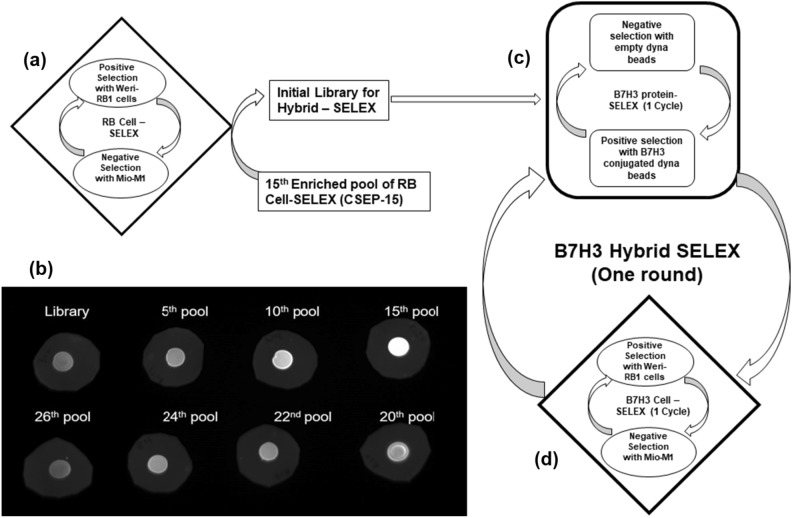
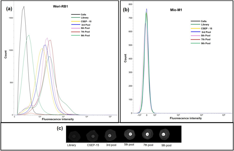
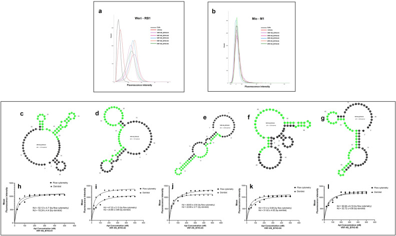
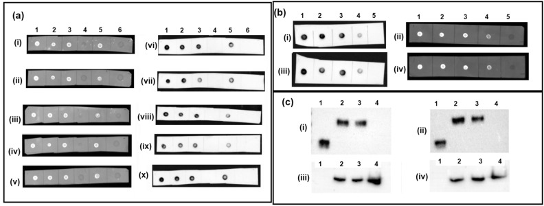
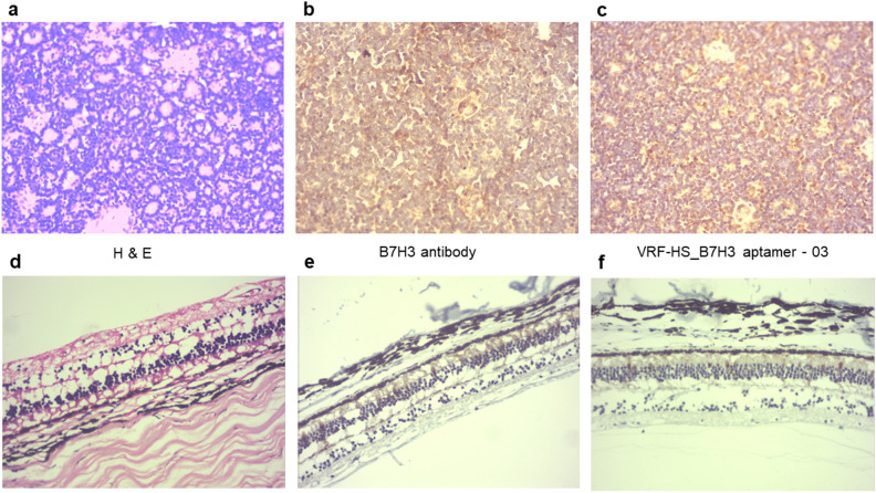
Antibodies have been extensively used in numerous applications within proteomics-based technologies, requiring high sensitivity, specificity, a broad dynamic range for detection, and precise, reproducible quantification. Seeking alternatives to antibodies due to several inherent limitations of antibodies is an area of active research of tremendous importance. Recently, aptamers have been receiving increasing attention, because they not only have all of the advantages of antibodies, but also have unique advantages, such as thermal stability, low cost, and unlimited applications. Aptamers are gaining importance in immunological studies and can potentially replace antibodies in immunoassays. B7H3, an immunoregulatory protein belonging to the B7 family, is an attractive and promising target due to its overexpression in several tumor tissues while exhibiting limited expression in normal tissues. This study employed hybrid-SELEX with next-generation sequencing to select ssDNA aptamers specifically binding to the B7H3 protein. These aptamers demonstrated versatility across various assays, including flow cytometry, dot-blot, and immunohistochemistry. Effective performance in sandwich dot-blot assays and western blot analysis suggests their potential for diagnostic applications and demonstrates their adaptability and cost-effectiveness in diverse protein detection techniques.


Conflict of interest statement

The authors declare no competing interests.

Figures

Figure 1
Figure 1
Schematic representation of our hybrid-SELEX method for selection of B7H3-specific ssDNA aptamer. (a) Retinoblastoma cell-SELEX to develop aptamers against retinoblastoma using Weri-RB1 cells and target cell and Mio-M1 as control cells. (b) We have screened the RB cell-SELEX enriched pools on recombinant B7H3 protein by dot-blot and chose the cell-SELEX enriched pool-15 (CSEP-15) as the starting library for the B7H3 hybrid SELEX. In our experiment, the hybrid-SELEX process is divided into (c) the B7H3 protein-based SELEX selection and (d) cell-based SELEX enrichment. The CSEP-15 is incubated with B7H3 protein immobilized on magnetic beads for positive selection and empty magnetic beads for counter selection for each cycle in protein-SELEX. The pool enriched from protein SELEX is incubated with Weri-RB1 for positive selection and Mio-M1 for counter selection in cell-SELEX. After 9 rounds of selection, the enriched aptamer pools were sequenced by NGS. SELEX, Systematic Evolution of Ligands by EXponential enrichment.
Figure 2
Figure 2
Monitoring the enrichment of the DNA libraries during hybrid-SELEX by flow cytometry and dot-blot. (a) Fluorescence intensities of target cells (Weri-RB1) incubated with FITC-labelled ssDNA pools from the initial library to the ninth-selection round. (b) Fluorescence intensities of negative control cells (Mio-M1) incubated with FITC-labelled ssDNA pools from the initial library to the ninth-selection round. (c) Represented is the dot-blot assay showing the fluorescence intensities of ssDNA pools from the initial library to the ninth-selection round, bound to the recombinant B7H3 protein.
Figure 3
Figure 3
Binding ability, secondary structures and dissociation constants of selected B7H3 aptamers. (a) Binding affinity of FITC-labelled aptamers VRF-HS_B7H3-01, VRF-HS_B7H3-02, VRF-HS_B7H3-03, VRF-HS_B7H3-04 and VRF-HS_B7H3-05 to Weri-RB1 cells assessed by flow cytometry. (b) Binding ability of FITC-labelled aptamers VRF-HS_B7H3-01, VRF-HS_B7H3-02, VRF-HS_B7H3-03, VRF-HS_B7H3-04 and VRF-HS_B7H3-05 to Mio-M1 cells assessed by flow cytometry. (c–g) Predicted secondary structures for five aptamer candidates, VRF-HS_B7H3-01, VRF-HS_B7H3-02, VRF-HS_B7H3-03, VRF-HS_B7H3-04 and VRF-HS_B7H3-05 selected for further. The presented predicted secondary structures were the ones with lowest ΔG. Constant sequence regions are highlighted in black, and green represents the random regions. (h–l) Binding curve of aptamers VRF-HS_B7H3-01, VRF-HS_B7H3-02, VRF-HS_B7H3-03, VRF-HS_B7H3-04 and VRF-HS_B7H3-05 with Weri-RB1 and Mio-M1 cells assessed by flow cytometry and recombinant B7H3 protein by dot-blot. Equilibrium dissociation constants (Kd) (nM) were calculated using GraphPad Prism 7, under the non-linear fit model, one-site non-competitive binding to fluorescent population ratio at used aptamer concentrations.
Figure 4
Figure 4
Affinity of B7H3 aptamers by Dot-blot and western blot analysis. (a) Dot-blot assay with (i–v) FITC labelled and (vi–x) biotin labelled aptamers to demonstrate the capability of the B7H3 aptamers to recognize their target immobilized on PVDF membranes; (i) & (vi)—VRF-HS_B7H3-01, (ii) & (vii)—VRF-HS_B7H3-02, (iii) & (viii)—VRF-HS_B7H3-03, (iv) & (viii)—VRF-HS_B7H3-04 and (v) & (x)—VRF-HS_B7H3-05; 1—B7H3 Recombinant protein, 2—RB tumor protein lysate, 3—Weri-RB1 protein lysate, 4—BSA, 5—Weri-RB1 secretome and 6—Secondary control. (b) Sandwich dot blot assay with (i–iii) biotin labelled and (ii–iv) FITC labelled aptamers to demonstrate the capability of aptamers to recognize different epitopes of their target immobilized on nitrocellulose membranes. (i) & (ii)—unlabelled VRF-HS_B7H3-01 is used as capture and FITC or biotin labelled VRF-HS_B7H3-03 is used for detection, (iii) & (iv) unlabelled VRF-HS_B7H3-03 is used as capture and FITC or biotin labelled VRF-HS_B7H3-01 is used for detection; 1—B7H3 Recombinant protein, 2—RB tumor protein lysate, 3—Weri-RB1 protein lysate, 4—Weri-RB1 secretome and 5—BSA. (c) Comparison of the specificity of B7H3 antibody to VRF-HS_B7H3-03 aptamer in a protein blot analysis (cropped image). Lane 1—B7H3 Recombinant protein, 2—RB tumor protein lysate, 3—Weri-RB1 protein lysate and 4—Mio-M1 protein lysate; (i) Probed with anti-B7H3 Rabbit monoclonal antibody, (ii) Probed with biotinylated VRF-HS_B7H3-03 aptamer and (iii) & (iv) Probed with GAPDH mouse monoclonal antibody. Results of an aptamer blot from a nonreducing SDS polyacrylamide gel in which B7H3 was clearly detected near 90 kDa similar to antibody. However, the recombinant protein manufacturer of the B7H3 (R&D systems) reported a molecular weight of 38–48 kDa for its product which is consistent with the strongly detected band’s weight (Full raw blot included in Supplementary data Fig. S5).
Figure 5
Figure 5
Immunohistochemistry of B7H3 antibody and VRF-HS_B7H3-03 to RB tumour sections and retina. (a,d) H and E staining, (b,e) IHC with B7H3 antibody, and (c,f) IHC with biotin-labelled VRF-HS_B7H3-03 of primary retinoblastoma tumour and retina respectively.

Similar articles

Cited by

References

    1. Ritz JB, et al. Use of monoclonal antibodies as diagnostic and therapeutic reagents in acute lymphoblastic leukemia. Cancer Res. 1981;41:4771–4775. - PubMed
    1. Marx V. Finding the right antibody for the job. Nat. Methods. 2013;10:703–707. doi: 10.1038/nmeth.2570. - DOI - PubMed
    1. Uhlen M, et al. A proposal for validation of antibodies. Nat. Methods. 2016;13:823–827. doi: 10.1038/nmeth.3995. - DOI - PMC - PubMed
    1. Brody EN, Gold L. Aptamers as therapeutic and diagnostic agents. J. Biotechnol. 2000;74:5–13. - PubMed
    1. Jayasena SD. Aptamers: An emerging class of molecules that rival antibodies in diagnostics. Clin. Chem. 1999;45:1628–1650. doi: 10.1093/clinchem/45.9.1628. - DOI - PubMed