

Intrinsic aerobic capacity modulates Alzheimer's disease pathological hallmarks, brain mitochondrial function and proteome during aging
- PMID: 38867031
- PMCID: PMC11336007
- DOI: 10.1007/s11357-024-01248-3
Intrinsic aerobic capacity modulates Alzheimer's disease pathological hallmarks, brain mitochondrial function and proteome during aging
Abstract
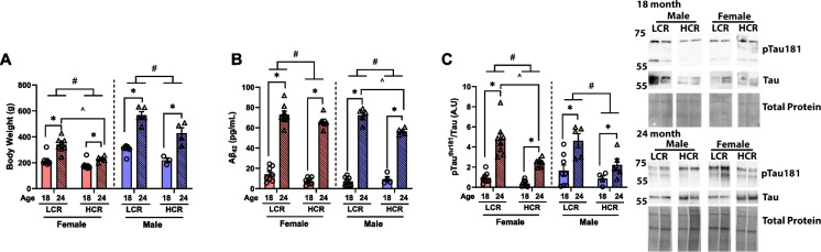
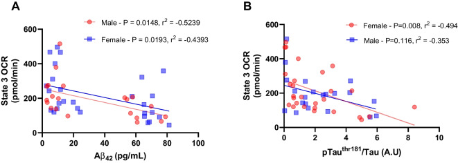
Low aerobic capacity is strongly associated with all-cause mortality and risk for Alzheimer's disease (AD). Individuals with early dementia and AD have lower aerobic capacity compared to age-matched controls. The mechanism by which aerobic capacity influences AD risk is unknown but is likely mediated by sexual dimorphism and tissue-level differences in mitochondrial energetics. Here, we used rats selectively bred for large differences in intrinsic aerobic exercise capacity. Brain tissue from 18-month and 24-month-old female and male low-capacity runner (LCR) and high-capacity runner (HCR) rats were analyzed for markers of mitochondrial function and AD-associated pathologies. LCR rats, irrespective of sex, exhibited a greater increase in brain amyloid beta (Aβ42) and tau hyperphosphorylation (pTauthr181/total tau) with aging. In female LCR rats, brain mitochondrial respiration at states 3, 4, and FCCP-induced uncoupling, when stimulated with pyruvate/malate, was reduced at 18 and 24 months, leading to lower ATP-linked mitochondrial respiration compared to mitochondria from HCR rats. Male LCR rats also showed reduced complex II-stimulated mitochondrial respiration (succinate + rotenone) at 24 months compared to HCR rats. Differences in mitochondrial respiration were associated with tau hyperphosphorylation and Aβ42 alterations in both HCR and LCR strains. Proteomic analysis unveiled a distinct difference in the mitochondrial proteome, wherein female LCR rats displayed diminished mitochondrial translation and oxidative phosphorylation (OXPHOS) proteins at 18 months compared to female HCR rats. Conversely, male LCR rats exhibited increased OXPHOS protein abundance but reduced tricarboxylic acid (TCA) cycle proteins compared to male HCR rats. These findings underscore a robust association between intrinsic aerobic exercise capacity, brain mitochondrial function, and AD pathologies during aging.
Keywords: Aerobic capacity; Aging; Alzheimer’s disease; Amyloid beta; Bioenergetics; Mitochondria; Tau.
© 2024. The Author(s).
Conflict of interest statement
The authors declare no competing interests.
Figures


References
MeSH terms
Substances
Grants and funding
- R01AG069781/AG/NIA NIH HHS/United States
- 1I01BX002567/VA Merit Review Grant
- P20GM144269/Kansas Center for Metabolism and Obesity Research Center
- P30 AG035982/AG/NIA NIH HHS/United States
- R01DK121497/DK/NIDDK NIH HHS/United States
- T32 AG078114/AG/NIA NIH HHS/United States
- P20 GM144269/GM/NIGMS NIH HHS/United States
- P20 GM121293/GM/NIGMS NIH HHS/United States
- T32AG078114/AG/NIA NIH HHS/United States
- I01 BX002567/BX/BLRD VA/United States
- P20 GM103418/GM/NIGMS NIH HHS/United States
- R00AG056600/AG/NIA NIH HHS/United States
- R01 DK121497/DK/NIDDK NIH HHS/United States
- R01 AG078186/AG/NIA NIH HHS/United States
- P30AG035982/Margaret "Peg" McLaughlin and Lydia A. Walker Opportunity Fund
- R01AG078186/AG/NIA NIH HHS/United States
- R24 GM137786/GM/NIGMS NIH HHS/United States
- T32 DK128770/DK/NIDDK NIH HHS/United States
LinkOut - more resources
Full Text Sources
Medical
