. 2024 Jun 12;14(1):13507.
doi: 10.1038/s41598-024-63942-8.

Neuroprotective effects of naltrexone in a mouse model of post-traumatic seizures

Affiliations

Neuroprotective effects of naltrexone in a mouse model of post-traumatic seizures

Saul Rodriguez et al. Sci Rep. .

Abstract

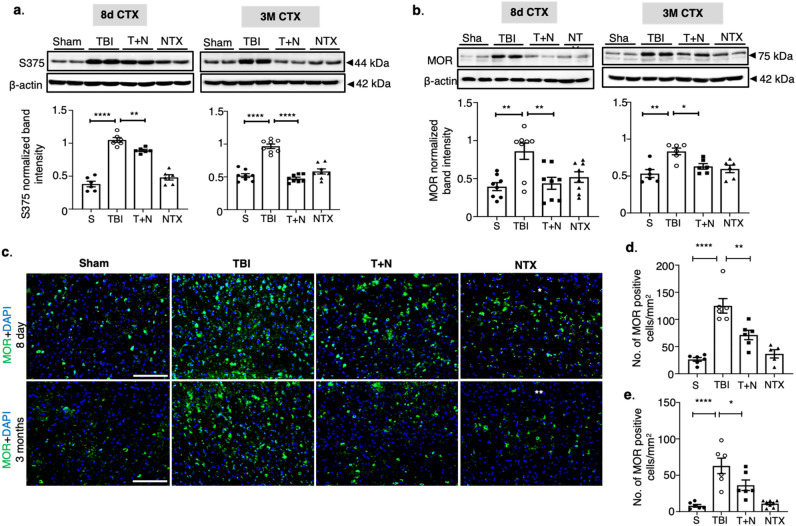
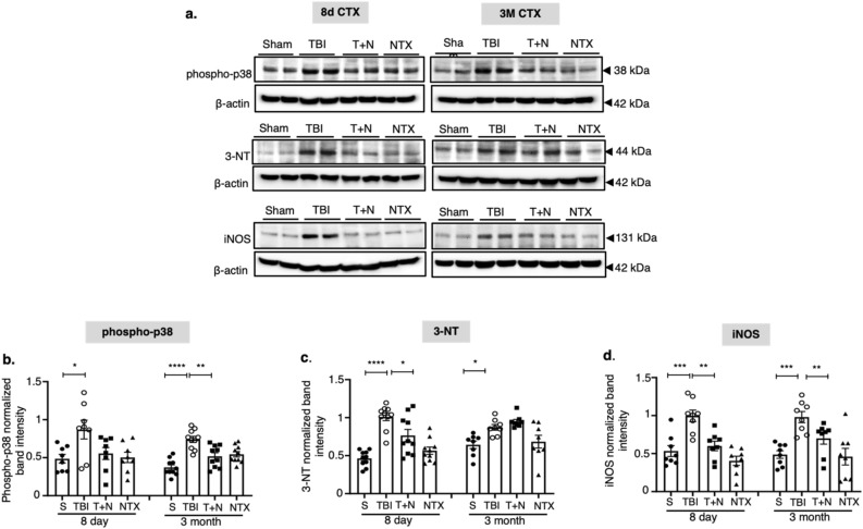
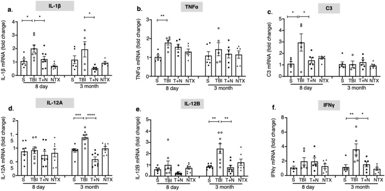
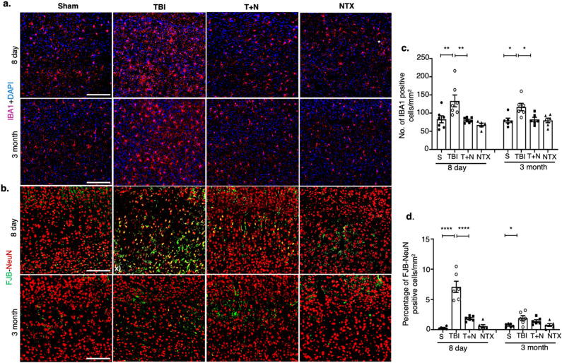
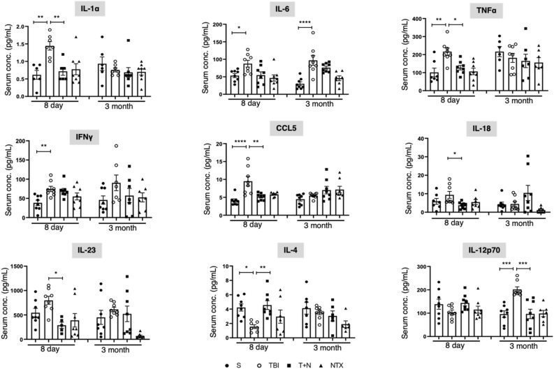
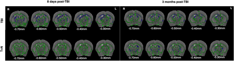
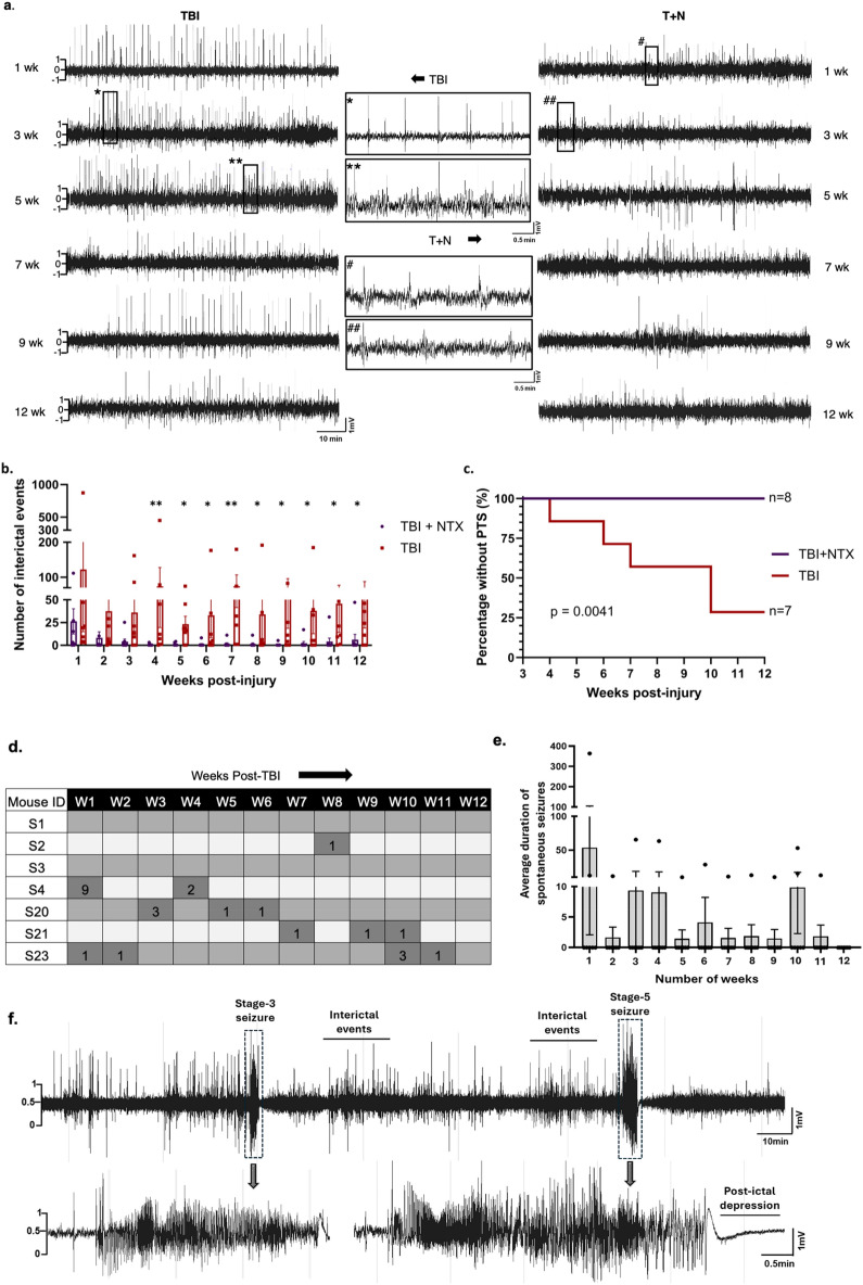
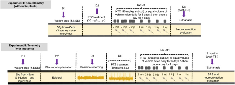
Traumatic Brain Injury (TBI) induces neuroinflammatory response that can initiate epileptogenesis, which develops into epilepsy. Recently, we identified anti-convulsive effects of naltrexone, a mu-opioid receptor (MOR) antagonist, used to treat drug addiction. While blocking opioid receptors can reduce inflammation, it is unclear if post-TBI seizures can be prevented by blocking MORs. Here, we tested if naltrexone prevents neuroinflammation and/or seizures post-TBI. TBI was induced by a modified Marmarou Weight-Drop (WD) method on 4-week-old C57BL/6J male mice. Mice were placed in two groups: non-telemetry assessing the acute effects or in telemetry monitoring for interictal events and spontaneous seizures both following TBI and naltrexone. Molecular, histological and neuroimaging techniques were used to evaluate neuroinflammation, neurodegeneration and fiber track integrity at 8 days and 3 months post-TBI. Peripheral immune responses were assessed through serum chemokine/cytokine measurements. Our results show an increase in MOR expression, nitro-oxidative stress, mRNA expression of inflammatory cytokines, microgliosis, neurodegeneration, and white matter damage in the neocortex of TBI mice. Video-EEG revealed increased interictal events in TBI mice, with 71% mice developing post-traumatic seizures (PTS). Naltrexone treatment ameliorated neuroinflammation, neurodegeneration, reduced interictal events and prevented seizures in all TBI mice, which makes naltrexone a promising candidate against PTS, TBI-associated neuroinflammation and epileptogenesis in a WD model of TBI.

Keywords: Mu-opioid receptors; Naltrexone; Neurodegeneration post-traumatic seizures; Neuroinflammation; Nitro-oxidative stress; Traumatic brain injury.


Conflict of interest statement

The authors declare no competing interests.

Figures

Figure 1
Figure 1
Naltrexone normalized MOR signaling in cortex following TBI. (a,b) Western blot analysis of phospho-MOR (S375) and MOR in the CTX tissue extract at 8 days and 3 months post-TBI. TBI increased both S375 phosphorylation (panel 1A) and MOR expression (1b) in the CTX at acute and chronic time-points compared to sham, and NTX treatment (T + N group) significantly reduced the effect of TBI. (c) MOR immunostaining in the CTX. Green represents MOR, and blue is DAPI (d,e). Quantification of MOR-positive cells (with DAPI) in the CTX at (d) 8 days and (e) 3 months post-TBI, both illustrating reduction of MOR-positive cells with NTX treatment. All groups were compared to each other using one-way ANOVA with Tukey’s post-hoc test; *p < 0.05, **p < 0.01, ****p < 0.0001; n = 6–8 (3–4 sections per animal). All the data is represented as standard error mean (SEM). Scale: all 100 μm. S sham, TBI traumatic brain injury, T + N TBI with naltrexone, NTX naltrexone only, without TBI, neocortex (CTX).
Figure 2
Figure 2
Phospho-MAPK levels and nitro-oxidative stress markers in the CTX. Western blot analysis of phospho-p38 (MAP kinase), 3-NT (a marker of protein nitrosylation), and iNOS from the CTX at 8 days and 3 months post-TBI (a). Increased levels of phospho-p38, 3-NT and iNOS were detected in the CTX at both time points compared to sham (b–d). NTX mitigated all biomarker levels when compared to the TBI group (except 3-NT at 3 months). *p < 0.05, **p < 0.01, ***p < 0.001, ****p < 0.0001; One-way ANOVA with Tukey’s post hoc test; n = 6–8. All the data is represented as standard error mean (SEM). S sham, TBI traumatic brain injury, T + N TBI with naltrexone, NTX naltrexone only, without TBI.
Figure 3
Figure 3
mRNA expression of inflammatory cytokines. mRNA levels were measured by qRT-PCR. IL-1β, TNFα, and complement protein C3 (encode inflammatory cytokines) were significantly higher at 8 days (a–c), whereas IL-12A, IL-12B and IFNγ mRNAs were increased at 3 months post-injury (d–f). NTX treatment attenuated IL-1β and C3 expression at 8 days, and decreased IL-12A and IL-12B at both time points (d,e), whereas IFNγ mRNA levels (f) were decreased at 3 months after TBI. *p < 0.05, **p < 0.01; One-way ANOVA with Tukey’s post hoc test; n = 5–8. All the data is represented as standard error mean (SEM). S sham, TBI traumatic brain injury, T + N TBI with naltrexone, NTX naltrexone only, without TBI.
Figure 4
Figure 4
TBI-induced microgliosis and neurodegeneration in the neocortex. (a) The total number of IBA1-positive cells were used to quantify microglia. IBA1 (pink) and DAPI (blue) immunostaining in panels in the CTX. Higher number of microglia cells were observed in TBI mice, which were significantly reduced after naltrexone treatment. (b) FJB-NeuN (green–red) in the cortex. Yellow staining represents co-localized cells showing degenerated or stressed neurons. (c) Count of IBA1-positive cells and quantification of FJB-positive cells (d) at 8 days and 3 months after TBI. *p < 0.05, **p < 0.01, ****p < 0.0001, One-way ANOVA with Tukey's post-hoc test; n = 6–8 (3 sections per animal). Data is represented as SEM. Scale, all 100 μm. Abbreviations: NTX, naltrexone treatment only, without TBI; T + N, naltrexone treatment, with TBI. S sham, TBI traumatic brain injury, T + N TBI with naltrexone, NTX naltrexone only, without TBI, FJB fluoro-jade-B, NeuN neuronal nuclear protein.
Figure 5
Figure 5
Multiplex cytokine profile for proinflammatory and anti-inflammatory cytokines. The effects of NTX treatment on cytokines and chemokine levels in the serum at 8 days and 3 months post-TBI. Most of the serum cytokine levels were higher in the TBI group at the acute phase, whereas IL-6 and IL-12 were higher during the chronic phase of injury. NTX normalized the serum levels of all the inflammatory cytokines and recovered anti-inflammatory IL-4 levels. *p < 0.05, **p < 0.01, **** < p < 0.001, ****p < 0.0001; One-way ANOVA with Tukey’s post hoc test; n = 6–8 per group. Data is represented as SEM. S sham, TBI traumatic brain injury, T + N TBI with naltrexone, NTX naltrexone only, without TBI.
Figure 6
Figure 6
Diffusion tensor imaging measurements of fractional anisotropy of TBI and TBI-naltrexone treated mice. t-statistical maps of whole brain diffusion tensor imaging showing differences in FA at 8 days and 3 months post-TBI. Images are overlaid onto the Waxholm template image with the mean FA skeleton shown in green. Statistics were calculated using a randomized algorithm from FMRIB software library (FSL), with 252 possible permutations and threshold-free cluster enhancement (TFCE). The images were thresholded at p < 0.05 (n = 5), with significant decreases in FA shown in blue. Abbreviations: diffusion tensor imaging (DTI); fractional anisotropy (FA); tract-based spatial statistics (TBSS). TBI traumatic brain injury, T + N TBI with naltrexone.
Figure 7
Figure 7
Naltrexone reduced interictal events and prevented post-traumatic seizures. (a) Segments of EEG traces showing interictal events between TBI and T + N groups. Asterisks and hashmarks represent the expanded EEG traces from the TBI and T + N groups, respectively. (b) The number of interictal events over 3-months was significantly reduced in the T + N group after 4 weeks and onward compared to the TBI group. (c) The proportion of mice with seizures following TBI + PTZ is significantly reduced in the T + N group compared to the TBI group. Week-wise representation of the number of spontaneous seizures shown by each mouse that sustained TBI (d), and the average duration of spontaneous seizures (e) during the 12 weeks of observation period. Dark grey columns with numbers in figure d represents the number of seizures each mouse (S1–S4; S20–S22) had in that week. The seizure severity stage type, i.e., stage 1–5 for individual mice for each week is illustrated in Supporting Table 5. (f) Representative EEG trace showing stage 3 and stage 5 seizure in mice. Stage 3 seizure was smaller in duration with lower spike amplitude, whereas stage 5 seizure was longer and had higher amplitude. Stage 5 seizure also showed post-ictal depression at the end of the seizure. One-way ANOVA Mann–Whitney test *p < 0.05, **p < 0.01, n = 7–8. Log rank P-value = 0.0041, n = 7–8 per group, all the data is represented as standard error mean (SEM). TBI traumatic brain injury, T + N TBI with naltrexone, PTS post traumatic seizures.
Figure 8
Figure 8
TBI model and naltrexone treatment regimen. Experimental design illustrating non-telemetry (experiment I, without implants) and telemetry (experiment II, with implants) groups, and detailing their respective treatment regimens and endpoints. For non-telemetry animals, traumatic brain injury was induced on day 1 (D1) and the PTZ-test was performed on day 2, whereas for telemetry animals, the PTZ-test was performed on day 5 post-TBI, i.e., after the electrode implantation (day 2). Naltrexone treatment was initiated 2 h after administration of the sub-convulsive dose of PTZ. The monitoring of the spontaneous seizures (experiment II) started three weeks after TBI. Animals were euthanized at 8 d (for experiment I) and 3 month (for experiment II) post-TBI.

Similar articles

Cited by

References

    1. Annegers JF, Hauser WA, Coan SP, Rocca WA. A population-based study of seizures after traumatic brain injuries. N. Engl. J. Med. 1998;338:20–24. doi: 10.1056/NEJM199801013380104. - DOI - PubMed
    1. Agrawal A, Timothy J, Pandit L, Manju M. Post-traumatic epilepsy: An overview. Clin. Neurol. Neurosurg. 2006;108:433–439. doi: 10.1016/j.clineuro.2005.09.001. - DOI - PubMed
    1. Ding, K., Gupta, P. K. & Diaz-Arrastia, R. in Translational Research in Traumatic Brain Injury Frontiers in Neuroscience (eds D. Laskowitz & G. Grant) (2016).
    1. Mukherjee S, et al. Neuroinflammatory mechanisms of post-traumatic epilepsy. J. Neuroinflammation. 2020;17:193. doi: 10.1186/s12974-020-01854-w. - DOI - PMC - PubMed
    1. Anwer F, et al. Post-traumatic seizures: A deep-dive into pathogenesis. Cureus. 2021;13:e14395. doi: 10.7759/cureus.14395. - DOI - PMC - PubMed