. 2024 Aug;31(8):1277-1285.
doi: 10.1038/s41594-024-01329-z. Epub 2024 Jun 12.

PRDX6 augments selenium utilization to limit iron toxicity and ferroptosis

Affiliations

PRDX6 augments selenium utilization to limit iron toxicity and ferroptosis

Hiroaki Fujita et al. Nat Struct Mol Biol. 2024 Aug.

Abstract

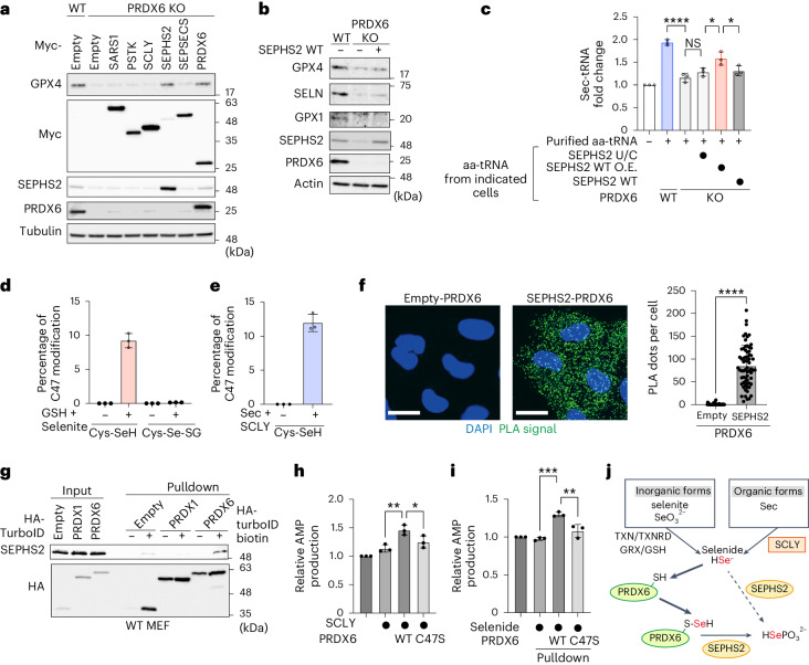
Ferroptosis is a form of regulated cell death induced by iron-dependent accumulation of lipid hydroperoxides. Selenoprotein glutathione peroxidase 4 (GPX4) suppresses ferroptosis by detoxifying lipid hydroperoxides via a catalytic selenocysteine (Sec) residue. Sec, the genetically encoded 21st amino acid, is biosynthesized from a reactive selenium donor on its cognate tRNA[Ser]Sec. It is thought that intracellular selenium must be delivered 'safely' and 'efficiently' by a carrier protein owing to its high reactivity and very low concentrations. Here, we identified peroxiredoxin 6 (PRDX6) as a novel selenoprotein synthesis factor. Loss of PRDX6 decreases the expression of selenoproteins and induces ferroptosis via a reduction in GPX4. Mechanistically, PRDX6 increases the efficiency of intracellular selenium utilization by transferring selenium between proteins within the selenocysteyl-tRNA[Ser]Sec synthesis machinery, leading to efficient synthesis of selenocysteyl-tRNA[Ser]Sec. These findings highlight previously unidentified selenium metabolic systems and provide new insights into ferroptosis.


Conflict of interest statement

The authors declare no competing interests.

Figures

Fig. 1
Fig. 1. Genome-wide CRISPR screening for iron-triggered ferroptosis.
a, WT or FBXL5 KO MEFs were treated with FAC (100 μg ml−1) for the indicated periods, and cell lysates were analyzed by immunoblotting. Data are representative of three independent experiments. b, Viability of WT or FBXL5 KO MEFs treated for 48 h with FAC (100 μg ml−1) in the presence of liproxstatin-1 (0, 1, 3 or 5 μM). c, Schematic showing the CRISPR–Cas9 screening strategy. d, Volcano plots showing the regulatory genes involved in iron-triggered ferroptosis. The P value and fold change were generated by the MAGeCK test. e, Viability of FBXL5 KO cells expressing sgEmpty or sgRNAs targeting ACSL4 or LPCAT3; cells were treated with FAC (100 μg ml−1) for 48 h. f, Viability of FBXL5 KO cells expressing sgEmpty, sgControl or sgRNAs targeting PDSS2 or FSP1; cells were treated with FAC (50 μg ml−1) for 30 h. Viability data (in b, e, f) are presented as the mean ± s.d. of three biological replicates. Source data
Fig. 2
Fig. 2. PRDX6 suppresses iron-triggered ferroptosis indirectly by augmenting expression of GPX4.
a, Immunoblot analysis of lysates from cells expressing sgEmpty, sgControl or sgRNAs targeting PRDX6. Data are representative of three independent experiments. bd, Viability of control cells or PRDX6 KO cells treated with FAC (50 or 100 μg ml−1) for 48 h (b), RSL3 (0.3 or 0.6 μM) for 24 h (c) or IKE (0.4 or 0.5 μM) for 24 h (d). e, Viability of control cells or PRDX6 KO cells stably expressing the GPX4 U/C mutant; cells were incubated with FAC (50 μg ml−1) for 48 h. f, Viability of PRDX6 KO cells stably expressing the indicated PRDX6 mutants incubated in the presence of FAC (50 μg ml−1) for 48 h. Immunoblot analysis of lysates from the indicated cells is also shown. PLA2, phospholipase A2. g, Measurement of GPX activity using recombinant proteins. Cumene hydroperoxide was used as a substrate. Data are presented as the mean ± s.e.m of three independent experiments. h, Immunoblot analysis of lysates from control or GPX4 KO cells stably expressing the Myc-PRDX6 or FSP1. Data are representative of two independent experiments. i, Continuous monitoring of the viability of the indicated cell lines after withdrawal of liproxstatin-1. Data are presented as the mean ± s.e.m of three biological replicates. j, Viability of the indicated cells in the presence of the indicated concentrations of IKE for 24 h. NS; not significant; nsP (IKE 0.75 μM, P = 0.8885; IKE 1 μM, P > 0.9999); two-way ANOVA. Viability data (in bf, j) are presented as the mean ± s.d. of three biological replicates. Source data
Fig. 3
Fig. 3. PRDX6 is involved in selenoprotein synthesis.
a, Co-essentiality network analysis of PRDX6 using FIREWORKS. b, Immunoblot analysis of lysates from cells expressing sgEmpty, sgControl or sgRNAs targeting PRDX6. Data are representative of three independent experiments. c, Immunoblot analysis of lysates from control or PRDX6 KO cells stably expressing the PRDX6 WT or C47S mutant. Data are representative of three independent experiments. d, Schematic showing the constructs of model selenoproteins (left). Immunoblot analysis of lysates from cells stably expressing Myc-GFP C70U or S175U in control cells or cells in which the indicated genes have been knocked out (middle). Quantification of the abundance of selenoproteins (right). Data are presented as the mean ± s.d. of three independent experiments. ***P < 0.001 (P = 0.0001); ****P < 0.0001; one-way ANOVA. Source data
Fig. 4
Fig. 4. PRDX6 augments efficient selenium utilization.
a, Immunoblot analysis of lysates from control or the indicated KO cells cultured for 32 h in the presence of sodium selenite (1 μM) or (Sec)2 (1 μM). Data are representative of two independent experiments. b,c, WT or PRDX6 KO cells were pretreated for 2 days with (Sec)2 (100 nM) or sodium selenite (100 nM). Then, cells were treated for 48 h with FAC (50, 100 μg ml−1) in the presence of (Sec)2 (100 nM) (b) or sodium selenite (100 nM) (c), and viability was measured. Viability data are presented as the mean ± s.d. of three biological replicates. df, Time course of selenoprotein expression by WT or PRDX6 KO MEFs in the presence of 50 nM (Sec)2 (d), 100 nM sodium selenite (e) or 0.5 μg ml−1 SELENOP (f). Data are representative of two (in f) or three (in d and e) independent experiments. g, Schematic showing the in vitro reconstruction system used to evaluate the amount of Sec-tRNA[Ser]Sec. h,i, aa-tRNAs, which were purified from the indicated cells treated (or not) for 105 min with 50 nM (Sec)2, followed by treatment for 15 min with cycloheximide, were added to wheat germ extract in the presence or absence of eEFSec and SBP2. Data of aa-tRNAs from WT or SEPHS2 KO cells (h), and WT or PRDX6 KO cells (i) are shown. Data are presented as the mean ± s.d. of three independent experiments. **P < 0.01 (WT ± eEFSec and SBP2, P = 0.0037; PRDX6 WT versus KO, P = 0.0031); ****P < 0.0001; two-way ANOVA. Source data
Fig. 5
Fig. 5. PRDX6 is a selenide carrier protein that enables efficient selenium utilization.
a, Immunoblot analysis of lysates from control or PRDX6 KO cells stably expressing enzymes involved in Sec-tRNA[Ser]Sec synthesis. Data are representative of three independent experiments. b, Immunoblot analysis of lysates from control or PRDX6 KO cells stably expressing SEPHS2 WT at endogenous levels. Data are representative of three independent experiments. c, aa-tRNAs, purified from the indicated cells treated with cycloheximide for 15 min, were added to wheat germ extract in the presence of eEFSec and SBP2. Data are expressed as the mean ± s.d. of three independent experiments. ****P < 0.0001; *P < 0.05 (SEPHS2 U/C versus SEPHS2 WT O.E., P = 0.0213; SEPHS2 WT O.E. versus SEPHS2 WT, P = 0.0422); NS (P = 0.6212); one-way ANOVA. d,e, Percentage of C47 modification as calculated by mass spectrometry analysis. PRDX6 was incubated with GSH and sodium selenite (d), or with Sec and SCLY (e) for 5 min before mass spectrometry analyses. Data are presented as the mean ± s.d of three biological replicates. f, The physical association between PRDX6 and SEPHS2 was detected in a PLA. Scale bars, 20 μm. Data are representative of three independent experiments. Quantification of the PLA dots is also shown. Data are presented as the mean ± s.d. (n = 42 cells (empty-PRDX6), n = 67 cells (SEPHS2-PRDX6) examined), ****P < 0.0001; unpaired two-sided t-test. g, MEFs expressing HA-TurboID-empty, -PRDX1 or -PRDX6 were cultured for 30 min with DMSO or 50 μM biotin. Cell lysates and pulldown samples were analyzed by immunoblotting. Data are representative of two independent experiments. h,i, PRDX6-mediated enhancement of selenophosphate-mediated synthesis of SEPHS2 was evaluated by measuring the AMP product. Data are presented as the mean ± s.d. of three independent experiments in which selenocysteine and SCLY were used as the selenium source; *P < 0.05 (P = 0.0423); **P < 0.01 (P = 0.004); one-way ANOVA (h) or selenium bound to PRDX6 was used as the selenium source; **P < 0.01 (P = 0.0018); ***P < 0.001 (P = 0.0001); one-way ANOVA (i). j, Schematic showing the role of PRDX6 as a selenide carrier. Source data
Extended Data Fig. 1
Extended Data Fig. 1. Characterization of iron-induced cell death in FBXL5 KO cells.
a, Phase-contrast images of cells after treatment with FAC (100 μg ml-1) for 48 h. Scale bars, 200 μm. Data are representative of three independent experiments. b, Fluorescence microscopy images of cells stained with a FerroOrange probe after treatment for 4 h with FAC (25 μg ml-1). Scale bars, 20 μm. Data are representative of three independent experiments. c, Flow cytometry analysis of lipid hydroperoxidation levels in WT or FBXL5 KO cells by C11-BODIPY staining after treatment with FAC (100 μg ml-1l) for 24 h. Data are representative of two independent experiments. d, WT MEFs were pretreated for 3 h with bafilomycin A1 (100 nM), followed by FAC (100 μg ml-1) for the indicated times. Cell lysates were analyzed by immunoblotting. Data are representative of two independent experiments. e, Viability of FBXL5 KO MEFs treated for 24 h with FAC (100 μg ml-1) in the presence of bafilomycin A1 (0, 10, 100 nM). Data are presented as the mean ± s.d. of three biological replicates. f, g, Immunoblot analysis of lysates from the indicated cells. Data (f) are representative of two independent experiments. Expression check of (g) is a single experiment. h, Volcano plots of iron-triggered ferroptosis screen, focusing on the mitochondrial electron transport chain (complex I and II: left, complex III and IV: right). The p value and fold change were generated by the MAGeCK test. Source data
Extended Data Fig. 2
Extended Data Fig. 2. Validation of hit genes, and identification of PRDX6 as a GPX4 regulator.
a, Hit suppressor genes [p-value cut off (p < 0.001)] were examined by gene ontology analysis. The p value was generated by the David gene ontology test. b, Negative score ranking of selenoproteins from the screening list. c, MAGeCK rank of suppressor genes. The p value and fold change were generated by the MAGeCK test. d, Immunoblot analysis of lysates from cells expressing sgRNAs targeting the indicated genes. Data are representative of two independent experiments. Source data
Extended Data Fig. 3
Extended Data Fig. 3. PRDX6 regulates ferroptosis by maintaining expression of GPX4.
a, Viability of FBXL5 KO cells or FBXL5/PRDX6 double knockout cells treated for 24 h with FAC (50 or 100 μg ml-1), and immunoblot analysis of lysates from the indicated cells. Viability data are presented as the mean ± s.d. of three biological replicates. b, WT or PRDX6 KO MEFs were treated with FAC (25 μg ml-1) for the indicated times, and cell lysates were analyzed by immunoblotting. Data are representative of two independent experiments. c, Flow cytometry analysis of lipid hydroperoxidation levels in WT and PRDX6 KO cells, as assessed by C11-BODIPY staining after treatment with FAC (50 μg ml-1) for 90 min. Data are representative of three independent experiments. d, Immunoblot analysis of lysates from control of PRDX6 KO cells expressing the GPX4 U/C mutant. Data are representative of two independent experiments. e, f, Viability of PRDX6 KO cells stably expressing the indicated PRDX6 mutants after incubation for 24 h in the presence of RSL3 (0.3 μM) (e) or IKE (0.3 μM) (f). Viability data are presented as the mean ± s.d. of three biological replicates. g, Measurement of GPX activity using recombinant proteins. Hydrogen peroxide (left) or tert-butyl hydroperoxide (right) were used as a substrate. Data are presented as the mean ± s.e.m of three independent experiments. h, Flow cytometry analysis of lipid hydroperoxidation after C11-BODIPY staining of WT or GPX4 KO cells stably expressing PRDX6 after removal of liiproxstatin-1 for 24 h. Data are representative of two independent experiments. i, Immunoblot analysis of lysates from the indicated cells. Data are representative of two independent experiments. Source data
Extended Data Fig. 4
Extended Data Fig. 4. PRDX6 regulates expression of selenoproteins.
a, Quantification of GPX4 mRNA levels by RT-qPCR. Data are expressed as the mean ± s.d of three biological replicates. ns; not significant; P (sgControl vs. sgPRDX6 #1 = 0.9523; sgControl vs. sgPRDX6 #2 = 0.9607); one-way ANOVA. b, c, Immunoblot analysis of lysates from control or PRDX6 KO cells incubated for 4 or 8 h in the presence of E64d/pep (10 μg ml-1) (b) or MG-132 (10 μM) (c). d, Immunoblot analysis of lysates from control or PRDX6 KO cells derived from the indicated human cancer cell lines (Hela, A549, H226, HepG2, NB-1). e, Immunoblot analysis of lysates from control cells or cells with KO of the indicated genes and stably expressing Myc-GFP WT. Data (b-e) are representative of two independent experiments. Source data
Extended Data Fig. 5
Extended Data Fig. 5. PRDX6 regulates selenium utilization efficiency.
a, Schematic showing selenium metabolism and the selenoprotein synthesis pathway. b, c, WT or PRDX6 KO cells were pretreated for 2 days with (Sec)2 (100 nM) or sodium selenite (100 nM). Then, cells were treated for 24 h with RSL3 (0.3 or 0.6 μM) (b) or IKE (0.3 or 0.4 μM) (c) in the presence of 100 nM (Sec)2 or 100 nM sodium selenite. d, Immunoblot analysis of lysates from control or PRDX6 KO Hela cells cultured for 48 h in the presence of sodium selenite (100 nM) or (Sec)2 (100 nM). e, WT or PRDX6 KO Hela cells were pretreated with (Sec)2 (100 nM) or sodium selenite (100 nM) for 2 days. Then, cells were treated for 48 h with FAC (100 or 200 μg ml-1) in the presence of (Sec)2 (100 nM) or sodium selenite (100 nM), and cell viability was measured. f, g, Immunoblot analysis of lysates from control or PRDX6 KO cells cultured for 32 h in the presence of the indicated concentrations of (Sec)2 (f) or sodium selenite (g). h, WT or PRDX6 KO cells were incubated for 24 h with 1% FBS in the presence of liproxstatin-1. Then, cells were treated with 50 nM (Sec)2 or 100 nM sodium selenite for the indicated times. Cell lysates were analyzed by immunoblotting. i, PRDX6 KO cells were preincubated for 36 h with 10 nM (Sec)2 or 40 nM sodium selenite. Then, WT or PRDX6 KO cells pretreated with selenium were exposed to 50 nM (Sec)2 or 200 nM sodium selenite for the indicated periods and the lysates were analyzed by immunoblotting. Data are representative of two (d, h, i) or three (f, g) independent experiments. Viability data (b, c, e) are presented as the mean ± s.d. of three biological replicates. Source data
Extended Data Fig. 6
Extended Data Fig. 6. Overexpression of SEPHS2 WT rescues the PRDX6 KO phenotype.
a, Immunoblot analysis of lysates from control or SEPHS2 KO cells stably expressing the SEPHS2 U/C mutant. b, Immunoblot analysis of lysates from control or PRDX6 KO cells stably expressing the SEPHS2 WT or U/C mutant. c, Immunoblot analysis of lysates from cells stably expressing Myc-GFP C70U or S175U in control or PRDX6 KO cells stably expressing the SEPHS2 WT or U/C mutant. Data (a-c) are representative of two independent experiments. d-f, Viability of the indicated cells in the presence of FAC (50 or 100 μg ml-1) for 48 h (d), or RSL3 (0.3 or 0.6 μM) (e) or IKE (0.3 or 0.45 μM) (f) for 24 h. Viability data are presented as the mean ± s.d. of three biological replicates. g, Viability of the indicated cells treated for 48 h with (Sec)2 (10 μM) and liproxstatin-1 (3 μM). Viability data are presented as the mean ± s.d. of three biological replicates. *P (PRDX6 WT vs. C47S = 0.0243; WT vs. sgSEPHS2 = 0.0238) < 0.05; one-way ANOVA. h, Coomassie-stained SDS-PAGE gel showing the recombinant PRDX6 WT or C47S mutant used for ICP-MS analysis. ICP-MS analysis of the amount of selenium bound to recombinant PRDX6 WT or C47S. Data are presented as the mean ± s.d. of three independent experiments. *P (=0.0141) < 0.05, ***P (=0.0003) < 0.001; one-way ANOVA. Source data
Extended Data Fig. 7
Extended Data Fig. 7. Mass spectrometry analysis of PRDX6 C47 modification.
a, b, Mass spectrum of PRDX6 C47. PRDX6 (75 μM) was incubated with GSH (300 μM) and sodium selenite (75 μM) (a), or with Sec (75 μM) and SCLY (1.5 μM) (b) for 5 min prior to mass spectrometry analyses.
Extended Data Fig. 8
Extended Data Fig. 8. Interaction between PRDX6 and SEPHS2 or SCLY.
a, The physical association between PRDX6 and SCLY was detected in a PLA assay. Scale bars, 20 μm. Data are representative of three independent experiments. Quantification of the PLA dots is also shown. Data are presented as the mean ± s.d. [n = 52 cells (Empty-PRDX6), n = 50 cells (SCLY-PRDX6) examined], ****P < 0.0001; unpaired two-sided t-test. b, c PRDX6 KO MEFs expressing HA-TurboID-empty, -PRDX1, or -PRDX6 were cultured for 30 min with DMSO or 50 μM biotin. Cell lysates and pulldown samples were analyzed by immunoblotting. The association between PRDX6 and SEPHS2 (b) or between PRDX6 and SCLY (c) was analyzed. Data are representative of two (c) or three (b) independent experiments. Source data
Extended Data Fig. 9
Extended Data Fig. 9. Expression of PRDX6 is associated with a poor prognosis for several cancers.
a, Expression profile of PRDX6 in several types of normal and tumor tissue. Data were obtained from TNMplot (https://tnmplot.com/analysis/), which is a web server that stores data regarding expression of normal and cancer-related genes. Those with higher values in tumor tissue compared to normal tissue and with significant differences (Mann-Whitney U test) are marked in red (P < 0.01). b, Kaplan-Meier survival curves comparing cancer samples showing high PRDX6 expression with samples showing low PRDX6 expression (curves were constructed using Kaplan-Meier plotter).
Extended Data Fig. 10
Extended Data Fig. 10. Expression of PRDX6 is important for survival of cancer cell lines.
a, Immunoblot analysis of lysates from WT or PRDX6 KO Panc-1 cells. b, Continuous monitoring of the viability of WT or PRDX6 KO Panc-1 cells in the presence or absence of sodium selenite (100 nM), (Sec)2 (100 nM), or liproxstatin-1 (1 μM). c, Immunoblot analysis of lysates from WT or PRDX6 KO MIA PaCa-2 cells. d, Continuous monitoring of the viability of WT or PRDX6 KO MIA PaCa-2 cells in the presence or absence of sodium selenite (100 nM), (Sec)2 (100 nM), or liproxstatin-1 (1 μM). e, Immunoblot analysis of lysates from indicated cell lines. f, Viability of indicated cell lines in the presence of FAC (50 or 100 μg ml-1) for 48 h. Viability data are presented as the mean ± s.d. of three biological replicates. g, Immunoblot analysis of lysates from WT or PRDX6 KO SK-N-DZ cells. h, Continuous monitoring of the viability of WT or PRDX6 KO SK-N-DZ cells in the presence or absence of sodium selenite (100 nM), (Sec)2 (100 nM), or liproxstatin-1 (1 μM). Viability data (b, d, h) are presented as the mean ± s.e.m of three biological replicates. Data (a, c, e, g) are representative of two independent experiments. Source data

References

    1. Jiang, X., Stockwell, B. R. & Conrad, M. Ferroptosis: mechanisms, biology and role in disease. Nat. Rev. Mol. Cell Biol.22, 266–282 (2021). 10.1038/s41580-020-00324-8 - DOI - PMC - PubMed
    1. Lei, G., Zhuang, L. & Gan, B. Targeting ferroptosis as a vulnerability in cancer. Nat. Rev. Cancer22, 381–396 (2022). 10.1038/s41568-022-00459-0 - DOI - PMC - PubMed
    1. Yang, W. S. et al. Regulation of ferroptotic cancer cell death by GPX4. Cell156, 317–331 (2014). 10.1016/j.cell.2013.12.010 - DOI - PMC - PubMed
    1. Ingold, I. et al. Selenium utilization by GPX4 is required to prevent hydroperoxide-induced ferroptosis. Cell172, 409–422.e21 (2018). 10.1016/j.cell.2017.11.048 - DOI - PubMed
    1. Friedmann Angeli, J. P. et al. Inactivation of the ferroptosis regulator Gpx4 triggers acute renal failure in mice. Nat. Cell Biol.16, 1180–1191 (2014). 10.1038/ncb3064 - DOI - PMC - PubMed