. 2024 Jun 12;25(1):239.
doi: 10.1186/s12931-024-02864-5.

Clinical, radiological and histopathological features of patients with familial pulmonary fibrosis

Affiliations

Clinical, radiological and histopathological features of patients with familial pulmonary fibrosis

Hanna Jaula et al. Respir Res. .

Abstract

Background: In familial pulmonary fibrosis (FPF) at least two biological relatives are affected. Patients with FPF have diverse clinical features.

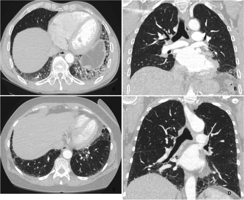
Research question: We aimed to characterize demographic and clinical features, re-evaluate high-resolution computed tomography (HRCT) scans and histopathology of surgical lung biopsies, assess survival and investigate the suitability of risk prediction models for FPF patients.

Study design: A retrospective cohort study.

Methods: FPF data (n = 68) were collected from the medical records of Oulu University Hospital (OUH) and Oulaskangas District Hospital between 1 Jan 2000 and 11 Jan 2023. The inclusion criterion was pulmonary fibrosis (PF) (ICD 10-code J84.X) and at least one self-reported relative with PF. Clinical information was gathered from hospital medical records. HRCT scans and histology were re-evaluated.

Results: Thirty-seven (54.4%) of the patients were men, and 31 (45.6%) were women. The mean ages of the women and men were 68.6 and 61.7 years, respectively (p = 0.003). Thirty-seven (54.4%) patients were nonsmokers. The most common radiological patterns were usual interstitial pneumonia (UIP) (51/75.0%), unclassifiable (8/11.8%) and nonspecific interstitial pneumonia (NSIP) (3/4.4%). Pleuroparenchymal fibroelastosis (PPFE) was observed as a single or combined pattern in 13.2% of the patients. According to the 2022 guidelines for idiopathic pulmonary fibrosis (IPF), the patients were categorized as UIP (31/45.6%), probable UIP (20/29.4%), indeterminate for UIP (7/10.3%) or alternative diagnosis (10/14.7%). The histopathological patterns were UIP (7/41.2%), probable UIP (1/5.9%), indeterminate for UIP (8/47.2%) and alternative diagnosis (1/5.9%). Rare genetic variants were found in 9 patients; these included telomerase reverse transcriptase (TERT, n = 6), telomerase RNA component (TERC, n = 2) and regulator of telomere elongation helicase 1 (RTEL1, n = 1). Half of the patients died (n = 29) or underwent lung transplantation (n = 5), with a median survival of 39.9 months. The risk prediction models composite physiology index (CPI), hazard ratio (HR) 1.07 (95.0% CI 1.04-1.10), and gender-age-physiology index (GAP) stage I predicted survival statistically significantly (p<0.001) compared to combined stages II and III.

Conclusions: This study confirmed the results of earlier studies showing that FPF patients' radiological and histopathological patterns are diverse. Moreover, radiological and histological features revealed unusual patterns and their combinations.

Keywords: Comorbidity; Familial pulmonary fibrosis; Histopathology; Interstitial lung disease; Radiology; Risk prediction model; Survival.


Conflict of interest statement

The authors declare no competing interests.

Figures

Fig. 1
Fig. 1
Radiologic UIP patterns according to the 2022 idiopathic pulmonary fibrosis guidelines. UIP = usual interstitial pneumonia
Fig. 2
Fig. 2
The most common comorbidities. HC = Hypercholesterolemia, CAD = Coronary artery disease, HT = Hypertension, Diabetes (type I and II), GERD = gastroesophageal reflux disease, CTD = Connective Tissue Disease, OSA = Obstructive Sleep Apnea, PE = pulmonary embolism
Fig. 3
Fig. 3
High resolution computed tomography (HRCT) scans of two patients with the TERC variant. A-B: HRCT scan of patient number 1 shows a left lung predominant usual interstitial pneumonia pattern with basal and peripheral distribution, reticular abnormalities, traction bronchiectasis and honeycombing. C-D: HRCT scan of patient number 2 reveals fine reticular abnormalities and traction bronchiectasis without honeycombing with basal and peripheral distribution
Fig. 4
Fig. 4
Histological images from a patient with a TERC variant (no 1). In the upper left lobe, there were separate subpleural or paraseptal areas similar to those of pleuroparenchymal fibroelastosis (A) with thick elastic fibers (B). There were also a few centrilobular areas with slightly fibrotic alveolar walls (C) and focal chronic inflammation (D), as well as peribronchiolar aggregations of fibromyxoid connective tissue and macrophages (E) and poorly formed granulomas (F). In the lower left lobe, fibrosis was more prominent, with centrilobular bridging fibrosis (G) and architectural remodeling (H) with occasional fibroblast foci (arrow)
Fig. 5
Fig. 5
Histological images of the right lower lobe of the other patient with a TERC variant (no 2). There was wide architectural remodeling resembling UIP (A) with numerous fibroblast foci (insert). However, unlike in UIP, most of the bronchi and peribronchial tissue were heavily inflamed (B), many showed constricting fibrosis (arrows) (C), and some were obliterated (D). UIP = usual interstitial pneumonia
Fig. 6
Fig. 6
Kaplan-Meier survival curves (A) GAP 1 (GAP stage I) vs. GAP 2 (combined GAP stages II and III). B CPI groups as median value 38 was set as the cut-off. C UIP vs. non-UIP group. D IPF vs. non-IPF group. CPI = composite physiology index, GAP = gender-age-physiology -index, IPF = idiopathic pulmonary fibrosis, UIP = usual interstitial pneumonia

References

    1. Zhang D, Newton CA. Familial Pulmonary Fibrosis: Genetic Features and Clinical Implications. Chest. 2021;160(5):1764-73. 10.1016/j.chest.2021.06.037. - PMC - PubMed
    1. Steele MP, Speer MC, Loyd JE, et al. Clinical and pathologic features of familial interstitial pneumonia. Am J Respir Crit Care Med. 2005;172(9):1146–1152. doi: 10.1164/RCCM.200408-1104OC/SUPPL_FILE/ONLINE_DATA_SUPPLEMENT_BLUE_200408_11040C_R3.PDF. - DOI - PMC - PubMed
    1. García-Sancho C, Buendía-Roldán I, Fernández-Plata MR, et al. Familial pulmonary fibrosis is the strongest risk factor for idiopathic pulmonary fibrosis. Respir Med. 2011;105(12):1902–1907. doi: 10.1016/j.rmed.2011.08.022. - DOI - PubMed
    1. Cutting CC, Bowman WS, Dao N, et al. Family History of Pulmonary Fibrosis Predicts Worse Survival in Patients With Interstitial Lung Disease. In: Chest. Vol 159. Elsevier Inc.; 2021:1913-1921. 10.1016/j.chest.2021.01.026. - PMC - PubMed
    1. Krauss E, Gehrken G, Drakopanagiotakis F, et al. Clinical characteristics of patients with familial idiopathic pulmonary fibrosis (f-IPF) BMC Pulm Med. 2019;19(1):130. doi: 10.1186/s12890-019-0895-6. - DOI - PMC - PubMed