. 2024 Jun 13;22(1):135.
doi: 10.1186/s12915-024-01925-x.

The venom and telopodal defence systems of the centipede Lithobius forficatus are functionally convergent serial homologues

Affiliations

The venom and telopodal defence systems of the centipede Lithobius forficatus are functionally convergent serial homologues

Vanessa Schendel et al. BMC Biol. .

Abstract

Background: Evolution of novelty is a central theme in evolutionary biology, yet studying the origins of traits with an apparently discontinuous origin remains a major challenge. Venom systems are a well-suited model for the study of this phenomenon because they capture several aspects of novelty across multiple levels of biological complexity. However, while there is some knowledge on the evolution of individual toxins, not much is known about the evolution of venom systems as a whole. One way of shedding light on the evolution of new traits is to investigate less specialised serial homologues, i.e. repeated traits in an organism that share a developmental origin. This approach can be particularly informative in animals with repetitive body segments, such as centipedes.

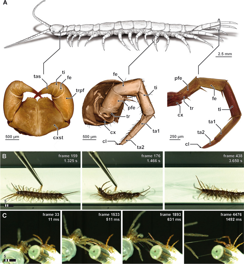
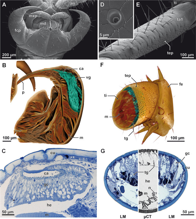
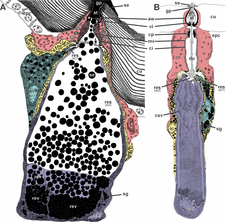
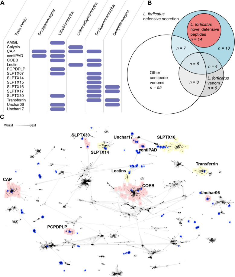
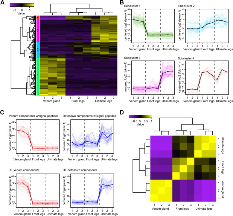
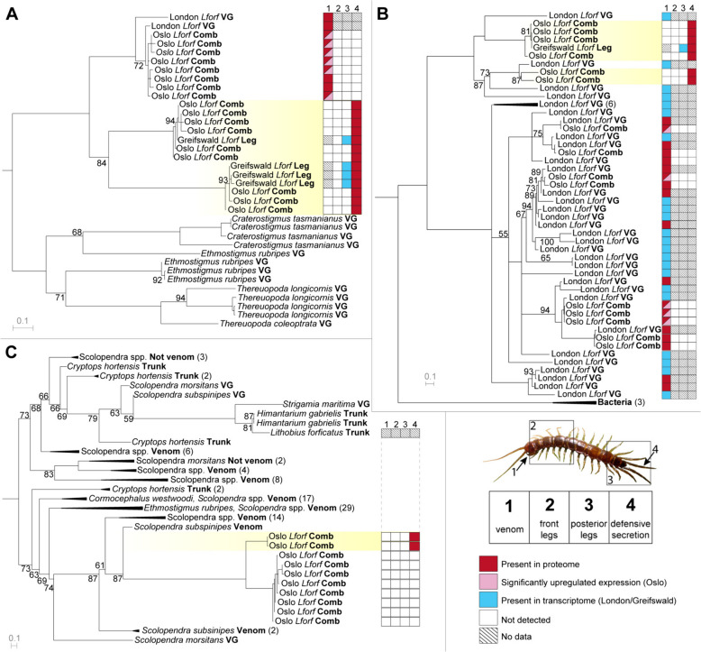
Results: Here, we investigate morphological and biochemical aspects of the defensive telopodal glandular organs borne on the posterior legs of venomous stone centipedes (Lithobiomorpha), using a multimethod approach, including behavioural observations, comparative morphology, proteomics, comparative transcriptomics and molecular phylogenetics. We show that the anterior venom system and posterior telopodal defence system are functionally convergent serial homologues, where one (telopodal defence) represents a model for the putative early evolutionary state of the other (venom). Venom glands and telopodal glandular organs appear to have evolved from the same type of epidermal gland (four-cell recto-canal type) and while the telopodal defensive secretion shares a great degree of compositional overlap with centipede venoms in general, these similarities arose predominantly through convergent recruitment of distantly related toxin-like components. Both systems are composed of elements predisposed to functional innovation across levels of biological complexity that range from proteins to glands, demonstrating clear parallels between molecular and morphological traits in the properties that facilitate the evolution of novelty.

Conclusions: The evolution of the lithobiomorph telopodal defence system provides indirect empirical support for the plausibility of the hypothesised evolutionary origin of the centipede venom system, which occurred through functional innovation and gradual specialisation of existing epidermal glands. Our results thus exemplify how continuous transformation and functional innovation can drive the apparent discontinuous emergence of novelties on higher levels of biological complexity.

Keywords: Arthropoda; Chilopoda; Epidermal exocrine glands; Evolution; Innovation; Myriapoda; Novelty; Telopodal glandular organs; Venom.


Conflict of interest statement

The authors declare that they have no competing interests.

Figures

Fig. 1
Fig. 1
Habitus and defensive display of Lithobius forficatus. A Habitus of L. forficatus and three typical, serially homologous appendages: forcipules (left), locomotory leg 10 (centre) and the ultimate legs (right). 3D volume renderings based on microCT analyses, not to scale. B Single frames from high-speed footage (compare Additional file 1) showing the stereotypical defensive display using the tip of a brush. The fast up and down movements of the ultimate legs might also induce or support the distribution of the secretion (compare Additional file 1). Upon contact (left) ultimate and penultimate legs soar up within a tenth of a second in order to present the ventral and medioventral faces of the legs towards the point of irritation (middle). The upright posture of the ultimate legs is maintained for a couple of seconds while the penultimate legs are lowered in order to assist in the escape movement (right). C Single frames from high-speed footage (compare Additional file 2) showing the staged encounter of L. forficatus (right) and a male lycosid spider (left). Already shortly before contact, L. forficatus raises leg pairs 13–15 (leg 12 and more anterior legs were strapped down; left and middle left). Upon contact, beads-on-a-string-like threads become visible, emanating from and connecting ultimate legs and the spider (middle right and right). cl claw, cx coxa, cxst coxosternite, fe femur, pfe prefemur, ta1 tarsus 1, ta2 tarsus 2, tas tarsungulum, ti tibia, tr trochanter, trpf trochanteroprefemur
Fig. 2
Fig. 2
The telopodal glandular organs share a common organisation with venom glands. Morphology and glandular histology of the forcipule (AC) and the ultimate leg (DG). SEM micrographs of the ventral head with (A) forcipules and (E) the ventral aspect of the right ultimate leg tarsus 1 (ta1). The lateral face of the tarsus is covered by trichoid sensilla of various lengths, while the medial face is covered by pores of telopodal gland units (tep). D Close-up of a pore of a telopodal gland unit showing its non-symmetrical morphology: the shallow part of the pore always faces towards the distal tip of the leg. B 3D volume rendering of the left forcipule with labelled venom gland (vg) and associated calyx (ca), and F of the proximal femur (fe) and distal tibia (ti) of the ultimate leg with telopodal gland units (tg; medial face to front). Glandular tissue in both appendages highlighted in turquoise. Note that critical point drying affected the structural integrity of the telopodal glandular tissue (compare also shrinkage in G). C Longitudinal section of the forcipule and calyx (ca) showing the densely packed venom gland units (gc) individually connected to the cuticular calyx. G Cross section of the ultimate leg tibia, medial face to top. Blue-stained aspects based on histological sections, grey-stained aspect based on microCT analysis. After chemical fixation and sectioning, the glandular epithelium takes up approx. 70% of the leg volume. an antenna, ca calyx, cu cuticle, fcp forcipule, fe femur, gc venom, gland units, he hemolymphatic space, m musculature, mxp maxillar palp, md mandible, n nerve, p pore of the venom duct, sc secretory cell, ta1 tarsus 1, tep telopodal gland pore, ti tibia, tg telopodal gland, vg venom gland. Scalebars in µm
Fig. 3
Fig. 3
Semi-schematic reconstructions of a telopodal gland unit and a venom gland unit of Lithobius forficatus, analysed by TEM. A Telopodal gland unit and B venom gland unit cut along the medio-longitudinal plane. Each four-cell gland is composed of a canal cell (red), an intermediary cell (yellow) and two different types of secretory cells (sc1: turquoise, sc2: violet). The sc2 in the telopodal gland unit (A) is shown in active secreting state indicated by remains of the ripped apical membrane (black arrow), the content of the distalmost reservoir vacuole is released into the conducting canal, the vacuole-enclosed space becomes part of the then massively enlarged reservoir of the sc2. The venom gland unit (B) is drawn in dormant state indicated by the intact apical membrane of the sc2 (white arrow) separating the duct from the central (reservoir) secretory vacuole containing the secretion to be discharged. Most basal components such as the extracellular matrix, tracheae or associated parts of the nervous system are not shown in this reconstruction. Details of the lamellar system of the surrounding cuticle are also omitted in B. at atrium, aw atrial wall, cc canal cell, ci cuticular intima (lining the conducting canal), cp cuticular pad, csv central secretory (reservoir) vacuole, cu cuticle, cys cytoplasmic sheath of the type 2 secretory cell, du conducting canal (= duct), epc epidermal cell, ic intermediary cell, gp gland pore, mv border of microvilli formed by canal cell (connected to brush of microtubules), res reservoir, rev reservoir vacuole, sc1 type 1 secretory cell, sc2 type 2 secretory cell, sd secretion droplet, sg secretory granule, se discharged (amorphous) secretion, vl cuticular valve
Fig. 4
Fig. 4
The telopodal defensive secretion shares compositional similarities with centipede venom. A 16 of the protein and peptide families identified in the defensive secretion are also found in the venoms of centipedes. Dark blue bars indicate previously identified venom components. B Total compositional overlap between protein and peptide families from all centipede venoms except L. forficatus venom, and L. forficatus telopodal defensive secretion, with the proportion of unidentifiable families from the defensive secretion shown in red. C Pairwise blastp-based clustering analysis of all centipede venom components (black dots) and telopodal defensive secretion components (blue stars) shows that the defensive components cluster with members of putative centipede venom toxin families. Families previously described from L. forficatus venom are highlighted in red, while families not previously described from L. forficatus venom are highlighted in yellow
Fig. 5
Fig. 5
The transcriptional divergence of forcipules and ultimate legs from anterior walking legs. A Hierarchical clustering of differentially expressed genes (P < .05, C = 4) and grouping at half the length of the resulting tree yields four subclusters. Median-centred log2 FPKM expression values are shown as heatmaps and subclusters are indicated by coloured bars on the left side of the map. B Each subcluster shows distinct expression patterns across the forcipules, anterior walking legs and ultimate legs. The colours of lines in each graph correspond to those of the subclusters indicated in A. C The expression patterns of all venom (red) and telopodal defensive secretion (blue) components with signal peptides (top) are similar to components with significant differential expression (bottom). D Hierarchical clustering of samples based on correlations of differentially expressed genes show that ultimate and anterior legs are most similar, while ultimate legs and forcipules are least similar
Fig. 6
Fig. 6
The molecular evolution of toxin-like telopodal defensive secretion components. A Reconstruction of the centipede CAP1 family by maximum likelihood (ML; under WAG + I + G4) shows components of the telopodal defensive secretion form two clades that probably represent separate paralogues and that are separate from the clade containing the likely ancestral L. forficatus venom components. B Reconstruction of the lithobiid PCPDPLP family by ML (under WAG + F + G4) shows that the venom and telopodal defensive secretion components form distinct clades but not whether this family was first recruited into the venom or telopodal defensive secretion. For phylogenetic tree without collapsed clades see Additional file 3, Fig. S17. C Reconstruction of the centipede SLPTX15 family by ML (under VT + I + I + R3) shows components of the telopodal defensive secretion do not group with the L. forficatus trunk sequence, but instead form a clade within venom SLPTX15 from Scolopendridae. For phylogenetic tree without collapsed clades, see Additional file 3, Fig. S18. Trees are shown as midpoint rooted, while bootstrap support values < 95 are shown at each node and nodes with support < 50 are collapsed. Sequences from this study contained in clades with identified components of the telopodal secretion are highlighted in yellow, while the presence and absence in proteomes and transcriptomes of each sequence from L. forficatus are indicated in the boxes behind each sequence name according to the key in the lower right panel. Tissue sources are indicated in bold for each sequence: VG indicates venom gland while Comb indicates transcriptome assembly from pooled tissues (see “Methods”). In addition, venom indicates sequences previously detected in venom while Not venom indicates sequences previously found to be part of non-venom orthogroups

Similar articles

Cited by

References

    1. Difrisco J, Love AC, Wagner GP, Wetenschappelijk F, John O. The hierarchical basis of serial homology and evolutionary novelty. J Morphol. 2023;284:e21531. doi: 10.1002/jmor.21531. - DOI - PubMed
    1. Hall BK, Kerney R. Levels of biological organization and the origin of novelty. J Exp Zool B Mol Dev Evol. 2012;318:428–437. doi: 10.1002/jez.b.21425. - DOI - PubMed
    1. Pigliucci M. What, if anything, is an evolutionary novelty? Philos Sci. 2008;75:887–898. doi: 10.1086/594532. - DOI
    1. Casewell NR, Wüster W, Vonk FJ, Harrison RA, Fry BG. Complex cocktails: the evolutionary novelty of venoms. Trends Ecol Evol. 2013;28:219–229. doi: 10.1016/j.tree.2012.10.020. - DOI - PubMed
    1. DiFrisco J, Wagner GP, Love AC. Reframing research on evolutionary novelty and co-option: character identity mechanisms versus deep homology. Semin Cell Dev Biol. 2023;145:3–12. doi: 10.1016/j.semcdb.2022.03.030. - DOI - PubMed

Substances

LinkOut - more resources