. 2024 Jun 12;24(1):207.
doi: 10.1186/s12935-024-03384-4.

TEX19 increases the levels of CDK4 and promotes breast cancer by disrupting SKP2-mediated CDK4 ubiquitination

Affiliations

TEX19 increases the levels of CDK4 and promotes breast cancer by disrupting SKP2-mediated CDK4 ubiquitination

Huantao Liu et al. Cancer Cell Int. .

Abstract

Background: Globally, breast cancer in women is the fifth leading cause of cancer death. There is an urgent need to explore the molecular mechanism of breast cancer proliferation and metastasis.

Method: TCGA database analysis was used to analyze genes expression in breast cancer and normal samples and the association between gene expression and prognosis. Immunohistochemical staining, qPCR and western blotting was sued to detected gene expression. The cell function tests were conducted to investigate the effects of TEX19 and CDK4 with abnormal expression on cell proliferation, migration, apoptosis, cell cycle, and colony formation. Bioinformatics analysis methods combined with CHX tracking experiment and Co-IP experiment were performed to screen and verify the downstream molecule and regulatory mechanism of TEX19. Besides, subcutaneous tumorigenesis model in nude mice was constructed.

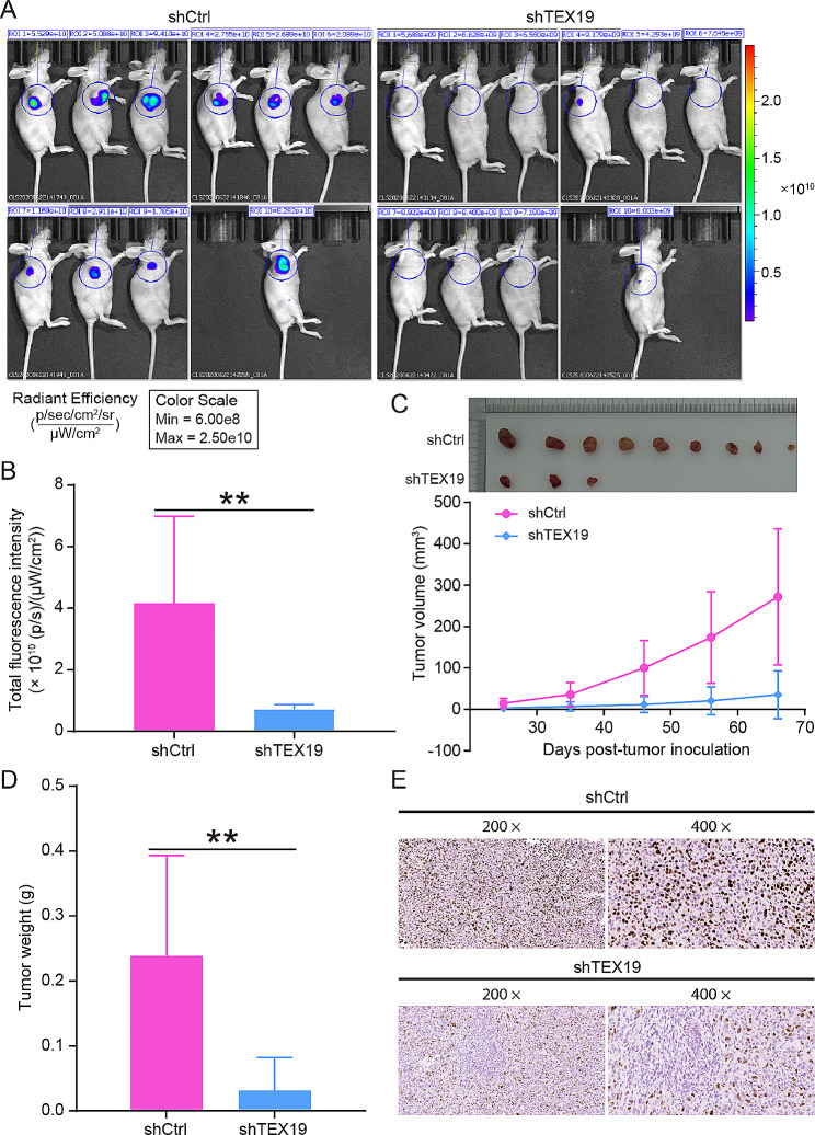
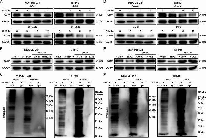
Results: TEX19 was significantly upregulated in breast cancer, and the TEX19 level was related to tumor invasion and prognosis. TEX19 knockdown inhibited the proliferation and migration of breast cancer cells, increased cell apoptosis, and blocked the cell cycle in the G2 phase. Besides, TEX19 suppressed the growth of tumors in the body. Mechanically, TEX19 upregulated the level of CDK4 protein, which depended on the E3 ubiquitin ligase SKP2. Specifically, TEX19 knockdown and SKP2 protein overexpression destroyed the stability of CDK4 protein and enhanced the ubiquitination of CDK4 protein. Additionally, CDK4 knockdown inhibited the proliferation, migration, and colony formation of breast cancer cells, and alleviated the promotion of TEX19 overexpression on the proliferation and migration of breast cancer cell.

Conclusion: TEX19 and CDK4 were upregulated in breast cancer, and TEX19 increased the level of CDK4 protein by influencing SKP2-mediated ubiquitination of CDK4, thereby promoting the progression of breast cancer.

Keywords: Breast cancer; CDK4; TEX19; Ubiquitination.


Conflict of interest statement

The authors declare no competing interests.

Figures

Fig. 1
Fig. 1
TEX19 was highly expressed in breast cancer. (A) The expression of TEX19 in the TCGA database was significantly higher in breast cancer than that in normal tissues. (B) The expression of TEX19 in the TCGA database was markedly different among different tumor stages of breast cancer patients. (C) The expression of TEX19 in the TCGA database was obviously different among different N grades of breast cancer. (D) The expression of TEX19 in the TCGA database was obviously different among different T grades of breast cancer. (E) The expression of TEX19 was divided into high expression group and low expression based on the optimal cut point, and then the difference of overall survival between the two groups was tested by log rank test. (F) The difference of progression-free survival between the high expression group of TEX19 or the low expression group of TEX19 was tested by log rank test. (G) qPCR and western blotting were performed to detect the mRNA and protein levels of TEX19 in MCF-10 A and breast cancer cell lines. (H) Immunohistochemical staining was carried out to assess the levels of TEX19 protein in breast cancer tissues and para-carcinoma tissues. The magnification: 200 × and 400 ×. (I) Kaplan-Meier analysis was used to evaluate the association between TEX19 levels and prognosis in breast cancer patients. *P < 0.05, ***P < 0.001
Fig. 2
Fig. 2
TEX19 knockdown inhibited proliferation and migration but induced apoptosis of MDA-MB-231 and BT549 cells. (A) The proliferation of MDA-MB-231 and BT549 cells was detected by Celigo cell counting assay after TEX19 knockdown, and it was found that TEX19 knockdown slowed down the cell proliferation rate. (B) Flow cytometry was used to determine the apoptosis of MDA-MB-231 and BT549 cells after TEX19 silence. (C) The effect of shTEX19 on cell cycle MDA-MB-231 and BT549 cells was demonstrated using Flow cytometry. (D) Transwell was performed to evaluate the migration ability of MDA-MB-231 and BT549 cells after TEX19 silence. (E) Wound healing was performed to evaluate the migration ability of MDA-MB-231 and BT549 cells after TEX19 silence. **P < 0.01, ***P < 0.001. These cell assays were repeated for 3 times
Fig. 3
Fig. 3
TEX19 downregulation suppressed the growth of tumors in vivo. (A-B) In vivo imaging showed the fluorescence intensity of tumors in nude mice, and the effects of TEX19 downregulation on tumor growth in vivo was analyzed. (C) Tumor volume growth curves in nude mice of shCtrl group and shTEX19 group. (D) Tumors taken from nude mice were weighted and found to be significantly lighter in the shTEX19 group compared with the shCtrl group. (E) The expression of Ki67 protein in solid tumor tissues in the shCtrl and shTEX19 groups was determined to assess the proliferation ability of tumor cells. The magnification: 200 × and 400 ×. **P < 0.01
Fig. 4
Fig. 4
TEX19 regulated CDK4 protein levels by interacting with SKP2. (A) IPA was used to conduct enrichment analysis of classical pathways to explore the enrichment of differential genes in classical pathways. (B) The molecular interaction network of TEX19, CDK4 and the classical pathways Cyclins and Cell Cycle Regulation, Estrogen-mediated S-phase Entry related proteins. (C) The expression of CDK4 in the TCGA database was significantly higher in breast cancer than that in normal tissues. (D) The difference of overall survival between the high expression group of CDK4 or the low expression group of CDK4 was tested by log rank test. (E) Immunohistochemical staining was carried out to assess the levels of CDK4 protein in breast cancer tissues and para-carcinoma tissues. The magnification: 200 × and 400 ×. (F) western blotting was conducted to detect the expression of related protein in the pathways (ERK, p-ERK, CCND1, CDK6, PIK3CA) in the MDA-MB-231 cells infected with shTEX19 lentivirus. (G) The ubiquitin ligase (E3) acting on CDK4 was predicted by UbiBrowser. (H) Co-IP assay was used to demonstrate the interaction between TEX19 and SKP2. (I) Co-IP assay was conducted to confirm the interaction between SKP2 and CDK4
Fig. 5
Fig. 5
TEX19 regulated the SKP2-involved ubiquitination of CDK4. (A) The MDA-MB-231 and BT549 cells were treated with or without knockdown of TEX19 with a protein synthesis inhibitor CHX, and then the levels of CDK4 protein were detected by western blotting after 0, 3, 6 and 12 h. (B) The proteasome inhibitor MG-132 was used to treat TEX19 knockdown MDA-MB-231 and BT549 cells, and the levels of CDK4 protein were detected by western blotting. (C) The TEX19 knockdown MDA-MB-231 and BT549 cells were treated with MG-132, and then the levels of CDK4 ubiquitination were assessed by Co-IP assay. (D) The breast cancer cells were treated with or without overexpression of SKP2 with CHX, and then the levels of CDK4 protein were detected by western blotting after 0, 3, 6 and 12 h. (E) MG-132 was treated SKP2 overexpression MDA-MB-231 and BT549 cells, and the levels of CDK4 protein were detected by western blotting. (F) The SKP2 overexpression breast cancer cells were treated with MG-132, and then the levels of CDK4 ubiquitination were assessed by Co-IP assay
Fig. 6
Fig. 6
TEX19 prompted the progression of breast cancer by interacting with CDK4. (A) The proliferation of MDA-MB-231 and BT549 cells was detected by CCK8 assay after genes overexpression or knockdown. (B) Transwell was performed to evaluate the migration ability of MDA-MB-231 and BT549 cells after genes overexpression or silence. (C) Colony formation assay was conducted to analyze the colony formation ability of MDA-MB-231 and BT549 cells after genes overexpression or silence. **P < 0.01, ***P < 0.001, compared to the NC group. #P < 0.05, ##P < 0.01, ###P < 0.001, compared to the TEX19 or shCDK4 group. These cell assays were repeated for 3 times

Similar articles

Cited by

References

    1. Sung H, Ferlay J, Siegel RL, Laversanne M, Soerjomataram I, Jemal A, et al. Global Cancer statistics 2020: GLOBOCAN estimates of incidence and Mortality Worldwide for 36 cancers in 185 countries. CA Cancer J Clin. 2021;71(3):209–49. doi: 10.3322/caac.21660. - DOI - PubMed
    1. Britt KL, Cuzick J, Phillips KA. Key steps for effective breast cancer prevention. Nat Rev Cancer. 2020;20(8):417–36. doi: 10.1038/s41568-020-0266-x. - DOI - PubMed
    1. Yin X, Wang P, Yang T, Li G, Teng X, Huang W, et al. Identification of key modules and genes associated with breast cancer prognosis using WGCNA and ceRNA network analysis. Aging. 2020;13(2):2519–38. doi: 10.18632/aging.202285. - DOI - PMC - PubMed
    1. Waks AG, Winer EP. Breast Cancer Treatment: a review. JAMA. 2019;321(3):288–300. doi: 10.1001/jama.2018.19323. - DOI - PubMed
    1. Liang Y, Zhang H, Song X, Yang Q. Metastatic heterogeneity of breast cancer: molecular mechanism and potential therapeutic targets. Semin Cancer Biol. 2020;60:14–27. doi: 10.1016/j.semcancer.2019.08.012. - DOI - PubMed

LinkOut - more resources