Review
. 2024 Jun 13;11(1):38.
doi: 10.1186/s40779-024-00541-8.

Digital in-line holographic microscopy for label-free identification and tracking of biological cells

Affiliations
Review

Digital in-line holographic microscopy for label-free identification and tracking of biological cells

Jihwan Kim et al. Mil Med Res. .

Abstract

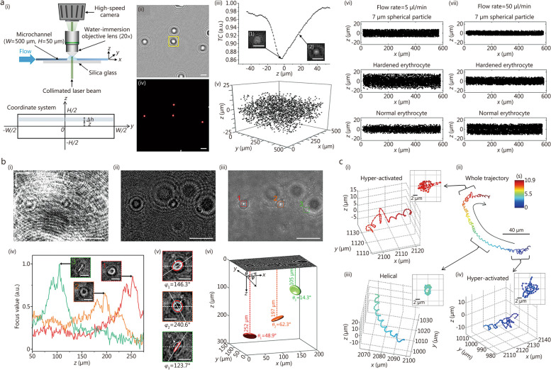
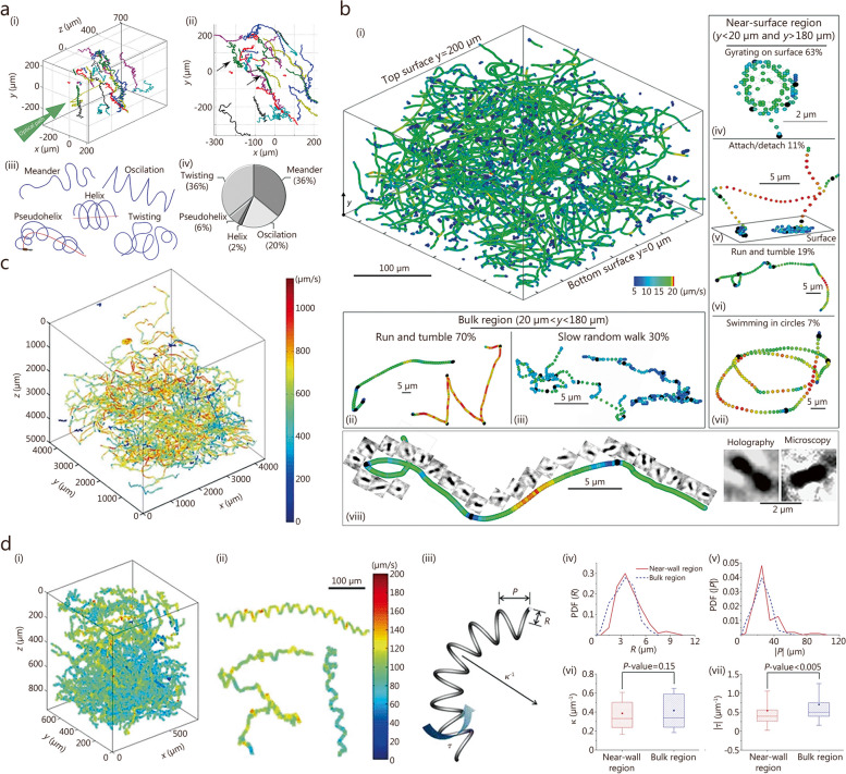
Digital in-line holographic microscopy (DIHM) is a non-invasive, real-time, label-free technique that captures three-dimensional (3D) positional, orientational, and morphological information from digital holographic images of living biological cells. Unlike conventional microscopies, the DIHM technique enables precise measurements of dynamic behaviors exhibited by living cells within a 3D volume. This review outlines the fundamental principles and comprehensive digital image processing procedures employed in DIHM-based cell tracking methods. In addition, recent applications of DIHM technique for label-free identification and digital tracking of various motile biological cells, including human blood cells, spermatozoa, diseased cells, and unicellular microorganisms, are thoroughly examined. Leveraging artificial intelligence has significantly enhanced both the speed and accuracy of digital image processing for cell tracking and identification. The quantitative data on cell morphology and dynamics captured by DIHM can effectively elucidate the underlying mechanisms governing various microbial behaviors and contribute to the accumulation of diagnostic databases and the development of clinical treatments.

Keywords: Artificial intelligence; Cell identification; Cell tracking; Digital in-line holographic microscopy (DIHM).


Conflict of interest statement

The authors declare that they have no competing interests.

Figures

Fig. 1
Fig. 1
Schematics of the optical configurations of digital in-line holographic microscopy using a point source (a) and a collimated beam (b)
Fig. 2
Fig. 2
Applications of digital in-line holographic microscopy (DIHM) to track human erythrocytes and spermatozoa. a Lateral migration of hardened and normal erythrocytes in viscoelastic flows under different microfluidic conditions. Experimental setup for the microfluidic measurement (i). Digital image processing procedure: background subtraction (ii, scale bar = 10 μm), depth localization using a Tamura coefficient (TC) focus function (iii, scale bar = 10 μm), in-plane positioning (iv, scale bar = 10 μm), and 3D spatial distributions of spherical particles, hardened erythrocytes, and normal erythrocytes measured using DIHM (v-vii). Reprinted from ref. [149], Copyright 2017. b Measurement of 3D locations and orientations of erythrocytes using DIHM and deep learning techniques. Digital image processing procedure: raw hologram (i, scale bar = 20 μm), background subtraction (ii, scale bar = 20 μm), projection (iii, scale bar = 20 μm), depth localization using a gradient focus function (iv, scale bar = 5 μm), in-plane angle measurement (v, scale bar = 5 μm), and 3D positions and orientations of erythrocytes measured using DIHM (vi). Reprinted with permission from ref. [151], Copyright 2023, Elsevier B.V. c Transitions between different swimming patterns of a human spermatozoon. Hyper-activated (i, iv) and helical patterns (iii) are observed in a whole trajectory of the human spermatozoon (ii). Reprinted from ref. [152], Copyright 2012
Fig. 3
Fig. 3
Applications of digital in-line holographic microscopy to track various unicellular microorganisms. a Trajectories of swimming Pseudomonas aeruginosa obtained by using DIHM (i, ii) and the corresponding statistical analysis of various swimming patterns, including meander, oscillation, helix, pseudohelix, and twisting patterns (iii, iv). Reprinted from ref. [158], Copyright 2014. b Various swimming patterns of Escherichia coli (E. coli) in near-surface and bulk regions. Trajectories of swimming E. coli (i, viii). Swimming patterns of E. coli in the bulk region: running and tumbling motions (ii) and slow random walk (iii). Swimming patterns of E. coli in the near-surface region: gyrating on a surface (iv), attaching and detaching motions (v), running and tumbling motions (vi), and swimming in circles (vii). Reprinted with permission from ref. [163], Copyright 2014, American Physical Society. c 3D trajectories of solitary and chain-forming Cochlodinium polykrikoides. Reprinted with permission from ref. [172], Copyright 2010, Springer-Verlag. d Trajectories of Prorocentrum minimum in helical motions (i-iii), obtained using DIHM. Probability density functions (PDFs) of helix parameters in the near and bulk regions: radius (R, iv) and pitch (P, v). Statistical differences in helix parameters between the near and bulk regions, represented as probability values (P-values): curvature (κ, vi) and torsion (τ, vii). Reprinted with permission from ref. [173], Copyright 2016, Springer-Verlag Berlin Heidelberg

References

    1. Stephens DJ, Allan VJ. Light microscopy techniques for live cell imaging. Science. 2003;300(5616):82–86. doi: 10.1126/science.1082160. - DOI - PubMed
    1. Ntziachristos V. Going deeper than microscopy: the optical imaging frontier in biology. Nat Methods. 2010;7(8):603–614. doi: 10.1038/nmeth.1483. - DOI - PubMed
    1. Spiller DG, Wood CD, Rand DA, White MR. Measurement of single-cell dynamics. Nature. 2010;465(7299):736–745. doi: 10.1038/nature09232. - DOI - PubMed
    1. Cierpka C, Kähler CJ. Particle imaging techniques for volumetric three-component (3D3C) velocity measurements in microfluidics. J Vis. 2012;15:1–31. doi: 10.1007/s12650-011-0107-9. - DOI
    1. Morris JD, Payne CK. Microscopy and cell biology: new methods and new questions. Ann Rev Phys Chem. 2019;70:199–218. doi: 10.1146/annurev-physchem-042018-052527. - DOI - PubMed

MeSH terms

LinkOut - more resources