. 2024 Jun 12;22(1):562.
doi: 10.1186/s12967-024-05338-w.

Dual anti-angiogenic and anti-inflammatory action of tRNA-Cys-5-0007 in ocular vascular disease

Affiliations

Dual anti-angiogenic and anti-inflammatory action of tRNA-Cys-5-0007 in ocular vascular disease

Yan Ma et al. J Transl Med. .

Abstract

Background: Intravitreal injections of angiogenesis inhibitors have proved efficacious in the majority of patients with ocular angiogenesis. However, one-fourth of all treated patients fail to derive benefits from intravitreal injections. tRNA-derived small RNA (tsRNA) emerges as a crucial class of non-coding RNA molecules, orchestrating key roles in the progression of human diseases by modulating multiple targets. Through our prior sequencing analyses and bioinformatics predictions, tRNA-Cys-5-0007 has shown as a potential regulator of ocular angiogenesis. This study endeavors to elucidate the precise role of tRNA-Cys-5-0007 in the context of ocular angiogenesis.

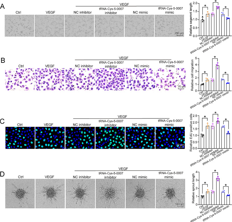
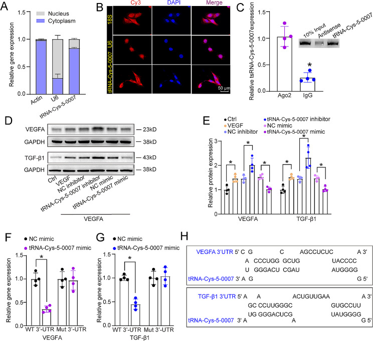
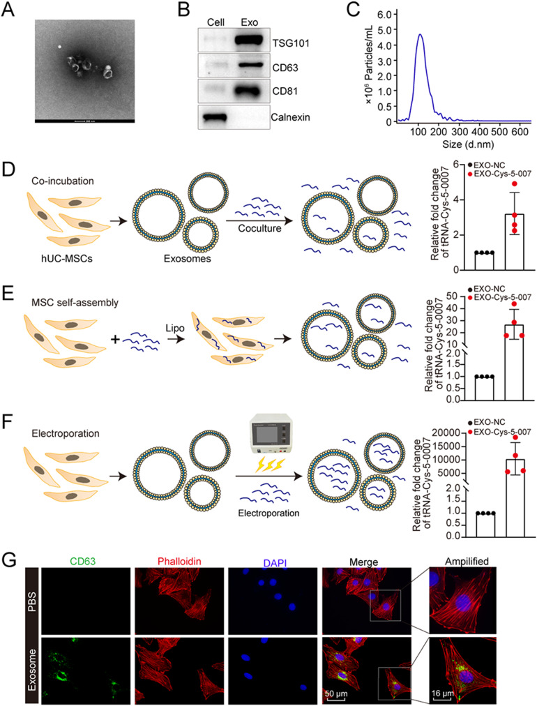
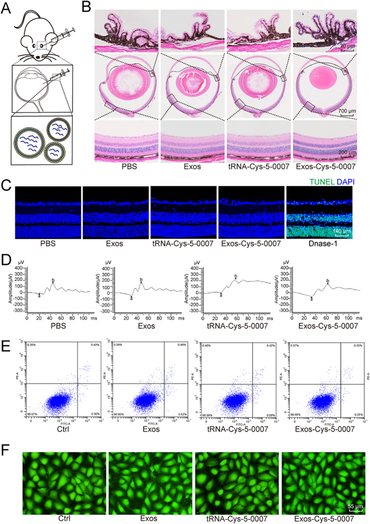
Methods: Quantitative reverse transcription PCR (qRT-PCR) assays were employed to detect tRNA-Cys-5-0007expression. EdU assays, sprouting assays, transwell assays, and Matrigel assays were conducted to elucidate the involvement of tRNA-Cys-5-0007 in endothelial angiogenic effects. STZ-induced diabetic model, OIR model, and laser-induced CNV model were utilized to replicate the pivotal features of ocular vascular diseases and evaluate the influence of tRNA-Cys-5-0007 on ocular angiogenesis and inflammatory responses. Bioinformatics analysis, luciferase activity assays, RNA pull-down assays, and in vitro studies were employed to elucidate the anti-angiogenic mechanism of tRNA-Cys-5-0007. Exosomal formulation was employed to enhance the synergistic anti-angiogenic and anti-inflammatory efficacy of tRNA-Cys-5-0007.

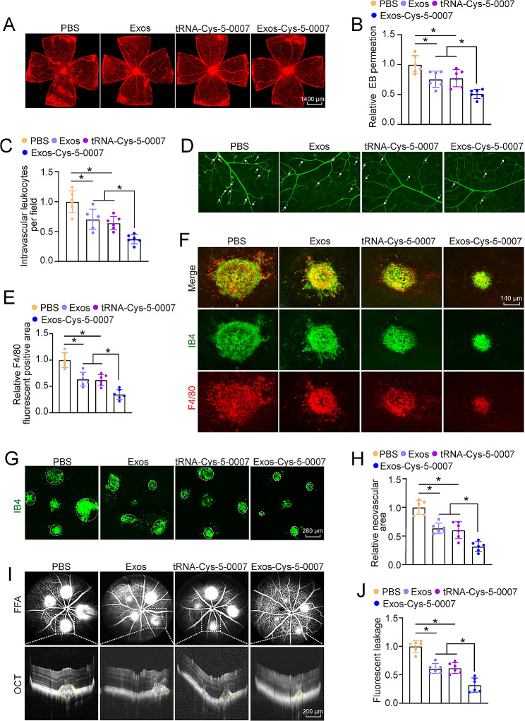
Results: tRNA-Cys-5-0007 expression was down-regulated under angiogenic conditions. Conversely, tRNA-Cys-5-0007 overexpression exhibited anti-angiogenic effects in retinal endothelial cells, as evidenced by reduced proliferation, sprouting, migration, and tube formation abilities. In diabetic, laser-induced CNV, and OIR models, tRNA-Cys-5-0007 overexpression led to decreased ocular vessel leakage, inhibited angiogenesis, and reduced ocular inflammation. Mechanistically, these effects were attributed to the targeting of vascular endothelial growth factor A (VEGFA) and TGF-β1 by tRNA-Cys-5-0007. The utilization of an exosomal formulation further potentiated the synergistic anti-angiogenic and anti-inflammatory efficacy of tRNA-Cys-5-0007.

Conclusions: Concurrent targeting of tRNA-Cys-5-0007 for anti-angiogenic and anti-inflammatory therapy holds promise for enhancing the effectiveness of current anti-angiogenic therapy.

Keywords: Anti-angiogenic therapy; Exosomal formulation; Ocular vascular disease; Transfer RNA-derived fragment.


Conflict of interest statement

The authors declare that they have no conflict of interest.

Figures

Fig. 1
Fig. 1
tRNA-Cys-5-0007 expression is reduced during ocular angiogenesis. (A-C) Schematic diagram depicting the experimental design of murine model of DR (A), OIR (B), and CNV (C). (D) qRT-PCR assays were performed to detect the expression of tRNA-Cys-5-0007 in DR model at 1 m, 3 m, and 6 m after STZ injection. (E) qRT-PCR assays were performed to detect the expression levels of tRNA-Cys-5-0007 in the murine model of OIR at P7, P12, and P17. (F) qRT-PCR assays were performed to detect the levels of tRNA-Cys-5-0007 expression in the RPE/choroid complex of C57BL/6J mice on day 3, 7, and 14 after laser photocoagulation. The data were presented as the means ± SD. n = 6 per group; *P < 0.05; Student’s t test
Fig. 2
Fig. 2
tRNA-Cys-5-0007 overexpression plays an anti-angiogenic and anti-inflammatory role during ocular angiogenesis. (A and B) Diabetic C57BL/6J mice (8-week-old, male) received intravitreous injections of PBS, scramble (Scr) agomir, tRNA-Cys-5-0007 agomir, Scr antagomir, tRNA-Cys-5-0007 antagomir. Wild-type C57BL/6J mice (WT) were taken as the controls. The mice were perfused with Evans blue dye for 30 min. The fluorescence signal of flat-mounted retina was detected by a fluorescence microscope. The representative images were shown. The infiltrated leukocytes in retinal vessels were detected by fluorescein-isothiocyanate (FITC)-coupled concanavalin A lectin perfusion assays. Adherent leukocytes (white arrows) were stained with FITC-conA (B). (C and D) CNV mice (8-week-old, male) received intravitreous injections of PBS, Scr agomir, tRNA-Cys-5-0007 agomir, Scr antagomir, tRNA-Cys-5-0007 antagomir, or left untreated. The RPE/choroid complexes were collected at day 14 and stained with IB-4 to label CNV lesions. The representative images were shown. Green staining indicated CNV lesions; dashed lines indicated CNV areas (C). On day 14 following laser injury, CNV lesions were stained with IB-4 (green) and F4/80 (red) to label CNV lesions and macrophages (D). The data were presented as the means ± SD. n = 6 mice per group; *P < 0.05; One-way ANOVA followed by Tukey’s multiple comparisons test
Fig. 3
Fig. 3
tRNA-Cys-5-0007 regulates endothelial angiogenic function. (A-D) HRVECs were transfected with negative control (NC) inhibitors, tRNA-Cys-5-0007 inhibitors, NC mimics, tRNA-Cys-5-0007 mimics, or left untreated (Ctrl) for 24 h. Representative images and quantification results of tube formation assays were shown (A). Representative images and quantification results of transwell migration assays were shown (B). Representative images and quantification of EdU proliferation assays were shown (C). Representative images and quantification results of endothelial cell spheroid‑based sprouting assays were shown (D). The data were presented as the means ± SD. n = 4 per group; *P < 0.05; One-way ANOVA followed by Tukey’s multiple comparisons test
Fig. 4
Fig. 4
tRNA-Cys-5-0007 regulates endothelial angiogenic effects by targeting VEGFA and TGF-β1. (A) The expression distribution of U6, actin, and tRNA-Cys-5-0007 was examined by qRT-PCRs in the nucleus fractions and cytoplasm fractions of HRVECs. (B) FISH assays were conducted to detect the cellular distribution of tRNA-Cys-5-0007. 18 S rRNA and U6 were detected as the cytoplasm control and nucleus control. (C) The fractions of HRVECs were immunoprecipitated using Ago2 or IgG antibody. The amounts of tRNA-Cys-5-0007 in immunoprecipitates were examined by qRT-PCRs. The biotinylated tRNA-Cys-5-0007 or its anti-sense RNA was incubated, targeted with streptavidin beads, and washed. Western blots were then conducted to detect the specific interaction between Ago2 and tRNA-Cys-5-0007. (D and E) The levels of VEGFA and TGF-β1 expression were examined by western blots in HRVECs following the transfection of negative control (NC) inhibitors, tRNA-Cys-5-0007 inhibitors, NC mimics, tRNA-Cys-5-0007 mimics, incubated with or without VEGF (Ctrl) for 24 h. (FH) The luciferase activity of wild-type VEGFA/TGF-β1 or mutant VEGFA/ TGF-β1 following the transfection with NC mimics or tRNA-Cys-5-0007 mimics for 24 h in HRVECs were detected. The data were presented as the means ± SD; n = 4 per group; *P < 0.05; Student’s t test
Fig. 5
Fig. 5
Exosomal preparation and optimization of tRNA-Cys-5-0007 loading in exosomes. (A) The morphology of purified exosomes from hUC-MSCs were observed by TEM. (B) Western blot analysis of exosome surface markers (TSG101, CD63, and CD81) and the negative exosome marker (calnexin) in hUC-MSCs and hUC-MSCs-Exos. (C) Diameter of hUC-MSCs-Exos were analyzed by the Nanoparticle tracking analysis (NTA) technology using ZetaView PMX 110. (D-F) Three different approaches (co-incubation, self-assembly, and electroporation) were used to load the negative control (NC) mimics or tRNA-Cys-5-0007 mimics into exosomes. The loading efficiency was detected by qRT-PCR assays. (G) Uptake of hUC-MSCs-derived exosomes by HRVECs. The cell nuclei were stained with DAPI (blue) and cytoskeletons were stained with Phalloidin (red). The data were presented as the means ± SD; n = 4 per group; *P < 0.05; Student’s t test
Fig. 6
Fig. 6
Safety evaluation of exosomal formulation of tRNA-Cys-5-0007 in vivo and in vitro. (A-D) The mice received intravitreal injections of PBS, exosomes, tRNA-Cys-5-0007, or Exos-Cys-5-0007 for 7 days. H&E staining was performed to detect the change of retinal structures. The representative images were shown (B, n = 5). TUNEL assays were conducted to detect retinal apoptosis along with the representative images (C, n = 5). ERG assays were conducted to detect the change of visual function (D, n = 5). (E and F) HRVECs were incubated with exosomes, tRNA-Cys-5-0007, Exos-Cys-5-0007, or PBS (Ctrl) for 24 h. Flow cytometry assays with Annexin V-FITC staining (E, n = 5) and PI/Calcein-AM double staining (F, n = 5) were conducted to detect the change of retinal apoptosis
Fig. 7
Fig. 7
Exosomal formulation enhances the synergistic anti-angiogenic and anti-inflammatory efficacy of tRNA-Cys-5-0007. (A-D) Diabetic C57BL/6J mice (8-week-old, male) received intravitreous injections of PBS, exosomes, tRNA-Cys-5-0007, or Exos-Cys-5-0007. They were perfused with Evans blue dye for 2 h. The fluorescence signal of flat-mounted retinas was detected by a fluorescence microscope. The representative images were shown (A and B). The infiltrations of leukocytes in retinal vessels were detected by fluorescein-isothiocyanate (FITC)-coupled concanavalin A lectin perfusion assays. Adherent leukocytes (white arrows) were stained with FITC-conA (C and D). (E-J) CNV mice (8-week-old, male) received intravitreous injections of exosomes, tRNA-Cys-5-0007, Exos-Cys-5-0007, or PBS. On day 14 after laser injury, CNV lesions were stained with IB-4 (green) and F4/80 (red) to label CNV lesions and macrophages (E and F, n = 5). RPE/choroid complexes were collected and stained with IB-4 to label CNV lesions. The representative images were shown. Green staining indicated CNV lesions; dashed line indicated CNV areas (G and H). FFA and OCT images of CNV mice following treatment with exosomes, tRNA-Cys-5-0007, Exos-Cys-5-0007, or PBS and quantitative fluorescence leakage areas for spots were shown. The non-CNV group was taken as the control group. The part circled in yellow delineated the bulges in subretinal spaces (I and J). The data were presented as the means ± SD. n = 6 mice per group; *P < 0.05; One-way ANOVA followed by Tukey’s multiple comparisons test

Similar articles

Cited by

References

    1. Li X, Carmeliet P. Targeting angiogenic metabolism in disease. Science. 2018;359:1335–6. doi: 10.1126/science.aar5557. - DOI - PubMed
    1. Flammer J, Konieczka K, Bruno RM, Virdis A, Flammer AJ, Taddei S. The eye and the heart. Eur Heart J. 2013;34:1270–8. doi: 10.1093/eurheartj/eht023. - DOI - PMC - PubMed
    1. Li X, Eriksson U. Novel VEGF family members: VEGF-B, VEGF-C and VEGF-D. Int J Biochem Cell Biol. 2001;33:421–6. doi: 10.1016/S1357-2725(01)00027-9. - DOI - PubMed
    1. Li X. VEGF-B: a thing of beauty. Cell Res. 2010;20:741–4. doi: 10.1038/cr.2010.77. - DOI - PMC - PubMed
    1. Arrigo A, Aragona E, Bandello F. VEGF-targeting drugs for the treatment of retinal neovascularization in diabetic retinopathy. Ann Med. 2022;54:1089–111. doi: 10.1080/07853890.2022.2064541. - DOI - PMC - PubMed

MeSH terms

LinkOut - more resources