

Giant complete hydatidiform mole: a case report and review of the literature
- PMID: 38867300
- PMCID: PMC11170884
- DOI: 10.1186/s13256-024-04474-7
Giant complete hydatidiform mole: a case report and review of the literature
Abstract
Background: This case describes the youngest patient documented in the literature who presented with a giant hydatidiform mole, effectively addressed through conservative treatment.
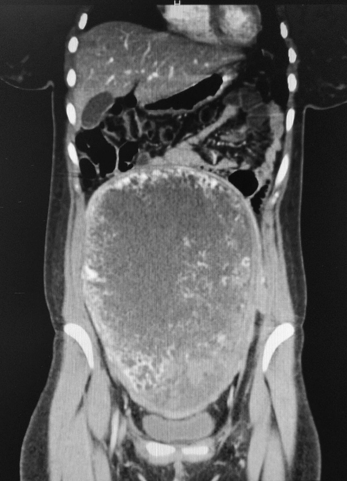
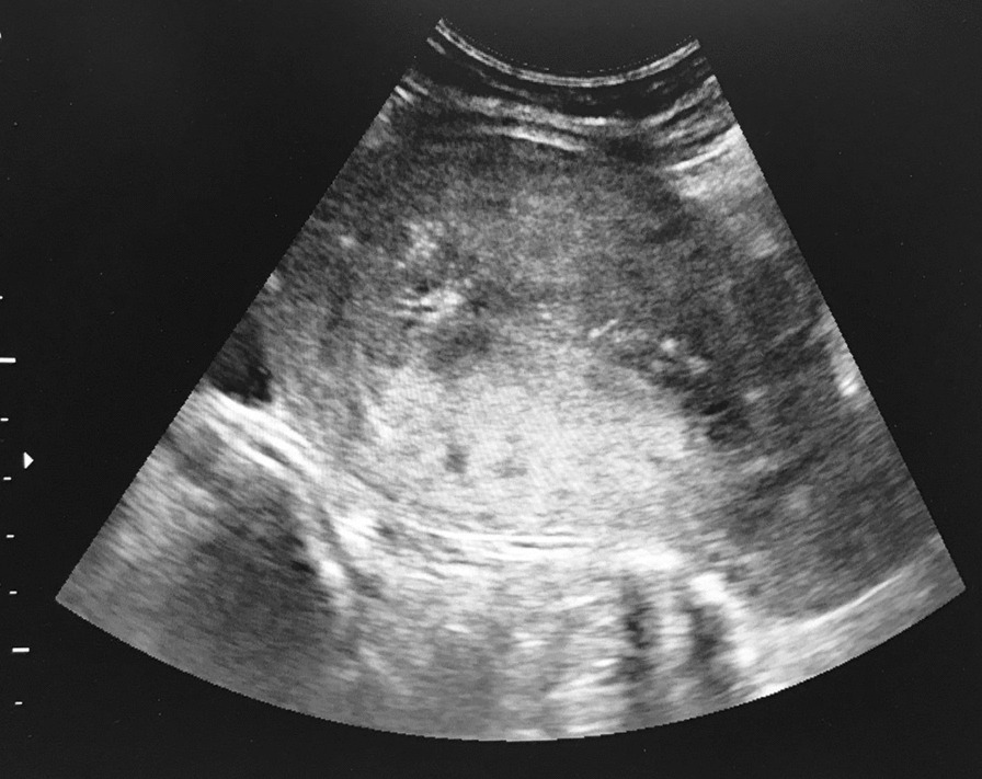
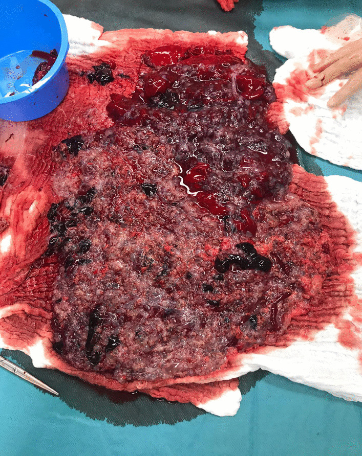
Case presentation: Our department received a 20-year-old Caucasian patient who was admitted due to significant metrorrhagia in an undisclosed pregnancy. During examination, we identified a massive, highly vascularized hydatidiform mole measuring 22 cm (cm). We performed a surgical dilatation and curettage. The anatomopathological findings confirmed the presence of a complete hydatidiform mole (CHM). Following the established guidelines, we conducted weekly monitoring of human chorionic gonadotropin (hCG). Unfortunately, the patient discontinued the follow-up and became pregnant again before achieving hCG negativation.
Conclusion: This case suggests that conservative treatment is a viable option regardless of the size of gestational trophoblastic disease (GTD), especially when the preservation of fertility is a crucial consideration, as effectively demonstrated in our case.
Keywords: Case report; Gestational trophoblastic disease; Human chorionic gonadotropin; Hydatidiform mole; Molar pregnancy.
© 2024. The Author(s).
Conflict of interest statement
The authors have no conflict of interest.
Figures
Similar articles
-
A Case of Rapid Transformation from Hydatidiform Mole to Invasive Mole: The Importance of β-hCG (Human Chorionic Gonadotropin) Serum Levels in Follow-Up Evaluation.Am J Case Rep. 2021 Jun 15;22:e931156. doi: 10.12659/AJCR.931156. Am J Case Rep. 2021. PMID: 34127641 Free PMC article.
-
Gestational trophoblastic neoplasia after spontaneous human chorionic gonadotropin normalization following molar pregnancy evacuation.Gynecol Oncol. 2015 Nov;139(2):283-7. doi: 10.1016/j.ygyno.2015.09.012. Epub 2015 Sep 14. Gynecol Oncol. 2015. PMID: 26383828
-
Gestational Trophoblastic Neoplasia After Human Chorionic Gonadotropin Normalization Following Molar Pregnancy: A Systematic Review and Meta-analysis.Obstet Gynecol. 2020 Jan;135(1):12-23. doi: 10.1097/AOG.0000000000003566. Obstet Gynecol. 2020. PMID: 31809433 Free PMC article.
-
Partial hydatidiform mole with false-negative urine human chorionic gonadatropin test in the emergency department.J Emerg Med. 2014 Mar;46(3):348-50. doi: 10.1016/j.jemermed.2013.08.053. Epub 2013 Nov 1. J Emerg Med. 2014. PMID: 24188605
-
Placental pathology casebook. Complete hydatidiform mole with coexistent term twin pregnancy.J Perinatol. 2001 Jan-Feb;21(1):72-5. doi: 10.1038/sj.jp.7200491. J Perinatol. 2001. PMID: 11268872 Review.
References
Publication types
MeSH terms
Substances
LinkOut - more resources
Full Text Sources
Medical
