. 2024 Jun 12;17(1):256.
doi: 10.1186/s13071-024-06327-0.

Alterations of plasma circulating microRNAs in BALB/c mice with Toxocara canis visceral and cerebral larva migrans

Affiliations

Alterations of plasma circulating microRNAs in BALB/c mice with Toxocara canis visceral and cerebral larva migrans

Yifan Yang et al. Parasit Vectors. .

Abstract

Background: Human toxocariasis is a neglected parasitic disease characterised by the syndromes visceral, cerebral, and ocular larva migrans. This disease is caused by the migrating larvae of Toxocara roundworms from dogs and cats, affecting 1.4 billion people globally. Via extracellular vesicles (EVs), microRNAs have been demonstrated to play roles in host-parasite interactions and proposed as circulating biomarkers for the diagnosis and follow-up of parasitic diseases.

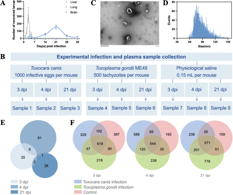
Methods: Small RNA-seq was conducted to identify miRNAs in the infective larvae of T. canis and plasma EV-containing preparations of infected BALB/c mice. Differential expression analysis and target prediction were performed to indicate miRNAs involved in host-parasite interactions and miRNAs associated with visceral and/or cerebral larva migrans in the infected mice. Quantitative real-time polymerase chain reaction (PCR) was used to amplify circulating miRNAs from the infected mice.

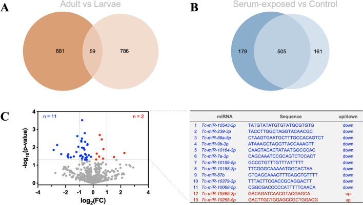
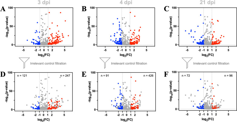
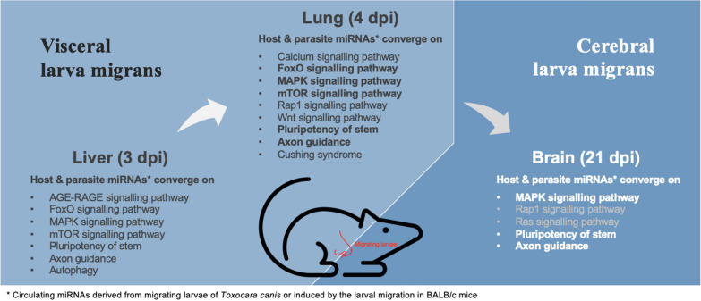
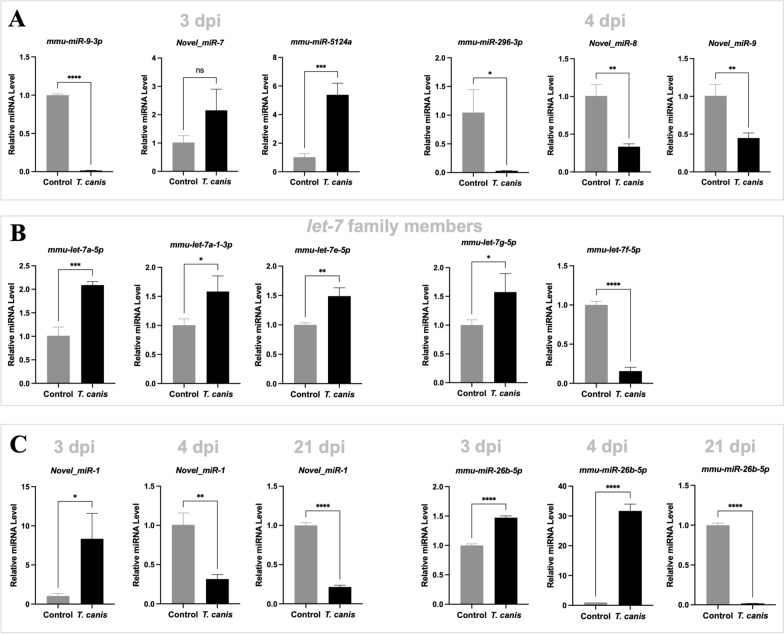
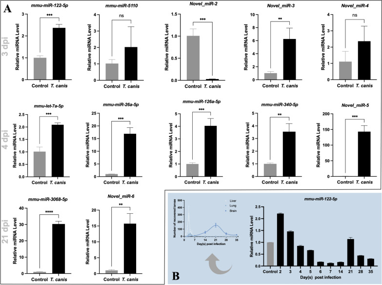
Results: This study reports host and parasite miRNAs in the plasma of BALB/c mice with visceral and cerebral larva migrans and demonstrates the alterations of these miRNAs during the migration of larvae from the livers through the lungs and to the brains of infected mice. After filtering unspecific changes in an irrelevant control, T. canis-derived miRNAs and T. canis infection-induced differential miRNAs are predicted to modulate genes consistently involved in mitogen-activated protein kinase (MAPK) signalling and pathways regulating axon guidance and pluripotency of stem in the infected mice with visceral and cerebral larva migrans. For these plasma circulating miRNAs predicted to be involved in host-parasite crosstalk, two murine miRNAs (miR-26b-5p and miR-122-5p) are experimentally verified to be responsive to larva migrans and represent circulating biomarker candidates for visceral and cerebral toxocariasis in BALB/c mice.

Conclusions: Our findings provide novel insights into the crosstalk of T. canis and the mammalian host via plasma circulating miRNAs, and prime agents and indicators for visceral and cerebral larva migrans. A deep understanding of these aspects will underpin the diagnosis and control of toxocariasis in humans and animals.

Keywords: Toxocara canis; Circulating miRNAs; Extracellular vesicles; Larva migrans; Toxocariasis.


Conflict of interest statement

No potential conflict of interest was reported by the authors.

Figures

Fig. 1
Fig. 1
An expanded repertoire of microRNAs for Toxocara canis. A Venn diagram of the number of miRNAs identified in previous work for adults and in the current work for the infective larvae of T. canis. B Venn diagram of the number of miRNAs identified in the murine serum-treated and untreated infective larvae of T. canis. C Volcano plot of differential miRNAs between the murine serum-treated and untreated infective larvae of T. canis. Down- and up-regulated miRNAs with a P value < 0.05 are indicated by blue and red dots, respectively. Differential miRNAs with a P value < 0.05 and a log2 fold change (FC) > 1 are indicated and listed in a table
Fig. 2
Fig. 2
MicroRNAs identified in the plasma extracellular vesicle (EV)-containing preparations of BALB/c mice with Toxocara canis larva migrans. A The number of T. canis larvae recovered from the livers, lungs and brains of infected mice at 1, 2, 3, 4, 5, 6, 7, 14, 21, 28 and 35 days post infection (dpi). B A schematic diagram showing the experimental design of T. canis infection and plasma sample collection. Specifically, plasma samples that were collected from mice (n  = 15) infected with T. canis at 3 dpi were pooled as sample 1. Those collected from mice (n  = 15) infected with T. canis at 4 dpi were pooled as sample 2. Those collected from mice (n = 15) infected with T. canis at 21 dpi were pooled as sample 3. In addition, plasma samples collected from mice (n = 15) infected with T. gondii at 3, 4 and 21 dpi were pooled as sample 4, 5 and 6, respectively. Plasma samples collected from uninfected mice (n = 15) at 3, 4 and 21 dpi (cf. infection groups) were pooled as sample 7, 8 and 9, respectively. C Transmission electron microscopy of EV-containing preparations (indicated by red arrows) isolated from the plasma of infected mice (collected at 3, 4 and 21 dpi). D Size distribution (50–150 nm) of EV-containing preparations from the plasma of infected mice. E Venn diagram of the numbers of T. canis-derived miRNAs (miRNAs that are mapped to the genome of T. canis and not present in the sera of irrelative and blank controls) sequenced in the EV-containing preparations of infected mice at 3, 4 and 21 dpi. F Venn diagram of the numbers of miRNAs identified in the pooled plasma sample of T. canis-infected mice, blank and irrelevant controls (infection with Toxoplasma gondii) at 3, 4 and 21 dpi
Fig. 3
Fig. 3
Alterations of microRNAs in the plasma extracellular vesicle (EV)-containing preparations of BALB/c mice with Toxocara canis infection. A, B, C Volcano plot of differential miRNAs identified in the plasma EV-containing preparations of T. canis-infected and uninfected mice at 3, 4 and 21 days post infection (dpi). D, E, F Volcano plot of differential miRNAs identified in the plasma EV-containing preparations of T. canis infected and uninfected mice at 3, 4 and 21 days post infection (dpi), after filtering the differential miRNAs induced by Toxoplasma gondii infection. Down- and up-regulated miRNAs with a P value < 0.05 and a log2 fold change (FC) > 1 are indicated by blue and red dots, respectively. The number of differential miRNAs is indicated
Fig. 4
Fig. 4
Pathways regulated by differential microRNAs in the plasma extracellular vesicle (EV)-containing preparations of BALB/c mice with toxocariasis. Differential miRNAs derived from T. canis and differential miRNAs induced by T. canis infection are predicted to target hundreds of genes in the infected mouse, respectively, which predominantly converge on AGE-RAGE, FoxO, MAPK and mTOR signalling pathways and pathways regulating autophagy, axon guidance and pluripotency of stem during the larvae migrating through the livers of mouse at 3 days post infection (dpi); on calcium, FoxO, MAPK, mTOR, Rap1 and Wnt signalling pathways and pathways regulating axon guidance, cushing syndrome and pluripotency of stem during the larvae migrating through the lungs of mouse at 4 dpi; on MAPK, Rap1, Ras signalling pathways and pathways regulating axon guidance and pluripotency of stem at 21 dpi. Specifically, FoxO, MAPK and mTOR signalling pathways and pathways regulating axon guidance and pluripotency of stem are consistently regulated by the differential microRNAs in the plasma EV-containing preparations of BALB/c mouse with visceral larva migrans, whereas the MAPK signalling pathway as well as pathways regulating axon guidance and pluripotency of stem are consistently regulated by the differential microRNAs in the plasma EV-containing preparations of BALB/c mouse with visceral and cerebral larva migrans
Fig. 5
Fig. 5
Experimental validation of differential microRNAs in the plasma extracellular vesicle (EV)-containing preparations of BALB/c mice induced by Toxocara canis infection. A Quantitative PCR amplification and the relative levels of selected miRNAs in the plasma sample of infected mice to those of uninfected mice at 3 or 4 days post infection (dpi). B Quantitative PCR amplification and the relative levels of let-7 family members in the plasma sample of infected mice to that of uninfected mice at 3 dpi or 4 dpi. C Quantitative PCR amplification and the relative levels of selected miRNAs in the plasma sample of infected mice versus those of uninfected mice at 3, 4 and 21 dpi. U6 is used as the reference control. An unpaired student’s t-test is used for the statistical analysis. *** indicates a P value < 0.001, ** indicates a P value < 0.01 and * indicates a P value < 0.01
Fig. 6
Fig. 6
Screening of microRNA biomarker candidates for Toxocara canis infection and tissue migration in BALB/c mice. A Quantitative PCR amplification and the relative levels of selected miRNAs in the plasma sample of infected mice to that of uninfected mice at 3, 4 and 21 days post infection (dpi). B Quantitative PCR amplification and the relative levels of miR-122-5p in the plasma sample of infected mice against that of uninfected mice at 2, 3, 4, 5, 6, 7, 14, 21, 28 and 35 dpi. The change of miR-122-5p abundance in the plasma of infected mice is consistent with the number of larvae recovered from the livers, lungs and brains of T. canis-infected mice. U6 is used as a reference control. An unpaired student’s t-test is used for the statistical analysis. *** indicates a P value < 0.001, ** indicates a P value < 0.01 and ns indicates no significance

Similar articles

Cited by

References

    1. Kerr-Muir MG. Toxocara canis and human health. BMJ. 1994;309:5–6. doi: 10.1136/bmj.309.6946.5. - DOI - PMC - PubMed
    1. Fisher M. Toxocara cati: an underestimated zoonotic agent. Trends Parasitol. 2003;19:167–170. doi: 10.1016/S1471-4922(03)00027-8. - DOI - PubMed
    1. Strube C, Heuer L, Janecek E. Toxocara spp infections in paratenic hosts. Vet Parasitol. 2013;193:375–389. doi: 10.1016/j.vetpar.2012.12.033. - DOI - PubMed
    1. Ma G, Holland CV, Wang T, Hofmann A, Fan CK, Maizels RM, et al. Human toxocariasis. Lancet Infect Dis. 2018;18:e14–e24. doi: 10.1016/S1473-3099(17)30331-6. - DOI - PubMed
    1. Cooper PJ. Toxocara canis infection: an important and neglected environmental risk factor for asthma? Clin Exp Allergy. 2008;38:551–553. doi: 10.1111/j.1365-2222.2008.02934.x. - DOI - PubMed

MeSH terms

LinkOut - more resources