. 2024 Jun 12;12(1):94.
doi: 10.1186/s40478-024-01806-y.

Cryo-EM structures reveal tau filaments from Down syndrome adopt Alzheimer's disease fold

Affiliations

Cryo-EM structures reveal tau filaments from Down syndrome adopt Alzheimer's disease fold

Ujjayini Ghosh et al. Acta Neuropathol Commun. .

Abstract

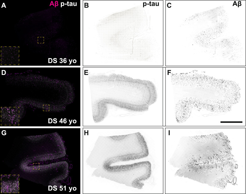
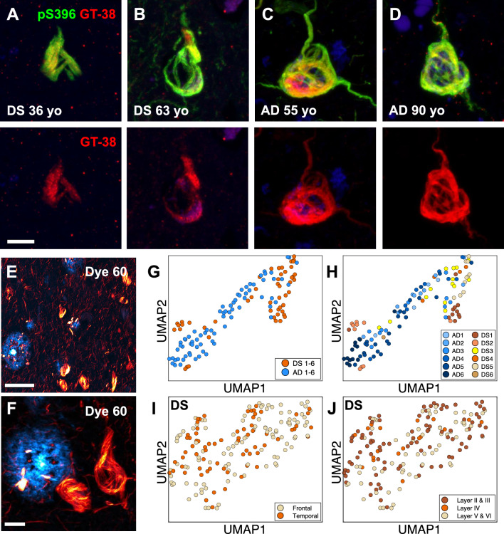
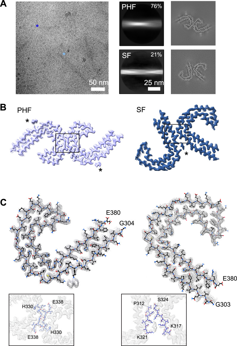
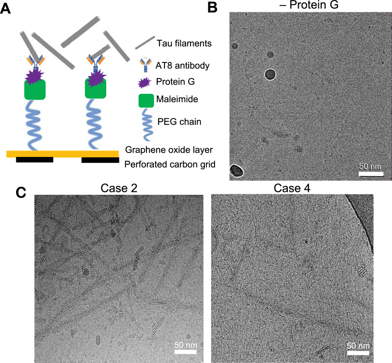
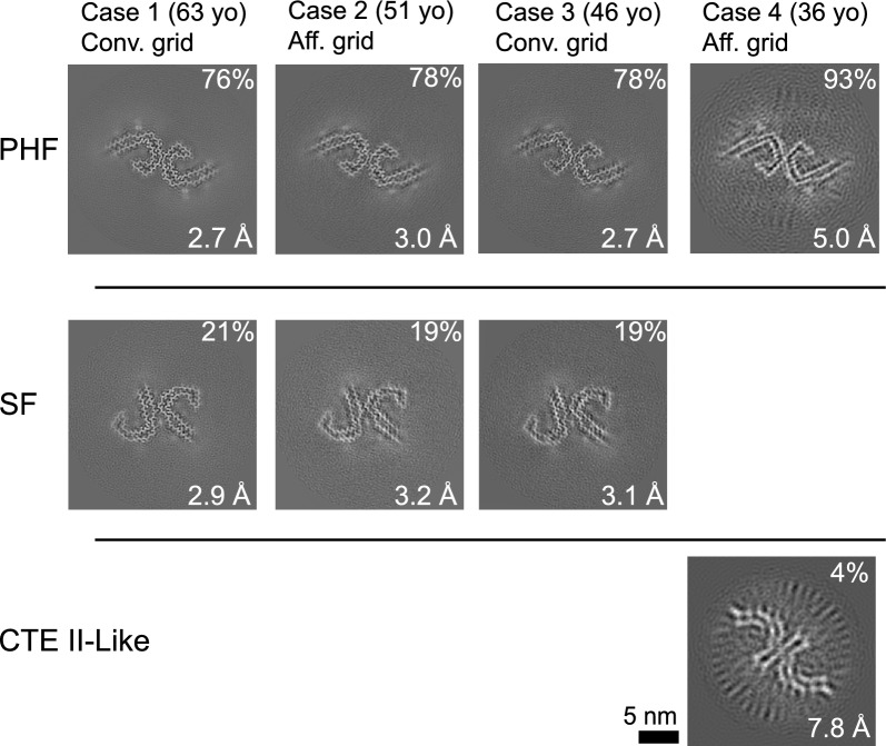
Down syndrome (DS) is a common genetic condition caused by trisomy of chromosome 21. Among their complex clinical features, including musculoskeletal, neurological, and cardiovascular disabilities, individuals with DS have an increased risk of developing progressive dementia and early-onset Alzheimer's disease (AD). This dementia is attributed to the increased gene dosage of the amyloid-β (Aβ) precursor protein gene, the formation of self-propagating Aβ and tau prion conformers, and the deposition of neurotoxic Aβ plaques and tau neurofibrillary tangles. Tau amyloid fibrils have previously been established to adopt many distinct conformations across different neurodegenerative conditions. Here, we report the characterization of brain samples from four DS cases spanning 36-63 years of age by spectral confocal imaging with conformation-specific dyes and cryo-electron microscopy (cryo-EM) to determine structures of isolated tau fibrils. High-resolution structures revealed paired helical filament (PHF) and straight filament (SF) conformations of tau that were identical to those determined from AD cases. The PHFs and SFs are made of two C-shaped protofilaments, each containing a cross-β/β-helix motif. Similar to filaments from AD cases, most filaments from the DS cases adopted the PHF form, while a minority (approximately 20%) formed SFs. Samples from the youngest individual with no documented dementia had sparse tau deposits. To isolate tau for cryo-EM from this challenging sample we used a novel affinity-grid method involving a graphene oxide surface derivatized with anti-tau antibodies. This method improved isolation and revealed that primarily tau PHFs and a minor population of chronic traumatic encephalopathy type II-like filaments were present in this youngest case. These findings expand the similarities between AD and DS to the molecular level, providing insight into their related pathologies and the potential for targeting common tau filament folds by small-molecule therapeutics and diagnostics.

Keywords: Alzheimer’s disease; Cryo–electron microscopy; Down syndrome; Protein conformation; Tauopathy.


Conflict of interest statement

S.B.P. is the founder of Prio-Pharma, which did not contribute financial or any other support to these studies. All other authors declare that they have no competing interests.

Figures

Fig. 1
Fig. 1
Fluorescence images of immunolabeled Aβ plaques and tau tangles in DS brain samples used for cryo–electron microscopy experiments. Formalin-fixed frontal cortex sections stained with antibodies against total Aβ (magenta) and the phosphorylated tau S262 epitope (white) in DS case 4, 36 years old (AC), DS case 3, 46 years old (DF), and DS case 2, 51 years old (GI). Individual fluorescence channels were converted to grayscale and inverted for phosphorylated tau (B, E, H) and Aβ (C, F, I). Scale bar = 2 mm. Inset dimensions = 0.6 mm2
Fig. 2
Fig. 2
In situ assays show DS tau conformation is similar to tau tangles in sporadic AD. AD Representative confocal images of tau tangles in DS brain fixed slices stained with antibodies targeting the phosphorylated tau S396 epitope (green) and the AD conformation-specific tau fold (GT-38; red). Scale bar = 10 μm. E, F Representative 40× low (E) and high (F) zoom confocal images of Aβ plaques (cyan) and tau tangles (red) in DS brain fixed slices stained with dye 60, a novel structure-sensitive dye used for EMBER amyloid-strain discrimination. Scale bar = 50 μm (E); scale bar = 10 μm (F). G, H EMBER analysis of tau tangles in brain slices from six DS cases compared with tau tangles in brain slices of six AD cases. EMBER data are plotted in UMAP as all DS cases versus all AD cases (G), or each individual DS case or AD case plotted separately (H). I, J EMBER analysis of tau tangles in brain slices from a different set of five DS cases. EMBER data are plotted in UMAP as tau tangles in frontal cortex versus temporal cortex in the same brain (I), or by cortical layers in both frontal and temporal cortices combined (J)
Fig. 3
Fig. 3
Cryo-EM structures of tau PHF and SF conformations from DS case 1. A Representative electron micrograph of amyloid filaments from the frontal cortex of DS case 1 showing PHF and SF segments (left image; light and dark blue asterisks, respectively) and corresponding 2D class averages (middle images) and cross-section projection views of the conformations (right images). B Final cryo-EM density maps of the PHF and SF at 2.7-Å and 2.9-Å resolution, respectively. Asterisks indicate extra densities previously observed in structures from AD, and boxes show each protofilament interface, enlarged in (C). C Single protofilament cryo-EM map (mesh) with the fitted atomic model for the PHF (left) and SF (right), including tau residues G304 for PHF (G303 for SF) to E380 and an enlarged view of the protofilament interface
Fig. 4
Fig. 4
GO-AT8 antibody affinity-grid assembly and isolation of tau filaments for cryo-EM. A Schematic showing the affinity-grid assembly procedure for the attachment of AT8 anti-tau antibodies to the GO grid surface. B Control cryo-EM micrograph image for which protein G was not included in the affinity-grid assembly prior to incubation with tau filaments and vitrification. C Representative cryo-EM micrograph images following GO-AT8 affinity-grid isolation of tau filaments from DS case 2 (left) and DS case 4 (right). Fewer filaments are observed in case 4 due to the low overall abundance of tau deposits
Fig. 5
Fig. 5
PHF and SF cross-section views of the tau filament cryo–electron microscopy structures determined from four DS cases. The relative percentage of the segments for the reconstructions is indicated with the final resolution and grid preparation method. In the 36 years old case, a minor percentage refined to show a conformation similar to CTE type II filaments (indicated). However, an atomic model was unable to be determined due to the low resolution and small dataset size

Update of

Similar articles

Cited by

References

    1. Vos T, Allen C, Arora M. Global, regional, and national incidence, prevalence, and years lived with disability for 310 diseases and injuries, 1990–2015: a systematic analysis for the Global Burden of Disease Study 2015. Lancet. 2016;388:1545–1602. doi: 10.1016/S0140-6736(16)31678-6. - DOI - PMC - PubMed
    1. Lott IT, Head E. Dementia in Down syndrome: unique insights for Alzheimer disease research. Nat Rev Neurol. 2019;15:135–147. doi: 10.1038/s41582-018-0132-6. - DOI - PMC - PubMed
    1. Mann DM, Esiri MM. The pattern of acquisition of plaques and tangles in the brains of patients under 50 years of age with Down’s syndrome. J Neurol Sci. 1989;89:169–179. doi: 10.1016/0022-510x(89)90019-1. - DOI - PubMed
    1. Davidson YS, Robinson A, Prasher VP, Mann DMA. The age of onset and evolution of Braak tangle stage and Thal amyloid pathology of Alzheimer’s disease in individuals with Down syndrome. Acta Neuropathol Commun. 2018;6:56. doi: 10.1186/s40478-018-0559-4. - DOI - PMC - PubMed
    1. Sleegers K, Brouwers N, Gijselinck I, et al. APP duplication is sufficient to cause early onset Alzheimer’s dementia with cerebral amyloid angiopathy. Brain. 2006;129:2977–2983. doi: 10.1093/brain/awl203. - DOI - PubMed