. 2024;44(7):289-301.
doi: 10.1080/10985549.2024.2356880. Epub 2024 Jun 12.

Phosphorylation of Orc6 During Mitosis Regulates DNA Replication and Ribosome Biogenesis

Affiliations

Phosphorylation of Orc6 During Mitosis Regulates DNA Replication and Ribosome Biogenesis

Fredy Kurniawan et al. Mol Cell Biol. 2024.

Abstract

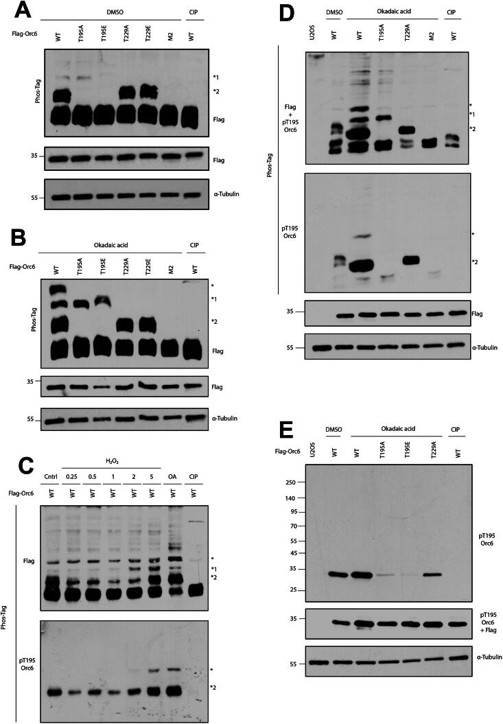
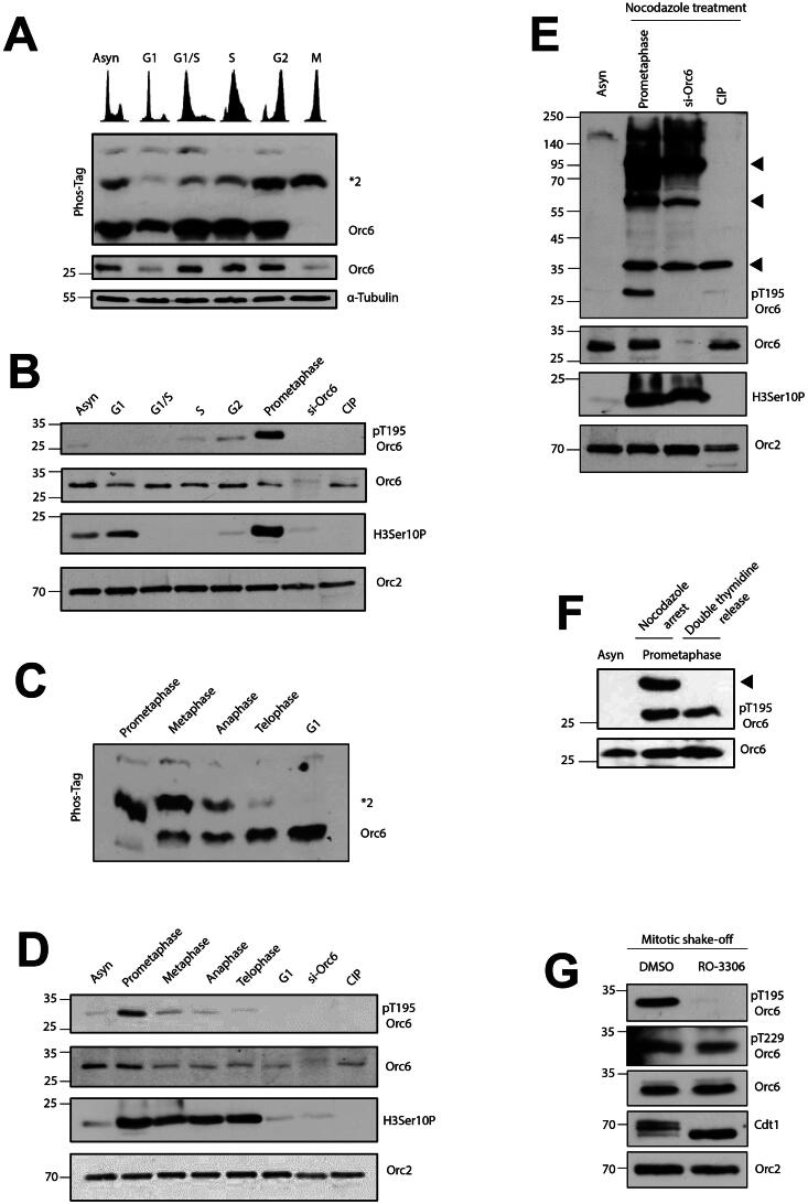
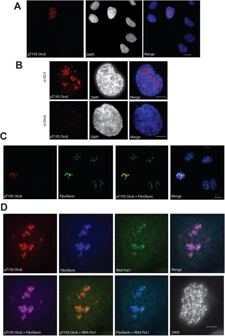
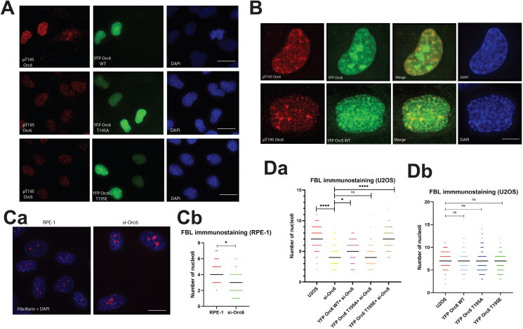
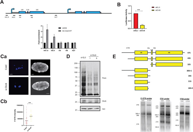
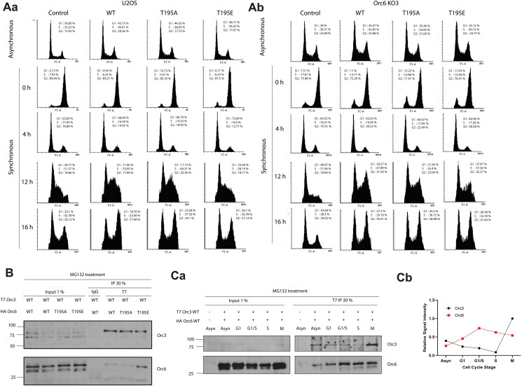
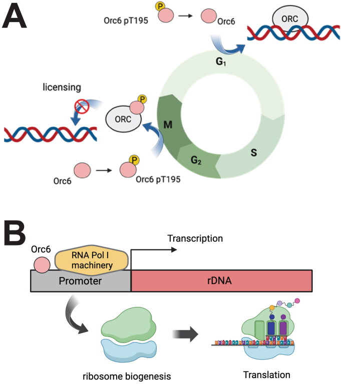
The human Origin Recognition Complex (ORC) is required not only for the initiation of DNA replication, but is also implicated in diverse cellular functions, including chromatin organization, centrosome biology, and cytokinesis. The smallest subunit of ORC, Orc6, is poorly conserved amongst eukaryotes. Recent studies from our laboratory have suggested that human Orc6 is not required for replication licensing, but is needed for S-phase progression. Further, ATR-dependent phosphorylation of Orc6 at T229 is implicated in DNA damage response during S-phase. In this study, we demonstrate that the CDK-dependent phosphorylation of Orc6 at T195 occurs during mitosis. While the phosphorylation at T195 does not seem to be required to exit mitosis, cells expressing the phosphomimetic T195E mutant of Orc6 impede S-phase progression. Moreover, the phosphorylated form of Orc6 associates with ORC more robustly, and Orc6 shows enhanced association with the ORC outside of G1, supporting the view that Orc6 may prevent the role of Orc1-5 in licensing outside of G1. Finally, Orc6 and the phosphorylated Orc6 localize to the nucleolar organizing centers and regulate ribosome biogenesis. Our results suggest that phosphorylated Orc6 at T195 prevents replication.

Keywords: Mitosis; Orc6; nucleolus; phosphorylation; replication.


Conflict of interest statement

No potential conflict of interest was reported by the authors.

Figures

Figure 1.
Figure 1.
Phosphorylation of Orc6 at threonine 195 (T195) is enhanced upon DNA damage. (A) Phos-tag gel of Orc6 phosphorylation in DMSO-treated cells. (B) Phos-tag gel of Orc6 phosphorylation in okadaic acid-treated (OA) cells. (C) Phos-tag gel of Orc6 phosphorylation in H2O2-treated cells. (D) Validation of Orc6 T195 antibody specificity in Phos-tag gel. Please note that parts of this figure (FLAG antibody and alpha-tubulin) is used in Figure 1A of Lin et al.). (E) Validation of Orc6 T195 antibody specificity in Western blot. *indicates Orc6 phosphorylated on both T195 and T229; *1 indicates T229 phosphorylation; *2 indicates T195 phosphorylation, M2 indicates both T195A and T229A mutation.
Figure 2.
Figure 2.
Orc6 is phosphorylated at T195 during mitosis. (A) Phos-tag gel of Orc6 phosphorylation in different cell cycle stages (B) Western blot showing Orc6 phosphorylation in different cell cycle stages (C) Phos-tag gel of Orc6 phosphorylation in different mitotic cell cycle stages. (D) Western blot analysis showing Orc6 phosphorylation at T195 in different mitotic cell cycle stages. (E) Western blot analysis showing Orc6 phosphorylation at T195 in nocodazole treated cells synchronized in prometaphase. (F) Western blot analysis of cells synchronized in prometaphase with both nocodazole and thymidine treatments show Orc6 phosphorylation at T195. (G) Loss of Orc6 T195 phosphorylation signal upon treatment with RO-3306, a CDK inhibitor in cells synchronized in mitosis. H3Ser10p serves as the marker for mitosis. Arrowhead indicates nonspecific bands. Cells were treated with calf intestinal phosphatase (CIP) and loss of phosphorylation-specific signal after CIP treatment confirms loss of phosphorylation.
Figure 3.
Figure 3.
Orc6 is localized at the nucleolus. (A) Immunostaining showing Orc6 T195 phosphorylation localizes to the nucleus (scale bar = 10 μm). (B) Validation of Orc6 pT195 antibody specificity in siGL3 and Orc6-depleted U2OS cell line (scale bar = 5 μm). (C) Orc6 pT195 colocalizes with fibrillarin, a nucleolar marker (scale bar = 10 μm). (D) Immunostaining of Orc6 pT195, fibrillarin, and RNA Pol I in the U2OS cell line (scale bar = 5 μm).
Figure 4.
Figure 4.
Depletion of Orc6 results in changes in nucleolar number. (A) Validation of Orc6 pT195 antibody in cells expressing YFP Orc6 WT/T195A/T195E (scale bar = 10 μm). (B) Immunostaining shows Orc6 pT195 in cells expressing YFP Orc6 WT in interphase (top) and mitosis (bottom) (scale bar = 5 μm). (Ca) Representative images of immunostaining of fibrillarin in wild-type and Orc6-depleted RPE-1 cell line (scale bar = 5 μm). (Cb) Quantification of nucleolar number in wild-type and Orc6-depleted RPE-1 cell line (n = 50). (Da) Quantification of nucleolar number in Orc6-depleted U2OS cells rescued with YFP Orc6 WT/T195A/T195E (n = 100). (Db) Quantification of nucleolar number in U2OS control and U2OS cells expressing YFP Orc6 WT/T195A/T195E (n = 100). Fibrillarin (FBL) is used as the nucleolar marker. ns P > 0.05,*P < .05, ****P < .0001, by unpaired Student’s two-tailed t-test.
Figure 5.
Figure 5.
Orc6 is involved in ribosome biogenesis. (A) HA ChIP-qPCR analysis at rDNA promoter regions in U2OS cells expressing HA Orc6 WT. (B) Luciferase activity of siGL3 and Orc6-depleted U2OS cells. *P < .05, ***P < .001, by unpaired Student’s two-tailed t test. (Ca) Representative images of RNA FISH between control and Orc6-depleted U2OS cells with 5’ETS probe. (Cb) Quantification of 5′ETS signal intensity in control and Orc6-depleted U2OS cells. (D) Western blot analysis of puromycin incorporation assay in siGL3 and Orc6-depleted U2OS cells. (E) Northern blot analysis of control and Orc6-depleted U2OS cells with probes targeting multiple rDNA promoter regions.
Figure 6.
Figure 6.
Phosphorylation of Orc6 T195 impacts its association with ORC and cell cycle progression. (A) Representative image of cell cycle profile between control (Aa) and Orc6 knockout (KO3, Ab) U2OS cells expressing wild-type (WT-Orc6), phospho-dead (Orc6-T195) or phosphomimetic (Orc6-T195E) mutants of Orc6. (B) Immunoprecipitation of U2OS cells expressing T7 Orc3 WT and HA Orc6 WT/T195A/T195E. (Ca) Immunoprecipitation of U2OS cells expressing T7 Orc3 WT and HA Orc6 WT in different stages of the cell cycle. (Cb) Relative signal intensity between Orc3 and Orc6 in different cell cycle stages.
Figure 7.
Figure 7.
Schematic representation of (A) the role of Orc6 phosphorylation at T195 during mitosis and our model on how it impacts ORC function, (B) the role of Orc6 in ribosome biogenesis. (Created with BioRender).

Similar articles

Cited by

References

    1. Bell SP, Stillman B.. Atp-dependent recognition of eukaryotic origins of DNA replication by a multiprotein complex. Nature. 1992;357:128–134. doi: 10.1038/357128a0. - DOI - PubMed
    1. Sasaki T, Gilbert DM.. The many faces of the origin recognition complex. Curr Opin Cell Biol. 2007;19:337–343. doi: 10.1016/j.ceb.2007.04.007. - DOI - PubMed
    1. Bell SP, Dutta A.. DNA replication in eukaryotic cells. Annu Rev Biochem. 2002;71:333–374. - PubMed
    1. Bernal JA, Venkitaraman AR.. A vertebrate n-end rule degron reveals that orc6 is required in mitosis for daughter cell abscission. J Cell Biol. 2011;192:969–978. doi: 10.1083/jcb.201008125. - DOI - PMC - PubMed
    1. Chesnokov IN, Chesnokova ON, Botchan M.. A cytokinetic function of drosophila orc6 protein resides in a domain distinct from its replication activity. Proc Natl Acad Sci USA. 2003;100:9150–9155. doi: 10.1073/pnas.1633580100. - DOI - PMC - PubMed

Publication types

Substances

LinkOut - more resources