Review
. 2024 Jun 12:7:0351.
doi: 10.34133/research.0351. eCollection 2024.

The Tricarboxylic Acid Cycle Metabolites for Cancer: Friend or Enemy

Affiliations
Review

The Tricarboxylic Acid Cycle Metabolites for Cancer: Friend or Enemy

Jie Wu et al. Research (Wash D C). .

Abstract

The tricarboxylic acid (TCA) cycle is capable of providing sufficient energy for the physiological activities under aerobic conditions. Although tumor metabolic reprogramming places aerobic glycolysis in a dominant position, the TCA cycle remains indispensable for tumor cells as a hub for the metabolic linkage and interconversion of glucose, lipids, and certain amino acids. TCA intermediates such as citrate, α-ketoglutarate, succinate, and fumarate are altered in tumors, and they regulate the tumor metabolism, signal transduction, and immune environment to affect tumorigenesis and tumor progression. This article provides a comprehensive review of the modifications occurring in tumor cells in relation to the intermediates of the TCA cycle, which affects tumor pathogenesis and current therapeutic strategy for therapy through targeting TCA cycle in cancer cells.


Conflict of interest statement

Competing interests: The authors declare that they have no competing interests.

Figures

Fig. 1.
Fig. 1.
Overview of glucose metabolism and OXPHOS. In the cytoplasm, glucose converts into pyruvate through glycolysis or synthesizes glycogen. Glucose is phosphorylated by HK to produce glucose-6-phosphate (G-6-P). The reaction is irreversible and consumes one molecule of ATP. G-6-P is converted to fructose-6-phosphate (F-6-P) catalyzed by phosphohexose isomerase or enters the pentose phosphate pathway (PPP). F-6-P converted to fructose-1,6-bisphosphate (F-1,6-BP) catalyzed by PFK1, which is also irreversible and consumes ATP. F-1,6-BP is catalyzed by aldolase to produce glyceraldehyde-3-phosphate and dihydroxyacetone phosphate, which are isomers that can be converted and supplemented by each other. Glyceraldehyde-3-phosphate is oxidized to 1,3-diphosphoglycerate by glyceraldehyde-3-phosphate dehydrogenase (GAPDH), and NAD+ accepts hydrogen and electrons as a coenzyme. Phosphoglycerate kinase catalyzes substrate-level phosphorylation of 1,3-diphosphoglycerate to produce ATP and 3-phosphoglycerate. 3-Phosphoglycerate converts to phosphoenolpyruvate (PEP) through 2-step reactions. PEP undergoes substrate-level phosphorylation to produce pyruvate and ATP catalyzed by PK, which is irreversible. Pyruvate can produce glucose in the opposite direction of glycolysis. There are only 3 differences in irreversible reactions. Pyruvate enters the mitochondria to transform into OAA and back into the cytoplasm to form PEP. F-6-P is catalyzed by FBP to produce G-6-P, which produces glucose catalyzed by glucose-6-phosphatase (G6Pase). Under hypoxia condition, pyruvate is catalyzed by LDH to produce lactate. Under normal oxygen conditions, pyruvate is transferred to the mitochondria to produce acetyl-CoA and then enters the TCA cycle. NADH and FADH2 gradually lose electrons through the electron respiratory chain and are oxidized to water. Complex I transfers electrons from NADH to ubiquinone, while complex II transfers electrons from succinate to ubiquinone. Complex III transfers electrons from reduced ubiquinone to cytochrome c and subsequently to oxygen through complex IV. The electron transport process is accompanied by the gradual release of energy, which drives the phosphorylation of ADP to ATP. The oxidation of NADH and FADH2 is coupled with the phosphorylation of ADP, which is called OXPHOS.
Fig. 2.
Fig. 2.
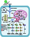
Citrate metabolism and signal transduction. In the mitochondria, citrate can be produced by CS-mediated condensation of OAA and acetyl-CoA and can also derive from glutamine. Citrate enters the cytoplasm via mCiC and antagonizes the glycolysis-related enzyme PFK1/2. Intracellular citrate can also be supplemented by glutamine. Citrate is cleaved to acetyl-CoA and OAA catalyzed by ACLY. Acetyl-CoA is involved in protein acetylation. In addition, acetyl-CoA can be converted to malonyl-CoA to participate in FAS or to HMG-CoA to enter cholesterol synthesis. OAA can be converted to aspartate to participate in nucleotide and polyamine synthesis, enter the gluconeogenesis process, or be converted to malate and returned to the mitochondrial. OAA competitively inhibits LDHA. Citrate can be transported to extracellular space by pmCiC, and CAFs in TME can also release citrate. Exogenous administration of high doses of citrate can activate caspase-8 and inhibit Mcl-1 and BCL-xL to induce apoptosis. Besides, citrate can activate autophagy and pyroptosis, inhibit angiogenesis, promote excess lipid biosynthesis, and induce cell senescence. Citrate binds to IGF1R, then down-regulates AKT, and up-regulates PTEN-eIF2α. High doses of citrate in TME can promote T cell infiltration and enhance the secretion of proinflammatory cytokines in macrophages.
Fig. 3.
Fig. 3.
Overview of α-KG biosynthetic pathways and impact on tumor cells. α-KG can be formed either by isocitrate in the TCA cycle or by glutamine in the cytoplasm or mitochondria via different pathways. IDH mutation leads to the accumulation of 2-HG. As oncometabolites, 2-HG, succinate, and fumarate are proved to be competitive inhibitors of αKGDDs. Increased oncometabolites inhibit PHD and subsequent HIF-1α degradation. Overexpressed HIF-1α and downstream genes promote tumors by facilitating angiogenesis, glycolysis, and EMT. Oncometabolites can inhibit DNA and histone demethylases, meanwhile inhibiting DNA repair. D-2-HG binds to DNMT1 and induces hypermethylation of the RIP3 promoter to inhibit necroptosis. In addition, D-2-HG reduces DEPTOR protein stability and activates mTOR. D-2-HG activates AMPK and inhibits mTOR, resulting in the decrease of Mcl-1. 2-HG binds to mutant p53 to reduce its degradation. D-2-HG exerts antitumor effects by inhibiting FTO and then MYC/CEBPA signaling, along with inhibiting glycolysis and ATP synthase. D-2-HG suppresses the antitumor effects of T cells, NK cells, and DCs in TME. α-KG up-regulates GLUT1 by activating IKKβ and NF-κB. Exogenous addition of α-KG results in pyroptosis, ferroptosis,and apoptosis of tumor cells. Besides, α-KG inhibits the WNT pathway and increases the expression of PD-L1, MHC-1, and differentiation-related genes. α-KG can promote T cells, inhibit Treg, and regulate macrophage M1/M2-related polarization. HRE, HIF-responsive element; NFAT, nuclear factor of activated T cells.
Fig. 4.
Fig. 4.
The role of succinate accumulation in tumors. SDH mutations lead to the truncation of TCA cycle, resulting in the accumulation of succinate. Subsequently, the increasing ROS activates NF-κB signaling and induces nuclear HIF-1α stabilization. In addition, SDH mutations also suppress p53 and promote EMT. The accumulation of succinate causes the down-regulation of KCNQ1 and promotes tumor progression. Extracellular succinate derived from tumor cells activates PI3K and HIF-1α by interacting with SUCNR1 on the surface of cancer cells, while inhibiting cGAS, resulting in decreased secretion of CCL5 and CXCL10 and inhibited recruitment of T cells. Succinate can also promote macrophage polarization to TAM and migration through the SUCNR1/PI3K/HIF-1α signaling pathway. TAM promotes the migration of cancer cells by secreting IL-6. Succinate inhibits the TCA cycle of T cells and the expression of cytokines such as IFN-γ and TNF-α. Tumor-derived lactate also inhibits the production of cytotoxic molecules such as IFN-γ and Granzyme B (GZMB) by affecting succinate secretion of T cells. Succinate promotes angiogenesis by activating the STAT3/ERK/VEGF pathway in endothelial cells through SUCNR1. Succinate acts on SUCNR1 on DC, resulting in enhanced DC migration and increased secretions of proinflammatory factors. Succinyl-CoA participates in the succinylation of lysine residues of proteins.
Fig. 5.
Fig. 5.
The role of fumarate accumulation in tumors. FH mutations in some tumors lead to fumarate accumulation, which can also be caused by up-regulation of mTOR. FH mutant tumors are characterized by increased glycolysis and ROS, accompany by overexpression of HIF-1α, Glut1, and LDHA. Increased glycolysis induces decreased levels of AMPK and p53, resulting in cytoplasmic iron deficiency and increased HIF-1α expression. FH-deficient cells participate in the biosynthesis and degradation of haem and rely on purine recycling for nucleotide biosynthesis. Excess fumarate can be used to generate argininosuccinate using the reverse activity of ASL with arginine. Fumarate releases mitochondrial DNA (mtDNA) into the cytoplasm via mitochondrial-derived vesicles and then triggers the activation of the cGAS/STING/TBK1 pathway. The accumulation of tumor-derived fumarate in the TME can succinate ZAP70, resulting in the inhibition of TCR signaling and CD8+ T cells. Increased fumarate causes high levels of protein succination. Succination of KEAP1 enhances NRF2 stability and promotes tumor progression through multiple pathways. Succination of IRP2, PTEN, GSDMD, GSH, and other proteins affects tumor cells by altering their downstream pathways. Succination of key proteins in the respiratory chain leads to the dysfunction of complex I, while fumarate accumulation can also directly inhibit complex II.

References

    1. Chandel NS. Glycolysis. Cold Spring Harb Perspect Biol. 2021;13(5):a040535. - PMC - PubMed
    1. Inigo M, Deja S, Burgess SC. Ins and outs of the TCA cycle: The central role of anaplerosis. Annu Rev Nutr. 2021;41:19–47. - PubMed
    1. Guertin DA, Wellen KE. Acetyl-CoA metabolism in cancer. Nat Rev Cancer. 2023;23(3):156–172. - PMC - PubMed
    1. Greene J, Segaran A, Lord S. Targeting OXPHOS and the electron transport chain in cancer; molecular and therapeutic implications. Semin Cancer Biol. 2022;86(Pt 2):851–859. - PubMed
    1. Icard P, Shulman S, Farhat D, Steyaert JM, Alifano M, Lincet H. How the Warburg effect supports aggressiveness and drug resistance of cancer cells? Drug Resist Updat. 2018;38:1–11. - PubMed

LinkOut - more resources