

Differential artery-vein analysis improves the OCTA classification of diabetic retinopathy
- PMID: 38867785
- PMCID: PMC11166441
- DOI: 10.1364/BOE.521657
Differential artery-vein analysis improves the OCTA classification of diabetic retinopathy
Abstract
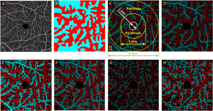
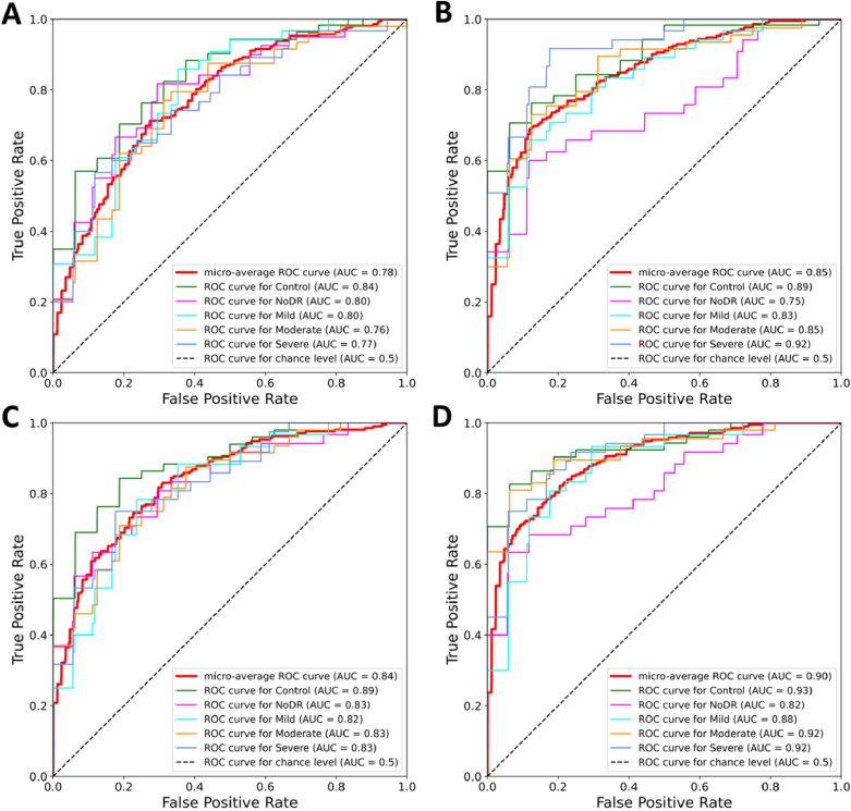
This study investigates the impact of differential artery-vein (AV) analysis in optical coherence tomography angiography (OCTA) on machine learning classification of diabetic retinopathy (DR). Leveraging deep learning for arterial-venous area (AVA) segmentation, six quantitative features, including perfusion intensity density (PID), blood vessel density (BVD), vessel area flux (VAF), blood vessel caliber (BVC), blood vessel tortuosity (BVT), and vessel perimeter index (VPI) features, were derived from OCTA images before and after AV differentiation. A support vector machine (SVM) classifier was utilized to assess both binary and multiclass classifications of control, diabetic patients without DR (NoDR), mild DR, moderate DR, and severe DR groups. Initially, one-region features, i.e., quantitative features extracted from the entire OCTA, were evaluated for DR classification. Differential AV analysis improved classification accuracies from 78.86% to 87.63% and from 79.62% to 85.66% for binary and multiclass classifications, respectively. Additionally, three-region features derived from the entire image, parafovea, and perifovea, were incorporated for DR classification. Differential AV analysis further enhanced classification accuracies from 84.43% to 93.33% and from 83.40% to 89.25% for binary and multiclass classifications, respectively. These findings highlight the potential of differential AV analysis in augmenting disease diagnosis and treatment assessment using OCTA.
© 2024 Optica Publishing Group.
Conflict of interest statement
No competing interest exists for any author.
Figures
Update of
- doi: 10.1364/opticaopen.25222430.
Similar articles
-
Differential artery-vein analysis in OCTA for predicting the anti-VEGF treatment outcome of diabetic macular edema.Biomed Opt Express. 2025 Apr 1;16(4):1732-1741. doi: 10.1364/BOE.557748. eCollection 2025 Apr 1. Biomed Opt Express. 2025. PMID: 40322014 Free PMC article.
-
An open-source deep learning network AVA-Net for arterial-venous area segmentation in optical coherence tomography angiography.Commun Med (Lond). 2023 Apr 17;3(1):54. doi: 10.1038/s43856-023-00287-9. Commun Med (Lond). 2023. PMID: 37069396 Free PMC article.
-
Identification of diabetic retinopathy classification using machine learning algorithms on clinical data and optical coherence tomography angiography.Eye (Lond). 2024 Oct;38(14):2813-2821. doi: 10.1038/s41433-024-03173-3. Epub 2024 Jun 13. Eye (Lond). 2024. PMID: 38871934
-
Optical coherence tomography (OCT) for detection of macular oedema in patients with diabetic retinopathy.Cochrane Database Syst Rev. 2015 Jan 7;1(1):CD008081. doi: 10.1002/14651858.CD008081.pub3. Cochrane Database Syst Rev. 2015. PMID: 25564068 Free PMC article.
-
Optical coherence tomography (OCT) for detection of macular oedema in patients with diabetic retinopathy.Cochrane Database Syst Rev. 2011 Jul 6;(7):CD008081. doi: 10.1002/14651858.CD008081.pub2. Cochrane Database Syst Rev. 2011. Update in: Cochrane Database Syst Rev. 2015 Jan 07;1:CD008081. doi: 10.1002/14651858.CD008081.pub3. PMID: 21735421 Updated.
Cited by
-
Differential artery-vein analysis in OCTA for predicting the anti-VEGF treatment outcome of diabetic macular edema.Biomed Opt Express. 2025 Apr 1;16(4):1732-1741. doi: 10.1364/BOE.557748. eCollection 2025 Apr 1. Biomed Opt Express. 2025. PMID: 40322014 Free PMC article.
-
OCTA-ReVA: an open-source toolbox for comprehensive retinal vessel feature analysis in optical coherence tomography angiography.Biomed Opt Express. 2024 Sep 25;15(10):6010-6023. doi: 10.1364/BOE.537727. eCollection 2024 Oct 1. Biomed Opt Express. 2024. PMID: 39421789 Free PMC article.
-
Deep learning segmentation of periarterial and perivenous capillary-free zones in optical coherence tomography angiography.J Biomed Opt. 2025 May;30(5):056005. doi: 10.1117/1.JBO.30.5.056005. Epub 2025 May 8. J Biomed Opt. 2025. PMID: 40342523 Free PMC article.
-
Advancing Diabetic Retinopathy Screening: A Systematic Review of Artificial Intelligence and Optical Coherence Tomography Angiography Innovations.Diagnostics (Basel). 2025 Mar 15;15(6):737. doi: 10.3390/diagnostics15060737. Diagnostics (Basel). 2025. PMID: 40150080 Free PMC article. Review.
-
Retinal vessel metric analysis of type 1 diabetes mellitus in OCT angiography.Front Med (Lausanne). 2025 Jun 13;12:1562809. doi: 10.3389/fmed.2025.1562809. eCollection 2025. Front Med (Lausanne). 2025. PMID: 40584707 Free PMC article.
References
Grants and funding
LinkOut - more resources
Full Text Sources