Cellular structural and functional imaging of donor and pathological corneas with label-free dual-mode full-field optical coherence tomography
- PMID: 38867788
- PMCID: PMC11166435
- DOI: 10.1364/BOE.525116
Cellular structural and functional imaging of donor and pathological corneas with label-free dual-mode full-field optical coherence tomography
Abstract
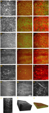
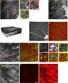
In this study, a dual-mode full-field optical coherence tomography (FFOCT) was customized for label-free static and dynamic imaging of corneal tissues, including donor grafts and pathological specimens. Static images effectively depict relatively stable structures such as stroma, scar, and nerve fibers, while dynamic images highlight cells with active intracellular metabolism, specifically for corneal epithelial cells. The dual-mode images complementarily demonstrate the 3D microstructural features of the cornea and limbus. Dual-modal imaging reveals morphological and functional changes in corneal epithelial cells without labeling, indicating cellular apoptosis, swelling, deformation, dynamic signal alterations, and distinctive features of inflammatory cells in keratoconus and corneal leukoplakia. These findings propose dual-mode FFOCT as a promising technique for cellular-level cornea and limbus imaging.
© 2024 Optica Publishing Group.
Conflict of interest statement
The authors declare that the research was conducted in the absence of any commercial or financial relationships that could be construed as a potential conflict of interest.
Figures


Similar articles
-
Full-field optical coherence tomography of human donor and pathological corneas.Curr Eye Res. 2015 May;40(5):526-34. doi: 10.3109/02713683.2014.935444. Epub 2014 Sep 24. Curr Eye Res. 2015. PMID: 25251769
-
New parameters in assessment of human donor corneal stroma.Acta Ophthalmol. 2017 Jun;95(4):e297-e306. doi: 10.1111/aos.13351. Epub 2017 Jan 30. Acta Ophthalmol. 2017. PMID: 28133954
-
Imaging Microscopic Features of Keratoconic Corneal Morphology.Cornea. 2016 Dec;35(12):1621-1630. doi: 10.1097/ICO.0000000000000979. Cornea. 2016. PMID: 27560027 Free PMC article.
-
[New diagnostic methods for imaging the anterior segment of the eye to enable treatment modalities selection].Nippon Ganka Gakkai Zasshi. 2011 Mar;115(3):297-322; discussion 323. Nippon Ganka Gakkai Zasshi. 2011. PMID: 21476312 Review. Japanese.
-
In vivo confocal microscopy and optical coherence tomography as innovative tools for the diagnosis of limbal stem cell deficiency.J Fr Ophtalmol. 2018 Nov;41(9):e395-e406. doi: 10.1016/j.jfo.2018.09.003. Epub 2018 Oct 24. J Fr Ophtalmol. 2018. PMID: 30458924 Review.
References
LinkOut - more resources
Full Text Sources
