

Application value of artificial intelligence algorithm-based magnetic resonance multi-sequence imaging in staging diagnosis of cervical cancer
- PMID: 38867922
- PMCID: PMC11167709
- DOI: 10.1515/biol-2022-0733
Application value of artificial intelligence algorithm-based magnetic resonance multi-sequence imaging in staging diagnosis of cervical cancer
Abstract
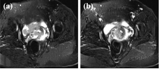
The aim of this research is to explore the application value of Deep residual network model (DRN) for deep learning-based multi-sequence magnetic resonance imaging (MRI) in the staging diagnosis of cervical cancer (CC). This research included 90 patients diagnosed with CC between August 2019 and May 2021 at the hospital. After undergoing MRI examination, the clinical staging and surgical pathological staging of patients were conducted. The research then evaluated the results of clinical staging and MRI staging to assess their diagnostic accuracy and correlation. In the staging diagnosis of CC, the feature enhancement layer was added to the DRN model, and the MRI imaging features of CC were used to enhance the image information. The precision, specificity, and sensitivity of the constructed model were analyzed, and then the accuracy of clinical diagnosis staging and MRI staging were compared. As the model constructed DRN in this research was compared with convolutional neural network (CNN) and the classic deep neural network visual geometry group (VGG), the precision was 67.7, 84.9, and 93.6%, respectively. The sensitivity was 70.4, 82.5, and 91.2%, while the specificity was 68.5, 83.8, and 92.2%, respectively. The precision, sensitivity, and specificity of the model were remarkably higher than those of CNN and VGG models (P < 0.05). As the clinical staging and MRI staging of CC were compared, the diagnostic accuracy of MRI was 100%, while that of clinical diagnosis was 83.7%, showing a significant difference between them (P < 0.05). Multi-sequence MRI under intelligent algorithm had a high diagnostic rate for CC staging, deserving a good clinical application value.
Keywords: artificial intelligence algorithm; cervical cancer staging; feature extraction; magnetic resonance multi-sequences.
© 2024 the author(s), published by De Gruyter.
Conflict of interest statement
Conflict of interest: Authors state no conflict of interest.
Figures


Similar articles
-
Multimodal MRI Analysis of Cervical Cancer on the Basis of Artificial Intelligence Algorithm.Contrast Media Mol Imaging. 2021 Nov 8;2021:1673490. doi: 10.1155/2021/1673490. eCollection 2021. Contrast Media Mol Imaging. 2021. PMID: 34858113 Free PMC article.
-
Magnetic Resonance Imaging Features on Deep Learning Algorithm for the Diagnosis of Nasopharyngeal Carcinoma.Contrast Media Mol Imaging. 2022 May 25;2022:3790269. doi: 10.1155/2022/3790269. eCollection 2022. Contrast Media Mol Imaging. 2022. PMID: 35677026 Free PMC article.
-
Diagnosis of Early Cervical Cancer with a Multimodal Magnetic Resonance Image under the Artificial Intelligence Algorithm.Contrast Media Mol Imaging. 2022 Mar 23;2022:6495309. doi: 10.1155/2022/6495309. eCollection 2022. Contrast Media Mol Imaging. 2022. PMID: 35386728 Free PMC article.
-
Image Features of Magnetic Resonance Imaging under the Deep Learning Algorithm in the Diagnosis and Nursing of Malignant Tumors.Contrast Media Mol Imaging. 2021 Aug 30;2021:1104611. doi: 10.1155/2021/1104611. eCollection 2021. Contrast Media Mol Imaging. 2021. PMID: 34548850 Free PMC article.
-
Multiparametric MRI in detection and staging of prostate cancer.Dan Med J. 2017 Feb;64(2):B5327. Dan Med J. 2017. PMID: 28157066 Review.
Cited by
-
Artificial intelligence radiomics in the diagnosis, treatment, and prognosis of gynecological cancer: a literature review.Transl Cancer Res. 2025 Apr 30;14(4):2508-2532. doi: 10.21037/tcr-2025-618. Epub 2025 Apr 27. Transl Cancer Res. 2025. PMID: 40386259 Free PMC article. Review.
References
-
- Shih IL, Yen RF, Chen CA, Cheng WF, Chen BB, Chang YH, et al. PET/MRI in cervical cancer: associations between imaging biomarkers and tumor stage, disease progression, and overall survival. J Magn Reson Imaging. 2021 Jan;53(1):305–18. 10.1002/jmri.27311. Epub 2020 Aug 14. PMID: 32798280. - DOI - PubMed
LinkOut - more resources
Full Text Sources
Research Materials