. 2024 May 29;10(11):e32027.
doi: 10.1016/j.heliyon.2024.e32027. eCollection 2024 Jun 15.

Cone-beam CT evaluation of post-extraction alveolar bone changes at the maxillary incisor sites in an East Asian population: A cross-sectional study

Affiliations

Cone-beam CT evaluation of post-extraction alveolar bone changes at the maxillary incisor sites in an East Asian population: A cross-sectional study

Fang Qu et al. Heliyon. .

Abstract

Objective: Understanding the characteristics of alveolar bone resorption in an East Asian population after maxillary incisor extraction and providing a reference for implant treatment plans.

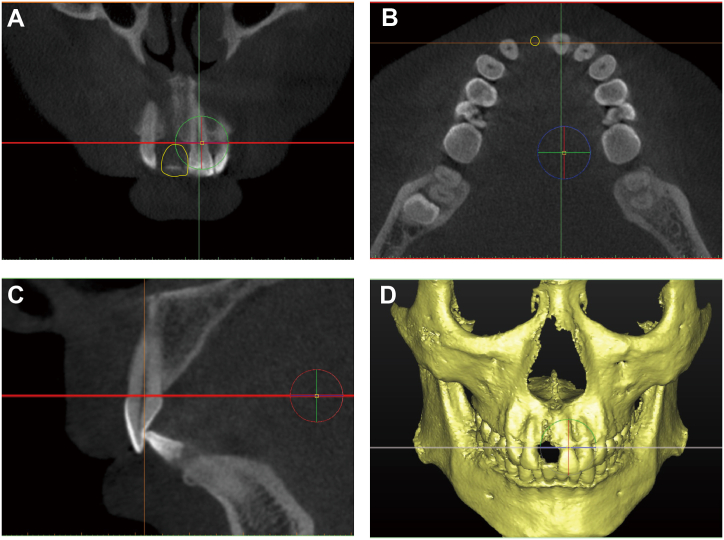
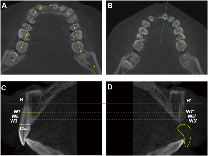
Study design: Cone-beam computerized tomography (CBCT) data of 125 East Asian patients with unilateral extraction of maxillary incisors for 3 months were collected. The alveolar bone width and height in the extraction sites were measured and compared with the corresponding contralateral sites.

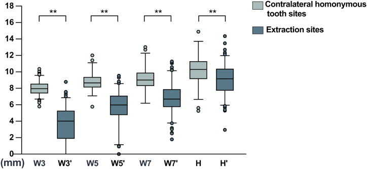
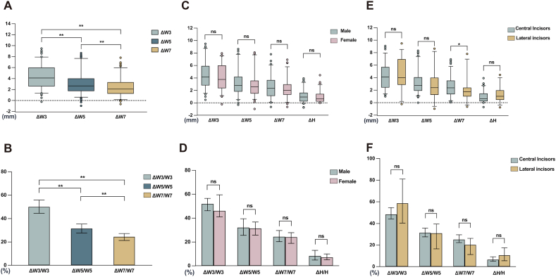
Results: The differences in alveolar bone width between the extraction site and contralateral site were as follows: 4.11 mm, 2.68 mm, and 2.09 mm (3 mm, 5 mm, 7 mm apical from CEJ of the contralateral tooth). Data are expressed as the median. The horizontal resorption ratio of alveolar bone was 49.94 %, 31.5 %, and 24.46 %. The difference in alveolar bone height was 0.78 mm. The vertical resorption ratio was 7.78 %. The resorption did not differ significantly between sexes and was not significantly affected by tooth positions.

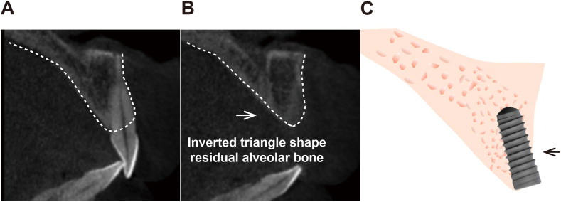
Conclusions: In the studied East Asian population, significant horizontal and vertical alveolar bone resorption occurs after natural healing of maxillary incisor extraction for 3 months. The closer to the alveolar ridge crest, the more significant the horizontal resorption, resulting in an "inverted triangle" shape residual alveolar bone.

Keywords: Alveolar bone resorption; Cone-beam computerized tomography (CBCT); East Asian; Maxillary incisor; Tooth extraction.


Conflict of interest statement

The authors declare that they have no known competing financial interests or personal relationships that could have appeared to influence the work reported in this paper.

Figures

Fig. 1
Fig. 1
Setting of the measurement reference planes A. Coronal plane. B. Axial plane. C. Sagittal plane. D. 3D reconstruction model.
Fig. 2
Fig. 2
Measurement of alveolar bone width and height A. The arch curve was drawn in Simplant software. B. A vertical line perpendicular to the arch curve at the contralateral homonymous tooth site was drawn, passing through its pulp chamber. C. Three horizontal lines passing 3 mm, 5 mm, and 7 mm apical from the CEJ of the contralateral homonymous tooth were drawn. The distance from the labial alveolar bone cortical surface to the palatal side was measured as the width of the alveolar bone. A horizontal line parallel to the CEJ was made at the apical point of the contralateral homonymous tooth, and the vertical distance from the farthest point of alveolar bone to this line was measured as the alveolar bone height. D. The three horizontal lines for width measurement and the horizontal line for height measurement were extended to the corresponding extraction site (CEJ: cemento-enamel junction. W3, W5 and W7: width measured at the planes 3 mm, 5 mm, and 7 mm apical from the CEJ of the contralateral homonymous tooth at the contralateral homonymous tooth site. W3′, W5′ and W7’: width measured at the planes 3 mm, 5 mm, and 7 mm apical from the CEJ of the contralateral homonymous tooth at the extraction site. H and H': alveolar bone height at the contralateral homonymous tooth site and the extraction site).
Fig. 3
Fig. 3
Alveolar bone width/height at the extraction sites and contralateral homonymous tooth sites. The results showed that the alveolar bone widths (W3′, W5′, W7′) and height (H′) at the extraction sites were significantly decreased compared to those at the contralateral homonymous tooth sites (W3, W5, W7, H) (W3, W5, W7 and W3′, W5′, W7’: widths measured at the planes 3 mm, 5 mm, 7 mm apical from the CEJ of the contralateral homonymous tooth at the contralateral homonymous tooth site and extraction site, respectively. H and H': alveolar bone height at the contralateral homonymous tooth site and the extraction site, respectively. **: p < 0.01).
Fig. 4
Fig. 4
Differences in the alveolar bone widths and horizontal resorption ratio at the extraction sites and the contralateral homonymous tooth sites A. There were significant differences between the differences in alveolar bone widths at different measuring planes apical from the CEJ of the contralateral homonymous tooth (ΔW3, ΔW5 and ΔW7). The value of ΔW3 was the largest, while the value of ΔW7 was the smallest. B. There were significant differences in the horizontal resorption ratio of alveolar bone at different measuring planes apical from the CEJ of the contralateral homonymous tooth (ΔW3/W3, ΔW5/W5, and ΔW7/W7). The ΔW3/W3 was the largest, and ΔW7/W7 was the smallest. C, D. The relevant data were greater in men than in women, but the differences were not statistically significant. E, F. The results showed that ΔW7 was significantly greater in the central incisors than in the lateral incisors (p < 0.05). The remaining data were not significantly different between the central and lateral incisors (ΔW3, ΔW5, ΔW7: difference between the widths of the alveolar bone at the contralateral homonymous tooth site and the extraction site measured at the planes 3 mm, 5 mm, and 7 mm apical from the CEJ of the contralateral homonymous tooth, respectively. ΔH: difference between the alveolar bone heights at the contralateral homonymous tooth site and the extraction site. ns: no statistical significance. *: p<0.05. **: p < 0.01).
Fig. 5
Fig. 5
Diagram of the inverted triangle shape residual alveolar bone after extraction in the maxillary incisor sites of the studied East Asian population A, B. The bone resorption after extraction of the maxillary incisor resulted in an inverted triangle shape residual alveolar bone, as visualized in CBCT. C. The inverted triangle shape residual alveolar bone may lead to a severe insufficiency of bone volume for dental implantation in this site.

Similar articles

Cited by

References

    1. Chen P., Yu S., Zhu G. The psychosocial impacts of implantation on the dental aesthetics of missing anterior teeth patients. Br. Dent. J. 2012;213:E20. doi: 10.1038/sj.bdj.2012.1090. - DOI - PubMed
    1. Araújo M.G., Silva C.O., Misawa M., et al. Alveolar socket healing: what can we learn? Periodontol 2000. 2015;68:122–134. doi: 10.1111/prd.12082. - DOI - PubMed
    1. Ten Heggeler J.M., Slot D.E., Van der Weijden G.A. Effect of socket preservation therapies following tooth extraction in non-molar regions in humans: a systematic review. Clin. Oral Implants Res. 2011;22:779–788. doi: 10.1111/j.1600-0501.2010.02064.x. - DOI - PubMed
    1. Couso-Queiruga E., Stuhr S., Tattan M., et al. Post-extraction dimensional changes: a systematic review and meta-analysis. J. Clin. Periodontol. 2021;48:126–144. doi: 10.1111/jcpe.13390. - DOI - PubMed
    1. Lombardo L., Coppola P., Siciliani G. Comparison of dental and alveolar arch forms between different ethnic groups. Int. Orthod. 2015;13:462–488. doi: 10.1016/j.ortho.2015.09.013. - DOI - PubMed

LinkOut - more resources