

Downregulation of hsa_circTLK1 represses non-small cell lung cancer progression by regulating miR-876-3p/SRSF7 axis
- PMID: 38868058
- PMCID: PMC11167351
- DOI: 10.1016/j.heliyon.2024.e31972
Downregulation of hsa_circTLK1 represses non-small cell lung cancer progression by regulating miR-876-3p/SRSF7 axis
Abstract
Background: This study clarified the expression of cicrTLK1 in non-small cell lung cancer (NSCLC) and explored its role in cancer growth, metastasis and immune escape, providing a potential molecular target and theoretical basis for NSCLC treatment.
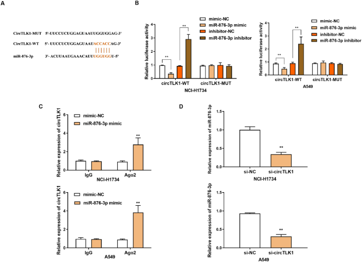
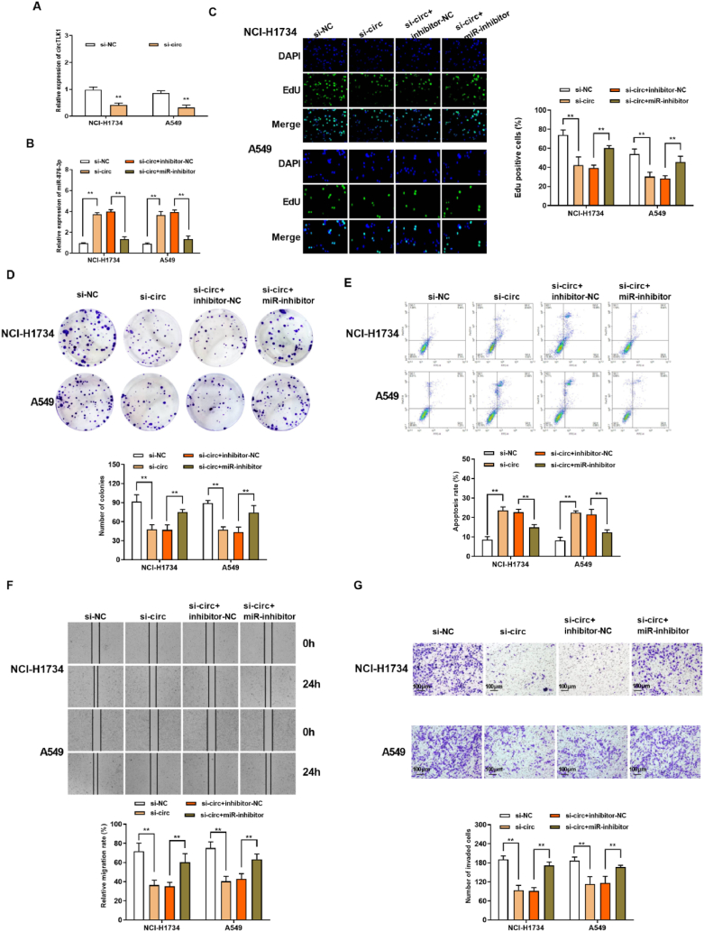
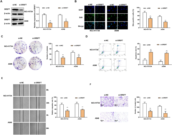
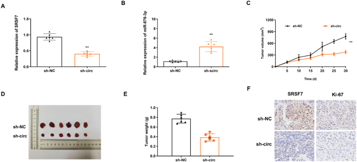
Methods: The expression levels of circTLK1, miR-876-3p and SRSF7 were determined by RT-qPCR assay. The localization of circTLK1 in NSCLC cells was determined by FISH assay. EdU and cell plate clone formation assay were applied to explore cell proliferation. Wound healing test and Transwell assay were applied to measure the migration and invasion ability. Cell apoptosis rate was detected by FCM assay. Western blotting assay was adopted to measure the protein expression of SRSF7. Dual-luciferase reporter gene assay was applied to assess the interaction between miR-876-3p and circTLK1, and between miR-876-3p and SRSF7. The ability of cirTLK1 to regulate tumor formation in vivo was examined by tumor transplantation experiments in nude mice.
Results: The relative expression of circTLK1 was increased in NSCLC cell lines. Knockdown of circTLK1 prohibited the proliferation, migration, and invasion, and promoted apoptosis rate, but miR-876-3p inhibitor reversed the effects of circTLK1 knockdown. In addition, silencing of circTLK1 overtly restrained the growth of transplanted tumors in vivo, and inhibited immune escape. In addition, circTLK1 interacted with miR-876-3p, and SRSF7 was concluded to be the target gene of miR-876-3p.
Conclusion: In this study, we researched the inhibitory effect of circTLK1knockdown on NSCLC progression and immune escape, and further elucidated the potential regulatory mechanism of circTLK1/miR876-3p/SRSF7 axis.
Keywords: SRSF7; hsa_circTLK1; miR-876-3p; non-small cell lung cancer.
© 2024 The Authors. Published by Elsevier Ltd.
Conflict of interest statement
The authors declare that they have no known competing financial interests or personal relationships that could have appeared to influence the work reported in this paper.
Figures


Similar articles
-
circTLK1 facilitates the proliferation and metastasis of renal cell carcinoma by regulating miR-495-3p/CBL axis.Open Life Sci. 2021 Apr 15;16(1):362-374. doi: 10.1515/biol-2021-0041. eCollection 2021. Open Life Sci. 2021. PMID: 33954256 Free PMC article.
-
Circ_0006006 facilitates non-small cell lung cancer progression by modulating miR-924/SRSF7 axis.J Gene Med. 2022 May;24(5):e3411. doi: 10.1002/jgm.3411. Epub 2022 Mar 21. J Gene Med. 2022. PMID: 35037349
-
Long non-coding RNA MALAT1 regulates proliferation, apoptosis, migration and invasion via miR-374b-5p/SRSF7 axis in non-small cell lung cancer.Eur Rev Med Pharmacol Sci. 2020 Feb;24(4):1853-1862. doi: 10.26355/eurrev_202002_20363. Eur Rev Med Pharmacol Sci. 2020. PMID: 32141554
-
Circ_0001715 Functions as a miR-1249-3p Sponge to Accelerate the Progression of Non-small Cell Lung Cancer via Upregulating the Level of FGF5.Biochem Genet. 2023 Oct;61(5):1807-1826. doi: 10.1007/s10528-023-10344-6. Epub 2023 Feb 21. Biochem Genet. 2023. PMID: 36808266
-
Circ_0074027 contributes to non-small cell lung cancer progression through positively modulating RHOA via sequestering miR-2467-3p.J Bioenerg Biomembr. 2021 Apr;53(2):223-233. doi: 10.1007/s10863-021-09876-6. Epub 2021 Mar 9. J Bioenerg Biomembr. 2021. PMID: 33687619
References
-
- Siegel R.L., Miller K.D., Fuchs H.E., Jemal A. Cancer statistics, 2022. CA A Cancer J. Clin. 2022;72(1):7–33. - PubMed
-
- Siegel R.L., Miller K.D., Wagle N.S., Jemal A. Cancer statistics. CA A Cancer J. Clin. 2023;73(1):17–48. 2023. - PubMed
-
- Sung H., Ferlay J., Siegel R.L., Laversanne M., Soerjomataram I., Jemal A., Bray F. Global cancer statistics 2020: GLOBOCAN Estimates of incidence and mortality worldwide for 36 cancers in 185 countries. CA A Cancer J. Clin. 2021;71(3):209–249. - PubMed
-
- Xing P.Y., Zhu Y.X., Wang L., Hui Z.G., Liu S.M., Ren J.S., Zhang Y., Song Y., Liu C.C., Huang Y.C., et al. What are the clinical symptoms and physical signs for non-small cell lung cancer before diagnosis is made? A nation-wide multicenter 10-year retrospective study in China. Cancer Med. 2019;8(8):4055–4069. - PMC - PubMed
LinkOut - more resources
Full Text Sources
