

Case Report: Reversal and subsequent return of optic disc cupping in a myocilin (MYOC) gene-associated severe Juvenile Open-Angle Glaucoma (JOAG) patient
- PMID: 38868171
- PMCID: PMC11167334
- DOI: 10.12688/f1000research.127871.1
Case Report: Reversal and subsequent return of optic disc cupping in a myocilin (MYOC) gene-associated severe Juvenile Open-Angle Glaucoma (JOAG) patient
Abstract
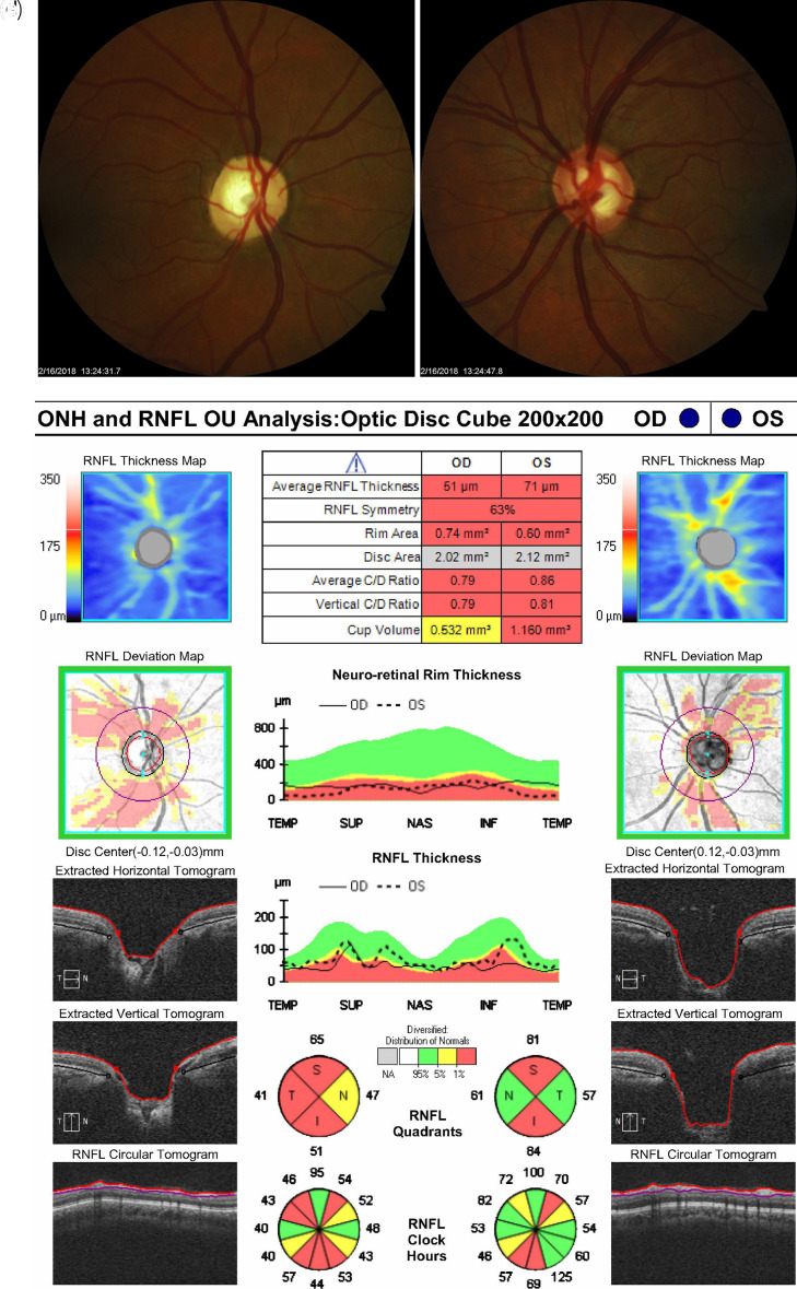
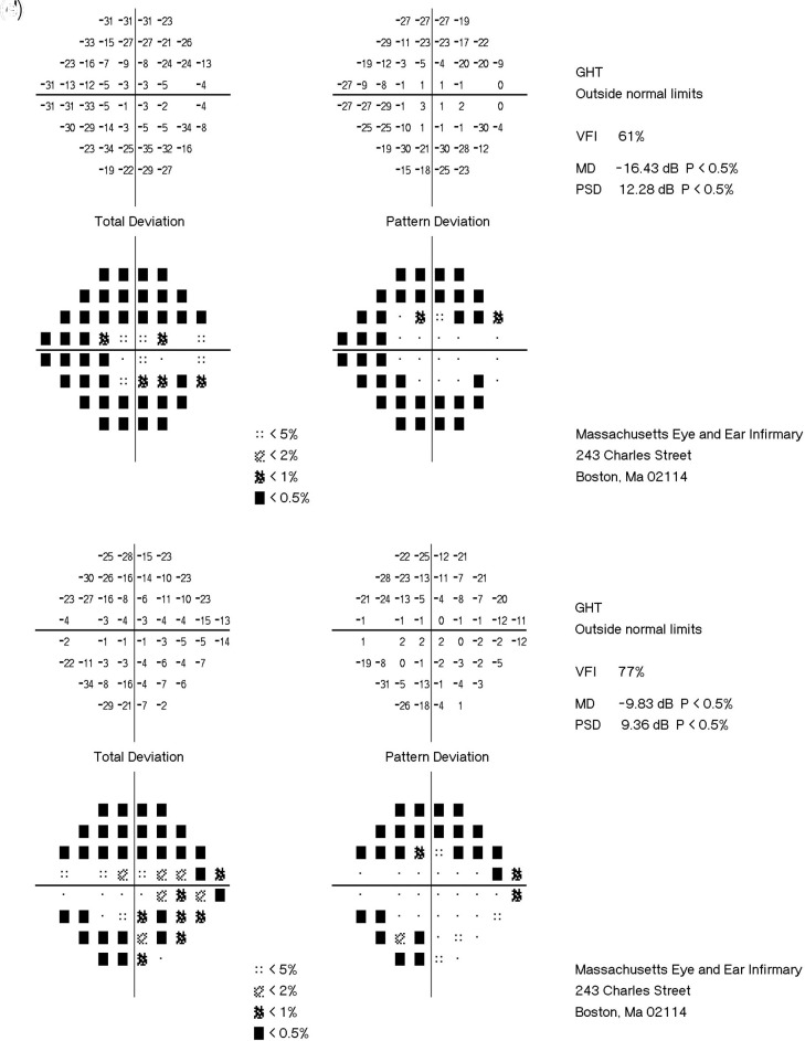
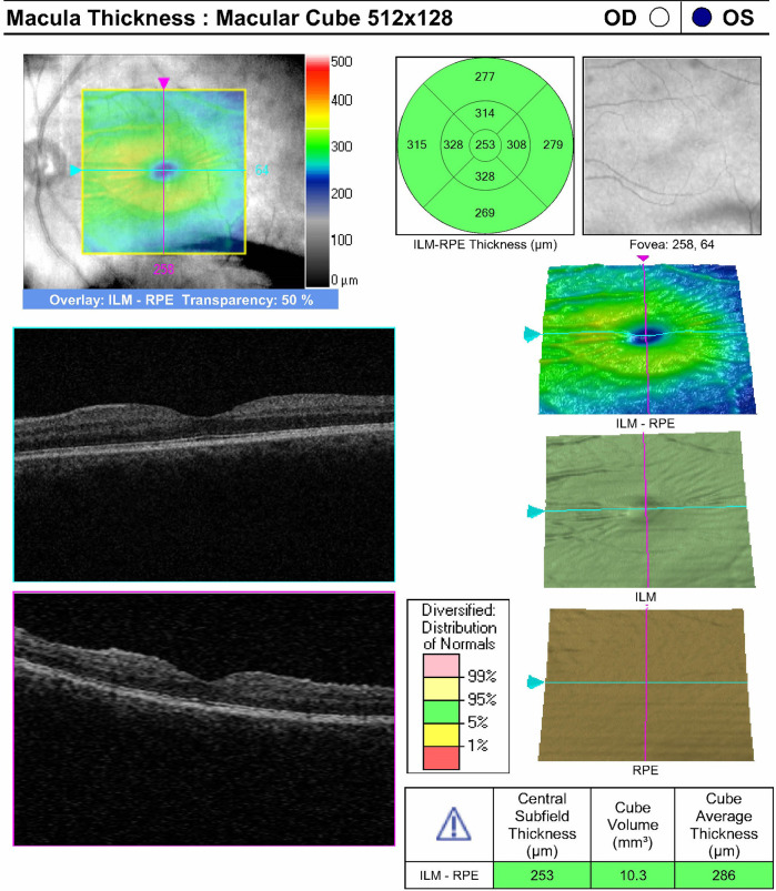
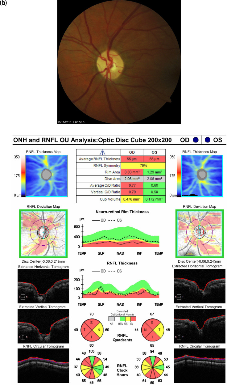
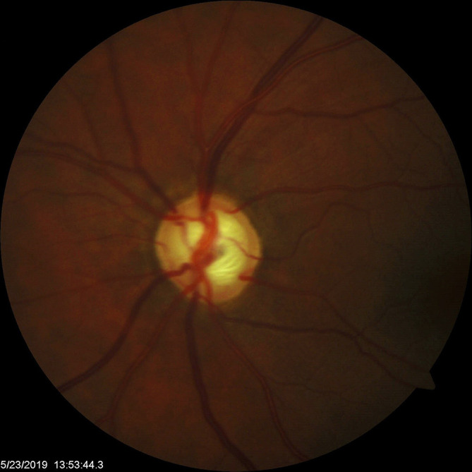
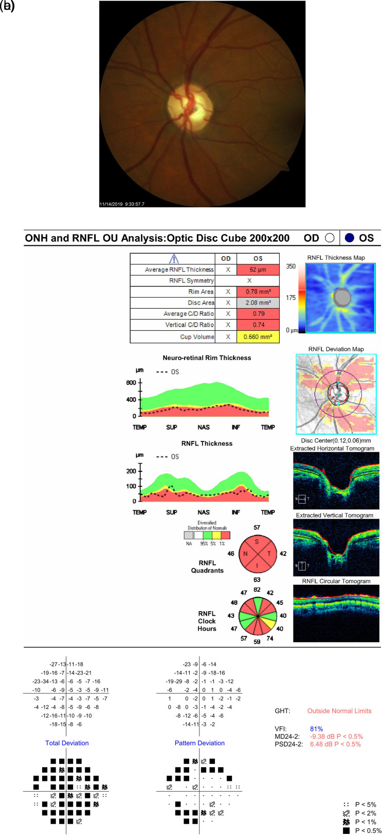
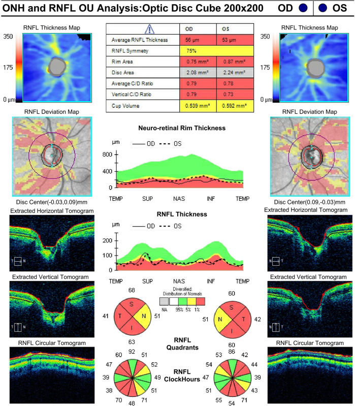
To our knowledge, this case report describes the first instance of reversal of glaucomatous optic nerve cupping in a young adult with a rare form of juvenile open-angle glaucoma (JOAG) associated with a novel variant of the myocilin gene (MYOC). This 25-year-old woman with severe-stage MYOC-associated JOAG presented with blurry vision and intermittent pain in her left eye. She had a strong family history of glaucoma in multiple first-degree relatives with an identified novel variant of MYOC. Examination revealed intraocular pressures (IOPs) of 10 mmHg OD and 46 mmHg OS, with cup-to-disc ratios of 0.90 and 0.80. The patient experienced substantial reversal of optic disc cupping OS following dramatic IOP reduction with trabeculectomy, and subsequently experienced a return of cupping after an IOP spike 15 months postoperatively. The reversal of cupping did not correspond to any changes in the patient's visual field. After an initial decrease in retinal nerve fiber layer (RNFL) thickness, RNFL remained stable for over 2 years after trabeculectomy as seen on Optical Coherence Tomography (OCT). This case suggests reversal of cupping can occur well into adulthood in a MYOC-associated JOAG patient, and it demonstrates the potential bidirectionality of this phenomenon. Moreover, it suggests that these structural changes may not correspond to any functional changes in visual fields or RNFL thickness.
Keywords: Juvenile open-angle glaucoma; Myocilin gene; Reversal of optic nerve head cupping; glaucoma filtration surgery.
Copyright: © 2022 El Helwe H et al.
Conflict of interest statement
Competing interests: Dr. David Solá-Del Valle receives Allergan (an AbbVie company) XEN Gel Stent lecture fees.
Figures
Similar articles
-
Gonioscopy-Assisted Transluminal Trabeculotomy for Myocilin-Associated Juvenile Open-Angle Glaucoma: A Case Series of 8 Eyes Over 2.2 to 4.1 Years.Ophthalmol Glaucoma. 2025 Apr 4:S2589-4196(25)00066-3. doi: 10.1016/j.ogla.2025.03.011. Online ahead of print. Ophthalmol Glaucoma. 2025. PMID: 40188877
-
Cupping reversal in pediatric glaucoma--evaluation of the retinal nerve fiber layer and visual field.Am J Ophthalmol. 2014 Nov;158(5):905-15. doi: 10.1016/j.ajo.2014.07.030. Epub 2014 Jul 25. Am J Ophthalmol. 2014. PMID: 25068638
-
Juvenile Glaucoma.2024 Aug 8. In: StatPearls [Internet]. Treasure Island (FL): StatPearls Publishing; 2025 Jan–. 2024 Aug 8. In: StatPearls [Internet]. Treasure Island (FL): StatPearls Publishing; 2025 Jan–. PMID: 32965934 Free Books & Documents.
-
Juvenile open-angle Glaucoma associated with Leber's hereditary optic neuropathy: a case report and literature review.BMC Ophthalmol. 2018 Dec 17;18(1):323. doi: 10.1186/s12886-018-0980-2. BMC Ophthalmol. 2018. PMID: 30558558 Free PMC article. Review.
-
Juvenile-onset open-angle glaucoma - A clinical and genetic update.Surv Ophthalmol. 2022 Jul-Aug;67(4):1099-1117. doi: 10.1016/j.survophthal.2021.09.001. Epub 2021 Sep 16. Surv Ophthalmol. 2022. PMID: 34536459 Free PMC article. Review.
References
Publication types
Associated data
LinkOut - more resources
Full Text Sources